

Superficial cortical landmarks for localization of the hippocampus: Application for temporal lobectomy and amygdalohippocampectomy
- PMID: 25709853
- PMCID: PMC4322378
- DOI: 10.4103/2152-7806.150663
Superficial cortical landmarks for localization of the hippocampus: Application for temporal lobectomy and amygdalohippocampectomy
Abstract
Background: Accessing the hippocampus for amygdalohippocampectomy and procedures such as depth electrode placement requires accurate knowledge regarding the location of the hippocampus.
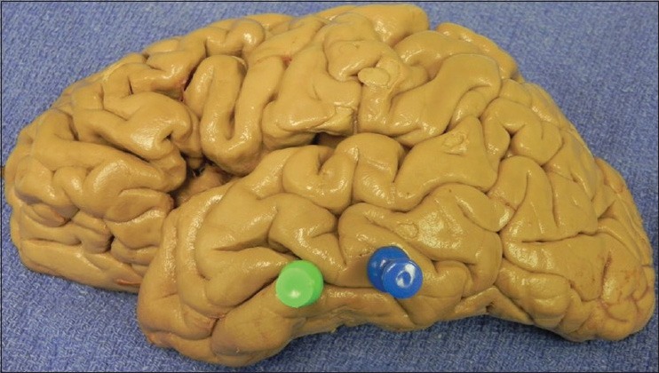
Methods: The authors removed 10 human cadaveric brains (20 sides) from their crania, noted relationships between the lateral temporal neocortex and underlying hippocampus, and measured the distance between the hippocampus and superficial landmarks.
Results: Mean distances were as follows: 3.8 cm from the tip of the temporal lobe to the head of the hippocampus; 6.5 cm from the tip of the temporal lobe to the junction of the fornix and hippocampus; and 3.5 cm between the tail and head of the hippocampus. The head of the hippocampus ranged from 0 to 5 mm inferior to the inferior temporal sulcus. The tail of the hippocampus ranged from 2.2 to 7 mm superior to the inferior temporal sulcus. In two specimens, the tail was deep to the superior temporal sulcus. Generally the length of the hippocampus was along the inferior temporal sulcus and inferior aspect of the middle temporal gyrus. The hippocampus tended to be more superiorly located and shorter in females and left sides, but this was not statistically significant.
Conclusions: Additional landmarks for localizing the underlying hippocampus may be helpful in temporal lobe surgery. Our study showed relatively constant anatomic landmarks between the hippocampus and overlying temporal cortex that may help localize the hippocampus during amygdalohippocampectomy and depth electrode implantation, verify the accuracy of image-guided methods, and used as adjuvant methodologies when these latter technologies are unavailable.
Keywords: Anatomy; epilepsy surgery; hippocampectomy; landmarks; neurosurgery; temporal lobe.
Figures


Similar articles
-
External cortical landmarks for localization of the hippocampus: Application for temporal lobectomy and amygdalohippocampectomy.Surg Neurol Int. 2018 Aug 22;9:171. doi: 10.4103/sni.sni_446_17. eCollection 2018. Surg Neurol Int. 2018. PMID: 30210904 Free PMC article.
-
External cortical landmarks and measurements for the temporal horn: Anatomic study with application to surgery of the temporal lobe.Surg Neurol Int. 2015 Feb 3;6:17. doi: 10.4103/2152-7806.150669. eCollection 2015. Surg Neurol Int. 2015. PMID: 25709854 Free PMC article.
-
Transcortical selective amygdalohippocampectomy technique through the middle temporal gyrus revisited: An anatomical study laboratory investigation.J Clin Neurosci. 2016 Dec;34:237-245. doi: 10.1016/j.jocn.2016.05.035. Epub 2016 Aug 4. J Clin Neurosci. 2016. PMID: 27499121
-
Selective amygdalohippocampectomy: the trans-middle temporal gyrus approach.Neurosurg Focus. 2008 Sep;25(3):E4. doi: 10.3171/FOC/2008/25/9/E4. Neurosurg Focus. 2008. PMID: 18759628 Review.
-
Selective Amygdalohippocampectomy.Neurosurg Clin N Am. 2016 Jan;27(1):1-17. doi: 10.1016/j.nec.2015.08.009. Neurosurg Clin N Am. 2016. PMID: 26615103 Review.
References
-
- Ayberk G, Yagli OE, Comert A, Esmer AF, Canturk N, Tekdemir I, et al. Anatomic relationship between the anterior sylvian point and the pars triangularis. Clin Anat. 2012;25:429–36. - PubMed
-
- Bronen RA, Cheung G. Relationship of hippocampus and amygdala to coronal MRI landmarks. Magn Reson Imaging. 1991;9:449–57. - PubMed
-
- Davies KG, Phillips BL, Hermann BP. MRI confirmation of accuracy of freehand placement of mesial temporal lobe depth electrodes in the investigation of intractable epilepsy. Br J Neurosurg. 1996;10:175–8. - PubMed
-
- Duvernoy HM. Berlin, Heidelberg, New York: Springer-Verlag; 2005. The Human Hippocampus.
-
- Naidich TP, Daniels DL, Haughton VM, Williams A, Pojunas K, Palacios E. Hippocampal formation and related structures of the limbic lobe: Anatomic-MR correlation. Part. 1. Surface features and coronal sections. Radiology. 1987;162:747–54. - PubMed
LinkOut - more resources
Full Text Sources
Other Literature Sources
