

Activated complement classical pathway in a murine model of oxygen-induced retinopathy
- PMID: 25709901
- PMCID: PMC4325235
- DOI: 10.3980/j.issn.2222-3959.2015.01.03
Activated complement classical pathway in a murine model of oxygen-induced retinopathy
Abstract
Aim: To investigate whether the complement system is involved in a murine model of oxygen-induced retinopathy (OIR).
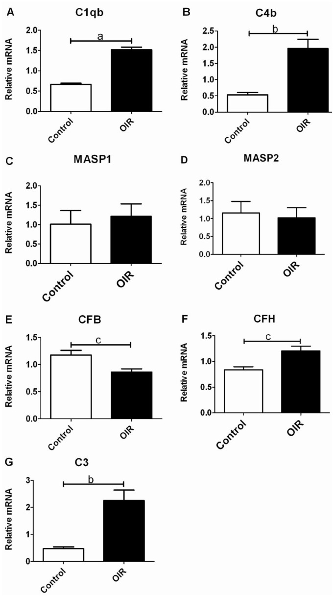
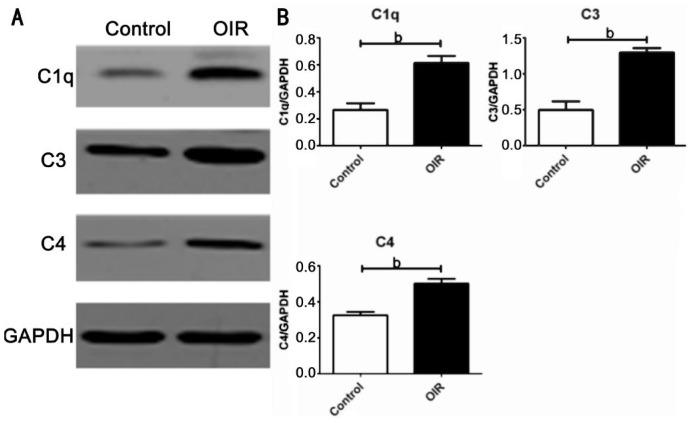
Methods: Forty C57BL/6J newborn mice were divided randomly into OIR group and control group. OIR was induced by exposing mice to 75%±2% oxygen from postnatal 7d (P7) to P12 and then recovered in room air. For the control group, the litters were raised in room air. At the postnatal 17d (P17), gene expressions of the complement components of the classical pathway (CP), the mannose-binding lectin (MBL) pathway and the alternative pathway (AP) in the retina were determined by quantitative real-time polymerase chain reaction (RT-PCR). Retinal protein expressions of the key components in the CP were examined by Western blotting.
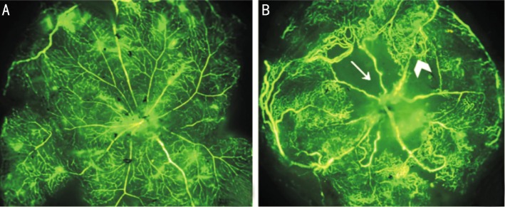
Results: Whole mounted retina in the OIR mice showed area of central hypoperfusion in both superficial and deep layers and neovascular tufts in the periphery. The expressions of C1qb and C4b genes in the OIR retina were significantly higher than those of the controls. The expression of retinal complement factor B (CFB) gene in OIR mice was significantly lower than those of the controls. However, the expressions of C3 and complement factor H (CFH) genes were higher. The protein synthesis of the key components involved in the CP (C1q, C4 and C3) were also significantly higher in OIR mouse retina. Although MBL-associated serine protease 1 (MASP1) and MASP2 were detected in both the OIR and the control groups, the expressions were weak and the difference between the two groups was not significant.
Conclusion: Our data suggest that the complement system CP is activated during the pathogenesis of murine model of OIR.
Keywords: classical pathway; complement activation; mouse; oxygen-induced retinopathy; retina.
Figures
Similar articles
-
[Alternation of retinal complement system in a mouse model of lipopolysaccharide- induced uveitis].Nan Fang Yi Ke Da Xue Xue Bao. 2014 Jul;34(8):1121-4. Nan Fang Yi Ke Da Xue Xue Bao. 2014. PMID: 25176078 Chinese.
-
Complement gene expression and regulation in mouse retina and retinal pigment epithelium/choroid.Mol Vis. 2011;17:1588-97. Epub 2011 Jun 14. Mol Vis. 2011. PMID: 21738388 Free PMC article.
-
Anti-angiogenic and anti-inflammatory effects of CD200-CD200R1 axis in oxygen-induced retinopathy mice model.Inflamm Res. 2019 Nov;68(11):945-955. doi: 10.1007/s00011-019-01276-2. Epub 2019 Aug 23. Inflamm Res. 2019. PMID: 31444514
-
CCR7/p-ERK1/2/VEGF signaling promotes retinal neovascularization in a mouse model of oxygen-induced retinopathy.Int J Ophthalmol. 2017 Jun 18;10(6):862-869. doi: 10.18240/ijo.2017.06.06. eCollection 2017. Int J Ophthalmol. 2017. PMID: 28730075 Free PMC article.
-
Spatial distribution of CD115+ and CD11b+ cells and their temporal activation during oxygen-induced retinopathy in mice.Graefes Arch Clin Exp Ophthalmol. 2018 Feb;256(2):313-323. doi: 10.1007/s00417-017-3845-0. Epub 2017 Nov 28. Graefes Arch Clin Exp Ophthalmol. 2018. PMID: 29185100
Cited by
-
Complement-Mediated Microglial Phagocytosis and Pathological Changes in the Development and Degeneration of the Visual System.Front Immunol. 2020 Sep 24;11:566892. doi: 10.3389/fimmu.2020.566892. eCollection 2020. Front Immunol. 2020. PMID: 33072106 Free PMC article. Review.
-
New insight into the role of the complement in the most common types of retinopathy-current literature review.Int J Ophthalmol. 2018 Nov 18;11(11):1856-1864. doi: 10.18240/ijo.2018.11.19. eCollection 2018. Int J Ophthalmol. 2018. PMID: 30450319 Free PMC article. Review.
References
-
- Smith LE, Wesolowski E, McLellan A, Kostyk SK, D'Amato R, Sullivan R, D'Amore PA. Oxygen-induced retinopathy in the mouse. Invest Ophthalmol Vis Sci. 1994;35(1):101–111. - PubMed
LinkOut - more resources
Full Text Sources
Miscellaneous