

Form-deprivation myopia induces decreased expression of bone morphogenetic protein-2, 5 in guinea pig sclera
- PMID: 25709905
- PMCID: PMC4325239
- DOI: 10.3980/j.issn.2222-3959.2015.01.07
Form-deprivation myopia induces decreased expression of bone morphogenetic protein-2, 5 in guinea pig sclera
Abstract
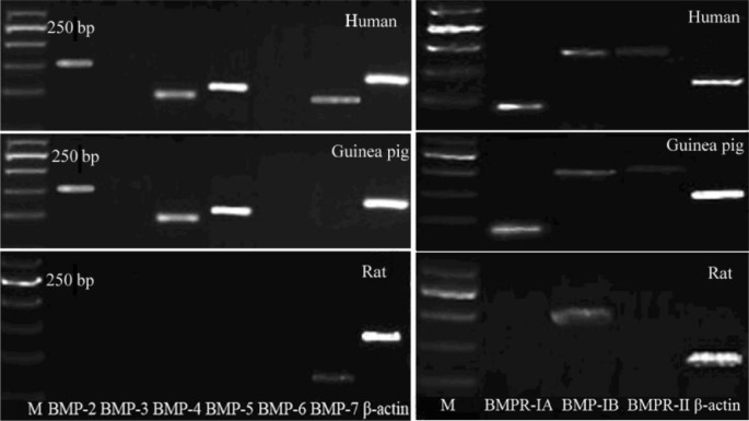
Aim: To identify the presence of various bone morphogenetic proteins (BMPs) and their receptors in normal sclera of human, rat and guinea pigs, and to determine whether their expression changed with form-deprivation myopia (FDM) in guinea pig sclera.
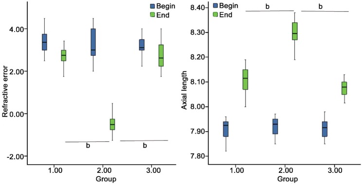
Methods: The expression of BMPs and BMP receptors were detected using reverse transcription polymerase chain reaction (RT-PCR) and immunofluorescence. Two-week-old guinea pigs were monocularly form-deprived with a translucent lens. After fourteen days induction of FDM, total RNA was isolated and subjected to RT-PCR to examine the changes of BMPs and BMP receptors in tissues from the posterior sclera. Western blotting analysis was used to investigate their changes in protein levels.
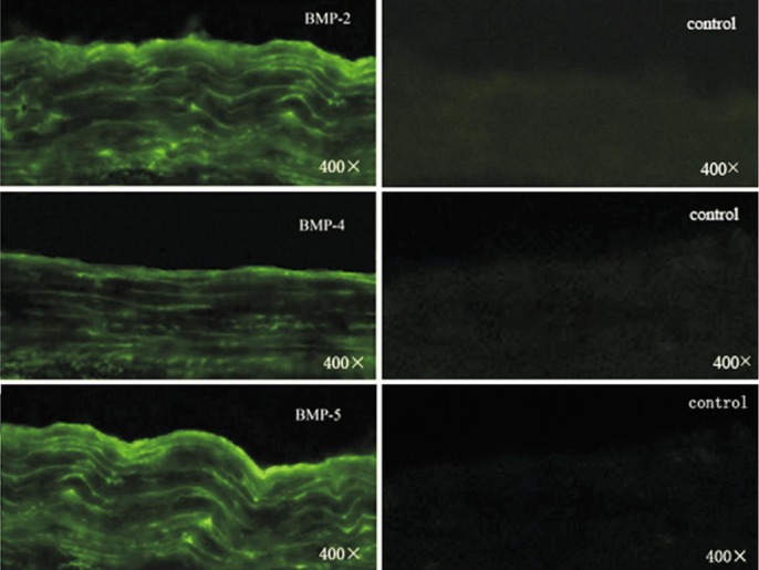
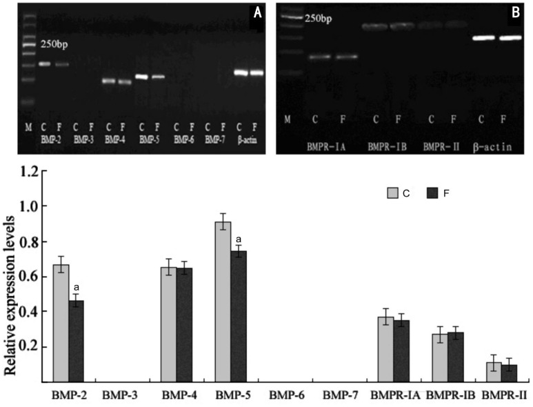
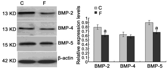
Results: Human sclera expressed mRNAs for BMP-2, -4, -5, -7, -RIA, -RIB and BMP-RII. Conversely, rat sclera only expressed mRNA for BMP-7 and BMP-RIB, while the expression of BMPs and BMP receptors in guinea pigs were similar to that of humans. Human sclera also expresses BMP-2, -4, -5,-7 in protein level. Fourteen days after the induction of myopia, significant decreased expressions for BMP-2 and BMP-5 in the posterior sclera of FDM-affected eyes (P<0.05 vs internal control eyes).
Conclusion: Various BMPs were expressed in human and guinea pig sclera. In the posterior sclera, expressions of BMP-2 and BMP-5 significantly decreased in FDM eyes. This finding indicates that various BMPs as components of the scleral cytokines regulating tissue homeostasis and provide evidence that alterations in the expression of BMP-2 and BMP-5 are associated with sclera remodeling during myopia induction.
Keywords: bone morphogenetic protein; form-deprivation myopia; guinea pig sclera; human sclera.
Figures
Similar articles
-
Role of bone morphogenetic proteins in form-deprivation myopia sclera.Mol Vis. 2011 Mar 8;17:647-57. Mol Vis. 2011. PMID: 21403850 Free PMC article.
-
Changes in muscarinic acetylcholine receptor expression in form deprivation myopia in guinea pigs.Mol Vis. 2007 Jul 20;13:1234-44. Mol Vis. 2007. PMID: 17679952
-
Dynamic Changes of AREG in the Sclera during the Development of Form-Deprivation Myopia in Guinea Pigs.Curr Eye Res. 2022 Mar;47(3):477-483. doi: 10.1080/02713683.2021.1998543. Epub 2021 Nov 12. Curr Eye Res. 2022. PMID: 34766531
-
Bone morphogenetic proteins.Growth Factors. 2004 Dec;22(4):233-41. doi: 10.1080/08977190412331279890. Growth Factors. 2004. PMID: 15621726 Review.
-
Regulation of scleral fibroblast differentiation by bone morphogenetic protein-2.Int J Ophthalmol. 2014 Feb 18;7(1):152-6. doi: 10.3980/j.issn.2222-3959.2014.01.28. eCollection 2014. Int J Ophthalmol. 2014. PMID: 24634882 Free PMC article. Review.
Cited by
-
Harnessing the potential of recombinant human BMP2: regulating scleral changes in myopic guinea pigs.Front Med (Lausanne). 2025 May 1;12:1526656. doi: 10.3389/fmed.2025.1526656. eCollection 2025. Front Med (Lausanne). 2025. PMID: 40375931 Free PMC article.
-
Single-Cell RNA Sequencing of Rabbit Sclera at Different Developmental Stages: Unveiling Scleral Cells Atlas and the Heterogeneity of Fibroblasts.Invest Ophthalmol Vis Sci. 2025 Jun 2;66(6):83. doi: 10.1167/iovs.66.6.83. Invest Ophthalmol Vis Sci. 2025. PMID: 40576432 Free PMC article.
-
Gene Expression Signatures of Contact Lens-Induced Myopia in Guinea Pig Retinal Pigment Epithelium.Invest Ophthalmol Vis Sci. 2022 Aug 2;63(9):25. doi: 10.1167/iovs.63.9.25. Invest Ophthalmol Vis Sci. 2022. PMID: 36006019 Free PMC article.
-
Dynamic changes of activator protein 1 and collagen I expression in the sclera of myopia guinea pigs.Int J Ophthalmol. 2019 Aug 18;12(8):1272-1276. doi: 10.18240/ijo.2019.08.06. eCollection 2019. Int J Ophthalmol. 2019. PMID: 31456916 Free PMC article.
-
Study of Vesicular Monoamine Transporter 2 in Myopic Retina Using [18F]FP-(+)-DTBZ.Mol Imaging Biol. 2018 Oct;20(5):771-779. doi: 10.1007/s11307-018-1183-1. Mol Imaging Biol. 2018. PMID: 29520623
References
-
- Morgan IG, Ohno-Matsui K, Saw SM. Myopia. Lancet. 2012;379(9827):1739–1748. - PubMed
-
- McBrien NA. Regulation of scleral metabolism in myopia and the role of transforming growth factor-beta. Exp Eye Res. 2013;114:128–140. - PubMed
-
- Rada JA, Shelton S, Norton TT. The sclera and myopia. Exp Eye Res. 2006;82(2):185–200. - PubMed
-
- McBrien NA, Gentle A. Role of the sclera in the development and pathological complications of myopia. Prog Retin Eye Res. 2003;22(3):307–338. - PubMed
-
- McBrien NA, Jobling AI, Gentle A. Biomechanics of the sclera in myopia: extracellular and cellular factors. Optom Vis Sci. 2009;86(1):E23–30. - PubMed
LinkOut - more resources
Full Text Sources