The effect of Kisspeptin-10 on mesenchymal stem cells migration in vitro and in vivo
- PMID: 25709985
- PMCID: PMC4333426
- DOI: 10.4103/2277-9175.149851
The effect of Kisspeptin-10 on mesenchymal stem cells migration in vitro and in vivo
Abstract
Background: Kisspeptins (kp) activate a receptor coupled to a Gαq subunit (GPR54 or KiSS-1R) receptor to perform a variety of functions, including inhibition of cell motility, chemotaxis, and metastasis. In this study we have investigated whether kp-10, the most potent member of the kisspeptin family, can modulate CXCR4 (C-X-C chemokine receptor type 4) expression and mesenchymal stem cells (MSCs) migration that may influence the development of tumors.
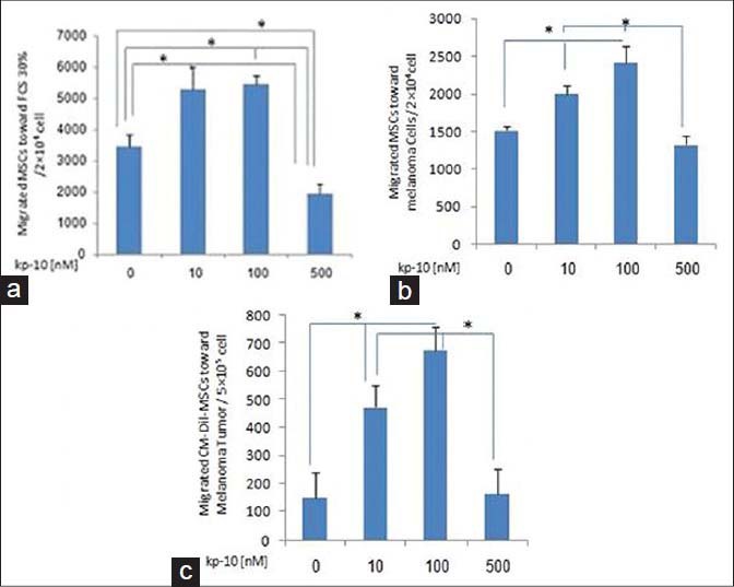
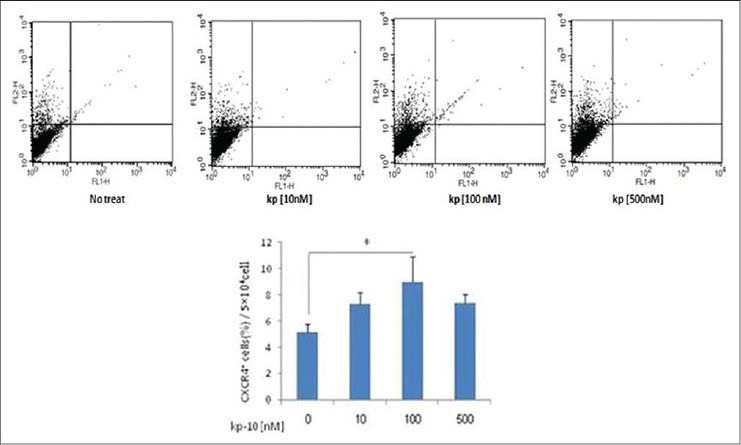
Materials and methods: We compared the directional migration of MSCs treated with 10-100 or 500 nM kp-10 for 24 hours and no treated cells using an in vitro transmembrane migration assay. In addition, Chloromethylbenzamido Dialkylacarbocyanine (CM-Dil) labeled adipose-derived mesenchymal stem cells treated with 10-100 or 500 nM kp-10 and no treated cells were transfused via the tail vein to the melanoma tumor bearing C57BL/6 mice. After 24 hours, the mice were scarified, the tumors were dissected, and the tumor cell suspensions were analyzed by flow cytometry for detection of CM-Dil(+) MSCs.
Results: We have found that kp-10 increased the MSCs migration at 100 nM, while it decreased the MSCs migration at 500 nM, both in vitro and in vivo, with a significant increase of CXCR4 expression at 100 nM kp-10 compared to the no treated cells, but it had no significant difference between the various concentrations of kp-10.
Conclusion: Thus, our data showed that kp-10 can differently affect MSCs migration in various concentrations, probably through different effects on CXCR4 expression in various concentrations.
Keywords: CXCR4; Kisspeptin-10; mesenchymal stem cell; migration.
Conflict of interest statement
Figures
Similar articles
-
Kisspeptin-10-induced signaling of GPR54 negatively regulates chemotactic responses mediated by CXCR4: a potential mechanism for the metastasis suppressor activity of kisspeptins.Cancer Res. 2005 Nov 15;65(22):10450-6. doi: 10.1158/0008-5472.CAN-05-1757. Cancer Res. 2005. PMID: 16288036
-
The effects of kisspeptin-10 on migration and proliferation of endothelial cell.Adv Biomed Res. 2015 Feb 11;4:41. doi: 10.4103/2277-9175.151250. eCollection 2015. Adv Biomed Res. 2015. PMID: 25789267 Free PMC article.
-
Potent Vasoconstrictor Kisspeptin-10 Induces Atherosclerotic Plaque Progression and Instability: Reversal by its Receptor GPR54 Antagonist.J Am Heart Assoc. 2017 Apr 14;6(4):e005790. doi: 10.1161/JAHA.117.005790. J Am Heart Assoc. 2017. PMID: 28411243 Free PMC article.
-
Intracellular signaling pathways activated by kisspeptins through GPR54: do multiple signals underlie function diversity?Peptides. 2009 Jan;30(1):10-5. doi: 10.1016/j.peptides.2008.07.025. Epub 2008 Aug 15. Peptides. 2009. PMID: 18775460 Review.
-
Roles of the kisspeptin/GPR54 system in pathomechanisms of atherosclerosis.Nutr Metab Cardiovasc Dis. 2020 Jun 9;30(6):889-895. doi: 10.1016/j.numecd.2020.02.017. Epub 2020 Mar 7. Nutr Metab Cardiovasc Dis. 2020. PMID: 32409274 Review.
Cited by
-
The sesquiterpene α-bisabolol in the adipocyte-cancer desmoplastic crosstalk: does it have an action on epithelial-mesenchymal transition mechanisms?Int J Clin Oncol. 2017 Apr;22(2):222-228. doi: 10.1007/s10147-016-1072-z. Epub 2016 Dec 9. Int J Clin Oncol. 2017. PMID: 27942879 Review.
-
Current status and future prospects of mesenchymal stem cell therapy for liver fibrosis.J Zhejiang Univ Sci B. 2016 Nov.;17(11):831-841. doi: 10.1631/jzus.B1600101. J Zhejiang Univ Sci B. 2016. PMID: 27819130 Free PMC article. Review.
-
KISS1 in breast cancer progression and autophagy.Cancer Metastasis Rev. 2019 Sep;38(3):493-506. doi: 10.1007/s10555-019-09814-4. Cancer Metastasis Rev. 2019. PMID: 31705228 Free PMC article. Review.
References
-
- Hung SC, Pochampally RR, Chen SC, Hsu SC, Prockop DJ. Angiogenic effects of human multipotent stromal cell conditioned medium activate the PI3K-Akt pathway in hypoxic endothelial cells to inhibit apoptosis, increase survival, and stimulate angiogenesis. Stem Cells. 2007;25:2363–70. - PubMed
-
- Deak E, Seifried E, Henschler R. Homing pathways of mesenchymal stromal cells (MSCs) and their role in clinical applications. Int Rev Immunol. 2010;29:514–29. - PubMed
LinkOut - more resources
Full Text Sources
Other Literature Sources