. 2015 Feb 24;10(2):e0118459.
doi: 10.1371/journal.pone.0118459. eCollection 2015.

Gene expression in obliterative bronchiolitis-like lesions in 2,3-pentanedione-exposed rats

Affiliations

Gene expression in obliterative bronchiolitis-like lesions in 2,3-pentanedione-exposed rats

Daniel L Morgan et al. PLoS One. .

Abstract

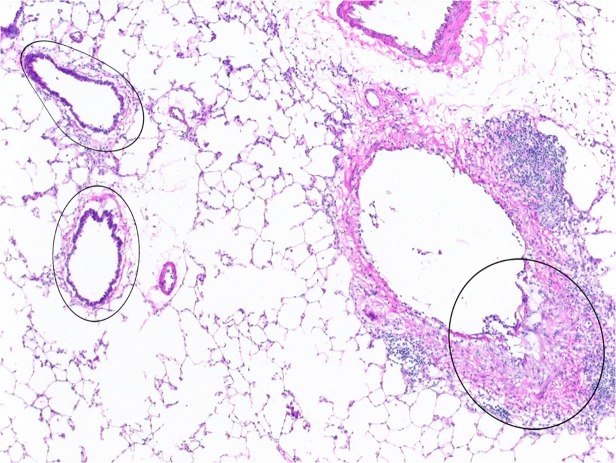
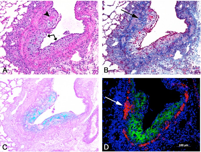
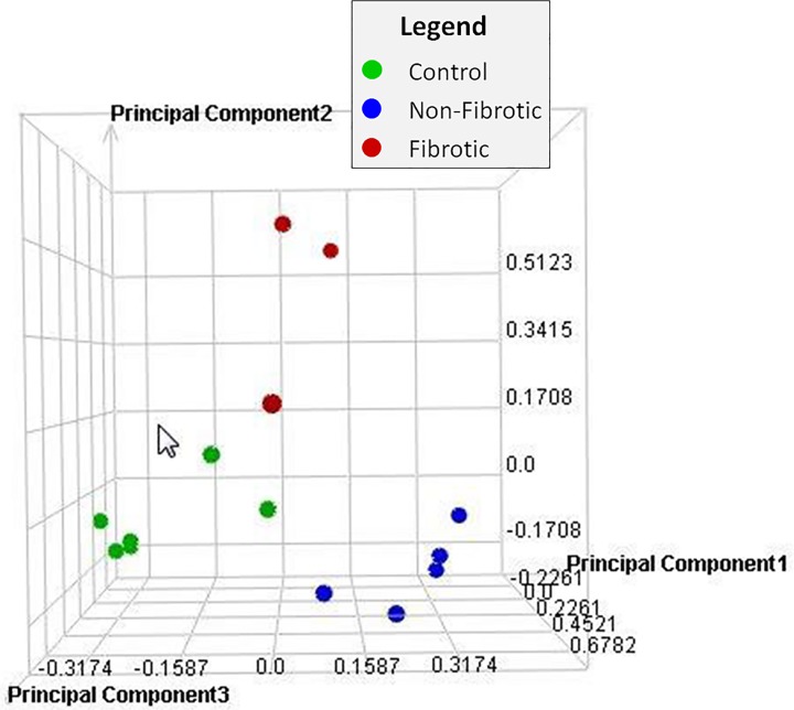
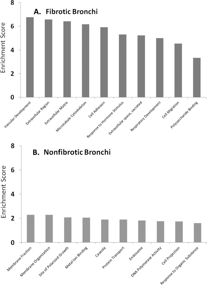
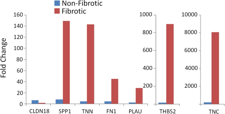
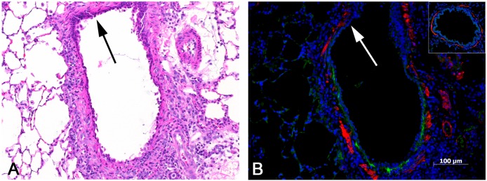
Obliterative bronchiolitis (OB) is an irreversible lung disease characterized by progressive fibrosis in the small airways with eventual occlusion of the airway lumens. OB is most commonly associated with lung transplant rejection; however, OB has also been diagnosed in workers exposed to artificial butter flavoring (ABF) vapors. Research has been limited by the lack of an adequate animal model of OB, and as a result the mechanism(s) is unclear and there are no effective treatments for this condition. Exposure of rats to the ABF component, 2,3-pentanedione (PD) results in airway lesions that are histopathologically similar to those in human OB. We used this animal model to evaluate changes in gene expression in the distal bronchi of rats with PD-induced OB. Male Wistar Han rats were exposed to 200 ppm PD or air 6 h/d, 5 d/wk for 2-wks. Bronchial tissues were laser microdissected from serial sections of frozen lung. In exposed lungs, both fibrotic and non-fibrotic airways were collected. Following RNA extraction and microarray analysis, differential gene expression was evaluated. In non-fibrotic bronchi of exposed rats, 4683 genes were significantly altered relative to air-exposed controls with notable down-regulation of many inflammatory cytokines and chemokines. In contrast, in fibrotic bronchi, 3807 genes were significantly altered with a majority of genes being up-regulated in affected pathways. Tgf-β2 and downstream genes implicated in fibrosis were significantly up-regulated in fibrotic lesions. Genes for collagens and extracellular matrix proteins were highly up-regulated. In addition, expression of genes for peptidases and peptidase inhibitors were significantly altered, indicative of the tissue remodeling that occurs during airway fibrosis. Our data provide new insights into the molecular mechanisms of OB. This new information is of potential significance with regard to future therapeutic targets for treatment.


Conflict of interest statement

Competing Interests: The authors have the following interests. These studies were conducted at the NIEHS inhalation facility under contract to Alion Science and Technology, Inc. There are no patents, products in development, or marketed products to declare. This does not alter the authors' adherence to all the PLOS ONE policies on sharing data and materials, as detailed online in the guide for authors.

Figures

Fig 1
Fig 1. Laser capture microdissection (LCM) of fibrotic and non-fibrotic bronchi.
Microdissection of fibrotic bronchial lesions, such as that indicated by the circled area on the right, was performed on frozen sections. Similarly, exposed but non-fibrotic bronchi, such as the two noted on the left, were microdissected and collected separately. RNA was subsequently extracted from both the fibrotic and the non-fibrotic bronchial specimens, and utilized for microarray and PCR analysis. 4x original objective magnification.
Fig 2
Fig 2. Bronchial fibrosis, intraluminal.
Serial sections of a fibrotic bronchus. A) A plaque-like proliferation of loose, myxoid, amphophilic connective tissue (arrow) projects into and partially obstructs the lumen of this bronchial branch. The fibrotic lesion is covered by an attenuated, regenerating epithelium, and contains focal deposits of eosinophilic fibrin (arrowhead). A mononuclear cell infiltrate is present in the adventitia. H&E. B) The fibrosis is largely devoid of mature collagen, except in the deeper portions of the lesion adjacent to the smooth muscle (arrow). Masson trichrome stain. C) The loose, organizing character of the lesion is highlighted by the blue-green web-like reticular pattern demonstrated by the alcian blue stain, indicative of the presence of acidic mucopolysaccharides. D) Immunofluorescent stains were used to visualize tenascin C and smooth muscle actin. Extensive deposition of tenascin C (in green) is noted throughout much of the fibrotic lesion. The muscle layer of the bronchial wall is demonstrated in red by the smooth muscle actin antibody; focally, the muscular wall appears to be thickened (arrow). All images, 10X original objective magnification.
Fig 3
Fig 3. Principal component analysis.
Principal Component Analysis (PCA) was performed on all samples and all probes to reduce the dimensionality of the data while preserving the variation in the data set. The PCA indicates that although there is some variability within groups, the samples separate by treatment (air or PD) and sample type (PD-exposed fibrotic; PD-exposed nonfibrotic; air-exposed nonfibrotic).
Fig 4
Fig 4. Enriched biological processes.
Differentially expressed transcripts from fibrotic bronchi (A) or exposed, non-fibrotic bronchi (B) were enriched for biological processes and cellular functions using the DAVID functional annotation tool. The combined enrichment score is shown on the y-axis for each of the top ten biological categories on the x-axis.
Fig 5
Fig 5. RT-PCR confirmation of microarray data.
Confirmation of the microarray results was obtained for a subset of genes by quantitative RT-PCR (qPCR). All changes in gene expression measured by qPCR were in agreement with the changes detected by microarray.
Fig 6
Fig 6. Inflamed, non-fibrotic bronchus with tenascin C expression.
A) This bronchus shows no evidence of fibrosis, but exhibits a mild infiltrate of mononuclear cells and is lined by an attenuated epithelium with a reactive and regenerative appearance. Absence of inflammation in the upper portion of bronchus (arrow). H&E, 10x original objective magnification. B) Early tenascin C expression is indicated by the linear green band located just beneath the epithelial lining. Note that there is no tenascin C expression in the upper portion of the bronchus (arrow), in which there is little or no inflammation and less attenuation of the epithelial lining (compare to A). The inset shows a bronchus from one of the control lungs, with absence of any tenascin C expression. Immunofluorescent stains were used to label tenascin C (green) and smooth muscle actin (red).

References

    1. Ryu JH, Myers J, Swenson SJ. Bronchiolar disorders. Am J Resp Crit Care Med. 2003;168(11):1277–92. - PubMed
    1. Kreiss K, Gomaa A, Kullman G, Fedan K, Simoes EJ, Enright PL. Clinical bronchiolitis obliterans in workers at a microwave-popcorn plant. N Engl J Med. 2002;347(5):330–8. . - PubMed
    1. Morgan DL, Jokinen MP, Johnson CL, Gwinn WM, Price HC, Flake GP. Bronchial fibrosis in rats exposed to 2,3-butanedione and 2,3-pentanedione. Toxicologist. 2012;126(1):866.
    1. Weng L, Dai H, Zhan Y, He Y, Stepaniants S, Bassett DE. Rosetta error model for gene expression analysis. Bioinformatics. 2006;22(9):1111–21. - PubMed
    1. Edgar R, Domrachev M, Lash AE. Gene Expression Omnibus: NCBI gene expression and hybridization array data repository. Nucleic Acids Research. 2002;30(1):207–10. - PMC - PubMed

Publication types

MeSH terms