. 2015 Mar 17;54(10):1976-87.
doi: 10.1021/bi501547k. Epub 2015 Mar 2.

Structural basis of activity against aztreonam and extended spectrum cephalosporins for two carbapenem-hydrolyzing class D β-lactamases from Acinetobacter baumannii

Affiliations

Structural basis of activity against aztreonam and extended spectrum cephalosporins for two carbapenem-hydrolyzing class D β-lactamases from Acinetobacter baumannii

Joshua M Mitchell et al. Biochemistry. .

Abstract

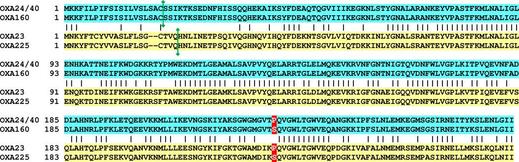
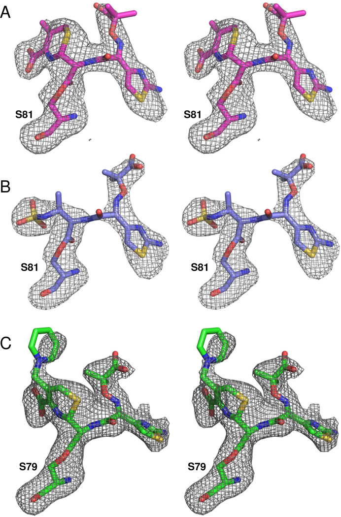
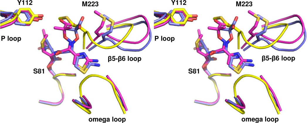
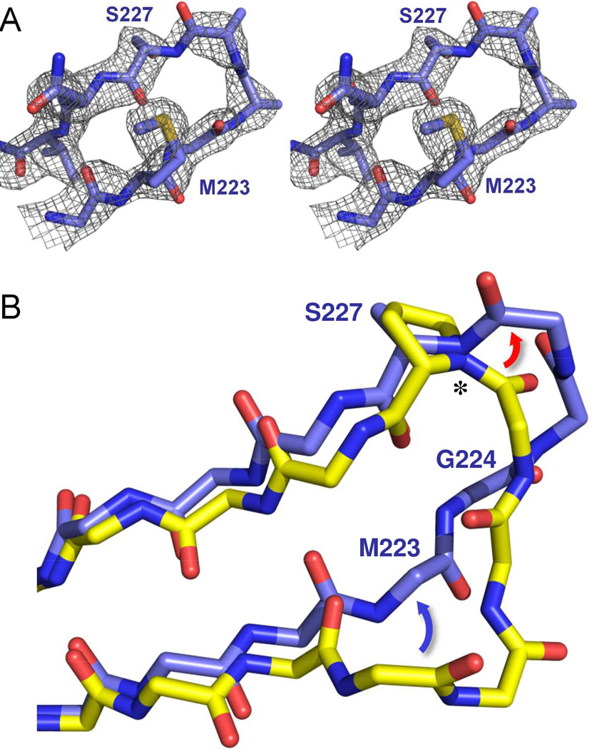
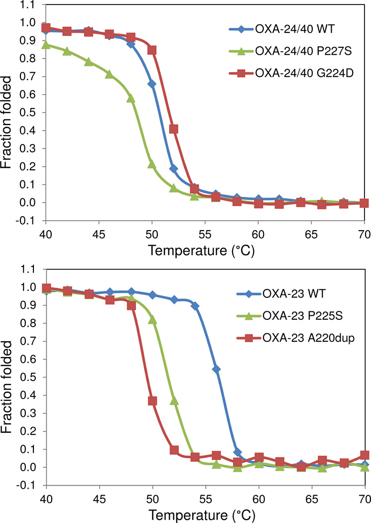
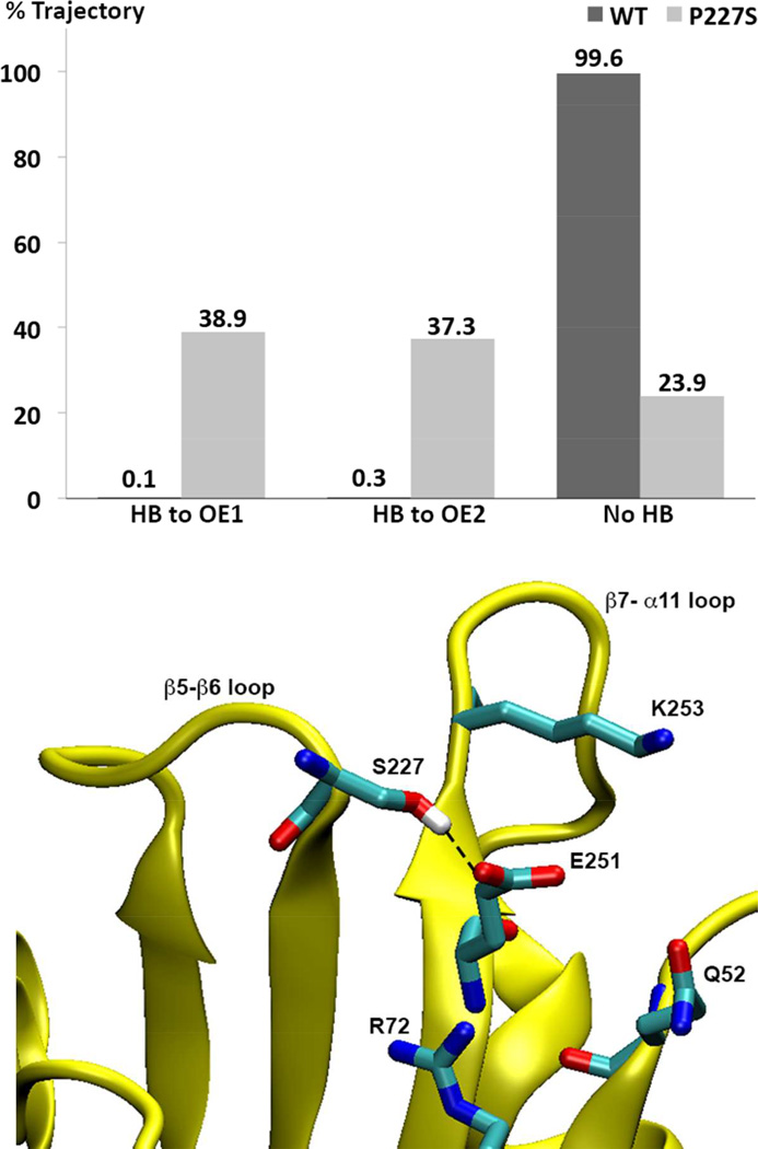
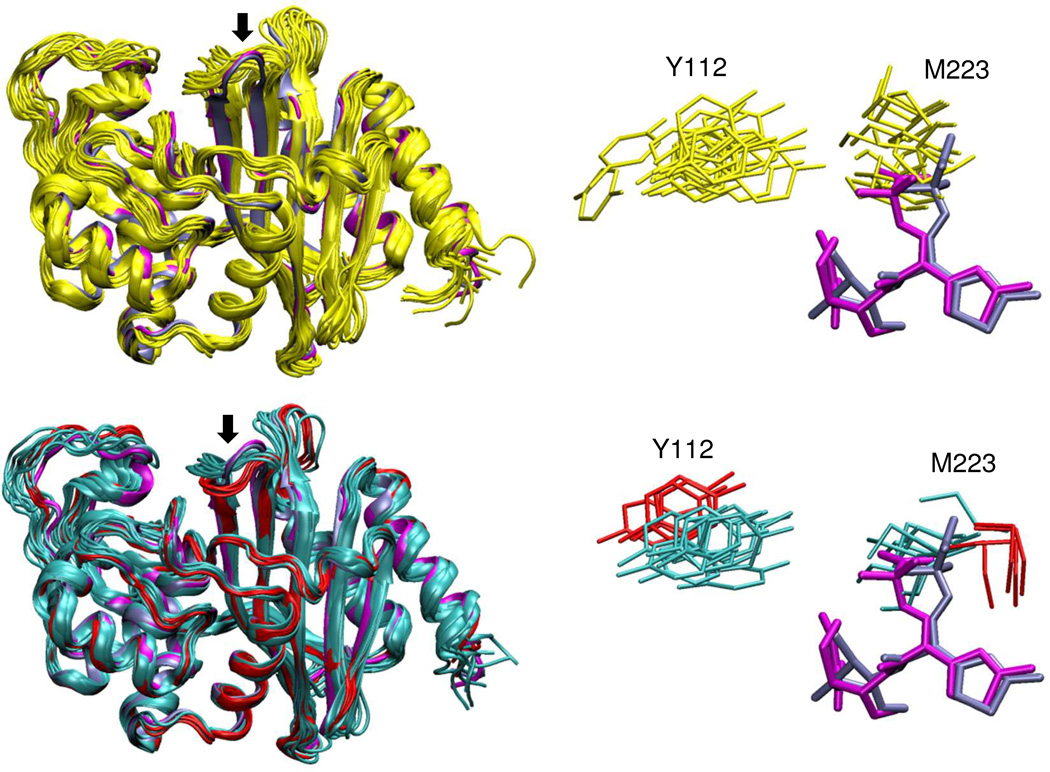
The carbapenem-hydrolyzing class D β-lactamases OXA-23 and OXA-24/40 have emerged worldwide as causative agents for β-lactam antibiotic resistance in Acinetobacter species. Many variants of these enzymes have appeared clinically, including OXA-160 and OXA-225, which both contain a P → S substitution at homologous positions in the OXA-24/40 and OXA-23 backgrounds, respectively. We purified OXA-160 and OXA-225 and used steady-state kinetic analysis to compare the substrate profiles of these variants to their parental enzymes, OXA-24/40 and OXA-23. OXA-160 and OXA-225 possess greatly enhanced hydrolytic activities against aztreonam, ceftazidime, cefotaxime, and ceftriaxone when compared to OXA-24/40 and OXA-23. These enhanced activities are the result of much lower Km values, suggesting that the P → S substitution enhances the binding affinity of these drugs. We have determined the structures of the acylated forms of OXA-160 (with ceftazidime and aztreonam) and OXA-225 (ceftazidime). These structures show that the R1 oxyimino side-chain of these drugs occupies a space near the β5-β6 loop and the omega loop of the enzymes. The P → S substitution found in OXA-160 and OXA-225 results in a deviation of the β5-β6 loop, relieving the steric clash with the R1 side-chain carboxypropyl group of aztreonam and ceftazidime. These results reveal worrying trends in the enhancement of substrate spectrum of class D β-lactamases but may also provide a map for β-lactam improvement.


Figures

Figure 1
Figure 1. The structures of ceftazidime and aztreonam
The structures of the two ligands used to produce acyl-intermediate structures in this study are shown. While ceftazidime (left) is a cephalosporin and aztreonam (right) is a monobactam, these drugs share an identical bulky oxyimino R1 side-chain connected the β-lactam ring.
Figure 2
Figure 2. Alignment of OXA-23, OXA-24/40 and two clinical variants containing a single P→S substitution
The protein sequences for OXA-24/40, OXA-160, OXA-23 and OXA-225 were aligned using ClustalW. Identical residues between the two subfamilies are indicated by vertical hashes, and the site of the P→S substitution is highlighted in red (P227S in OXA-24/40; P225S in OXA-23). The site of proteolytic processing to generate the mature secreted protein is shown by a vertical green line.
Figure 3
Figure 3. Ligand electron density
Fo-Fc omit maps (contoured to 3.0 σ) were calculated to show the structures of the ligand connected as an acyl-intermediate to the nucleophilic serine (S81 in OXA-160 or S79 in OXA-225). (A) OXA-160 V130D bound to ceftazidime (magenta; PDB 4X56). (B) OXA-160 V130D bound to aztreonam (blue; PDB 4X53). (C) OXA-225 K82D bound to ceftazidime (green; PDB 4X55). Figures were made with Pymol.
Figure 4
Figure 4. Comparison of acyl-enzyme intermediates in the active site of OXA-160
Stereoview of the structures of OXA-160 V130D with ceftazidime (magenta; PDB 4X56) and aztreonam (blue; PDB 4X53) bound. To generate the structural alignment, key active site residues from OXA-160 V130D/ceftazidime (S81, S128, K218 and G220) were superposed with the same residues of OXA-24/40 K84D (yellow; PDB 3PAE) using the align function of PyMOL generating a relative mean square deviation (RMSD) of 0.174 Å. This process was repeated for OXA-160 V130D/aztreonam and OXA-24/40 K84D (RMSD, 0.321 Å).
Figure 5
Figure 5. Comparison of the ceftazidime-bound structures of OXA-225 and OXA-160
A stereoview of the aligned structures of OXA-225 K82D (green; PDB 4X55) and OXA-160 V130D with ceftazidime bound as an acyl-enzyme intermediate (magenta; PDB 4X56). In OXA-225, ceftazidime is able to rotate higher in the active site, relieving steric clash with the omega loop in the area of L166. Alignment was carried out as described in Figure 4.
Figure 6
Figure 6. Structure of the β5-β6 loop in OXA-160
(A) An Fo-Fc omit map of the β5-β6 loop (residues 222–229) contoured at 3.0 σ (PDB 4X53). (B) Effect of the P→S mutation on the trajectory of the β5-β6 loop. OXA-24/40 K84D/doripenem (3PAE, yellow) was aligned with OXA-160 V130D/aztreonam (blue; PDB 4X53) as described in Figure 4. Only the main-chain atoms are shown for clarity, with the exception of P/S227. The largest difference between OXA-24/40 and OXA-160 is in the region of G222-T226, with the area of bridge residue M223 moving away from the active site (blue arrow) and the region of T226 extending further from the surface of the enzyme (red arrow) to form a turn. The deviation is accompanied by a large change in the φ torsion angle (*) for residue 227.
Figure 7
Figure 7. Thermal denaturation curves for OXA-23, OXA-24/40 and several of their variants
Unfolding was monitored by slowly raising the temperature of each protein solution and monitoring by circular dichroism. The P→S variant lowered the melting temperature in both the OXA-24/40 background (top panel) and the OXA-23 background (lower panel).
Figure 8
Figure 8. Prevalence of hydrogen bonding between S227 and E251 in OXA-160 and OXA-24/40
Upper panel: For OXA-160 (OXA-24/40 P227S), the hydroxyl oxygen atom of S227 remains within 3.2 Å distance from E251 Oε1 and Oε2 atoms for 38.9% and 37.3% of the time, respectively. In contrast, the corresponding atoms in P227 (Cγ) and E251 in the WT enzyme remain greater than 3.2 Å apart 99.6% of the time. Hydrogen bonding was determined over the 38 ns after equilibration based on the following geometric criteria: D-A < 3.2 Å, H-A < 2.2 Å and DHA > 120°. When hydrogen bonded, the average D-A distance in OXA-160 is 2.7 Å (with an entire trajectory average of 3.8 Å). Lower Panel: A trajectory snapshot of a conformation that displays a typical hydrogen bond between S227 and E251.
Figure 9
Figure 9. Comparison of conformational diversity in OXA-160 and OXA-24/40
Left panels: representative conformers observed in molecular dynamics simulations for OXA-24/40 (yellow) and OXA-160 (two clusters shown in cyan and red). The two OXA-160 structures from this study (PDB 4X53 and 4X56, blue and magenta respectively) are included in the alignment (ligands not shown). The β5-β6 loop is marked with an arrow in both structures. Right panels: Side-chains for the bridge residues Y112 and M223 from the same representative structures. Also shown are ceftazidime (magenta) and aztreonam (blue) from the two OXA-160 X-ray structures after alignment of those structures with the simulation conformers.

Similar articles

Cited by

References

    1. Kamolvit W, Derrington P, Paterson DL, Sidjabat HE. A case of IMP-4, OXA-421, OXA-96 and CARB-2-producing Acinetobacter pittii ST119 in Australia. J. Clin. Microbiol. 2014 in press. - PMC - PubMed
    1. Evans BA, Hamouda A, Amyes SG. The rise of carbapenem-resistant Acinetobacter baumannii. Curr. Pharm. Des. 2013;19:223–238. - PubMed
    1. Golemi D, Maveyraud L, Vakulenko S, Samama JP, Mobashery S. Critical involvement of a carbamylated lysine in catalytic function of class D β-lactamases. Proc. Natl. Acad. Sci. U.S.A. 2001;98:14280–14285. - PMC - PubMed
    1. Sun T, Nukaga M, Mayama K, Braswell EH, Knox JR. Comparison of β-lactamases of classes A and D: 1.5-Å crystallographic structure of the class D OXA-1 oxacillinase. Protein Sci. 2003;12:82–91. - PMC - PubMed
    1. Santillana E, Beceiro A, Bou G, Romero A. Crystal structure of the carbapenemase OXA-24 reveals insights into the mechanism of carbapenem hydrolysis. Proc. Natl. Acad. Sci. U.S.A. 2007;104:5354–5359. - PMC - PubMed

Publication types

LinkOut - more resources