. 2015 Mar 25;137(11):3933-45.
doi: 10.1021/jacs.5b00669. Epub 2015 Mar 11.

Chemical structure and properties of interstrand cross-links formed by reaction of guanine residues with abasic sites in duplex DNA

Affiliations

Chemical structure and properties of interstrand cross-links formed by reaction of guanine residues with abasic sites in duplex DNA

Michael J Catalano et al. J Am Chem Soc. .

Abstract

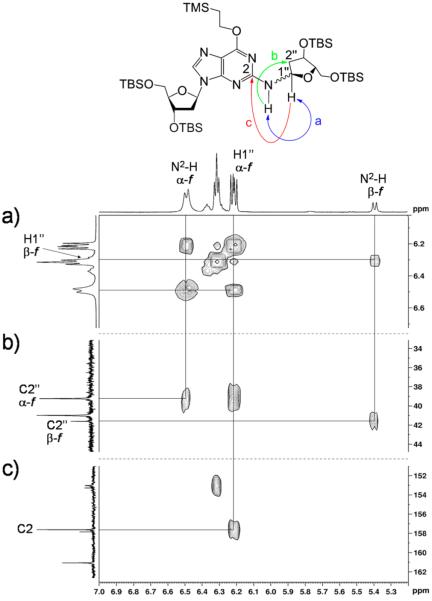
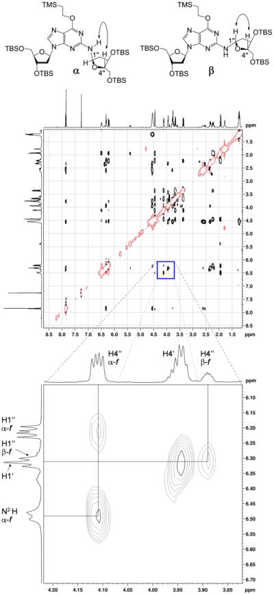
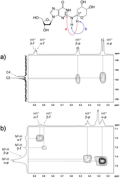
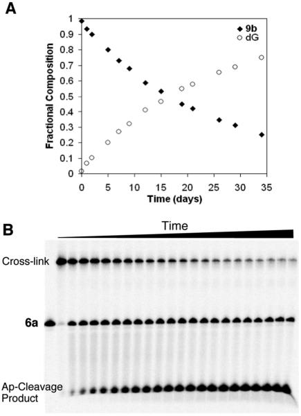
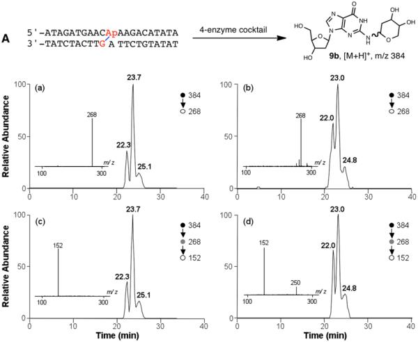
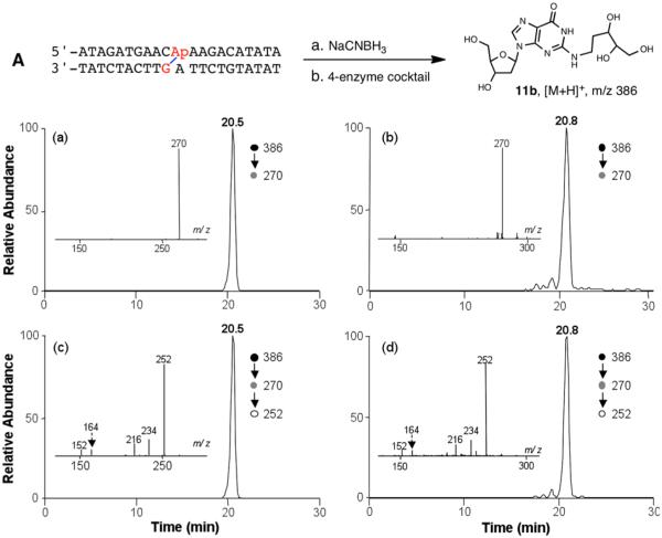
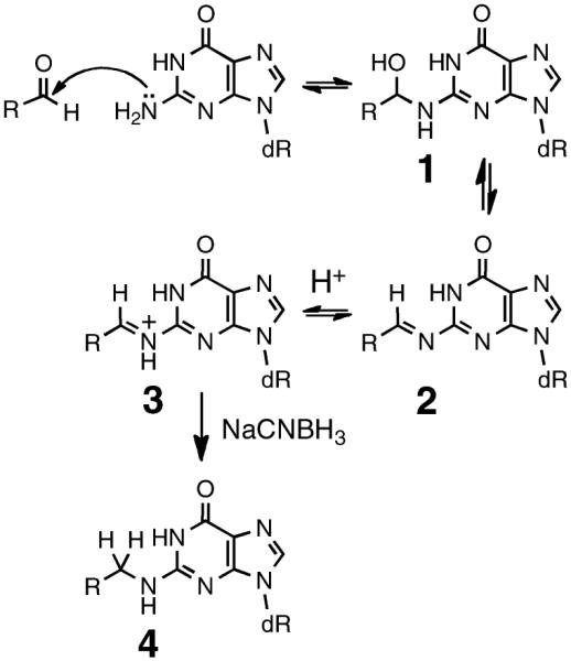
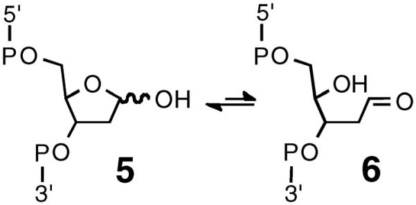
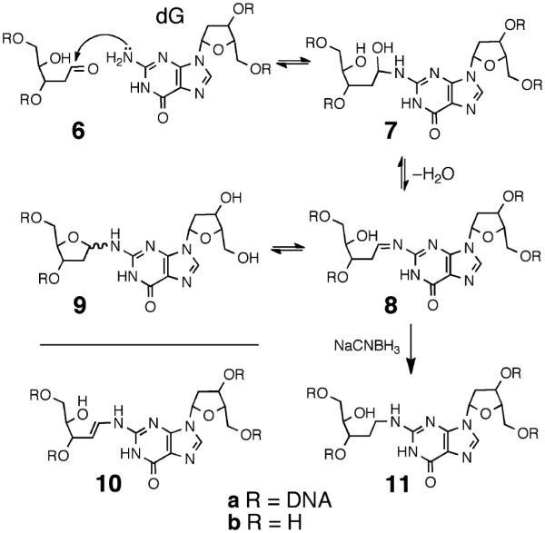
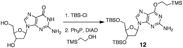
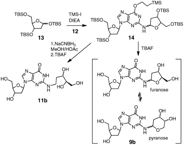
A new type of interstrand cross-link resulting from the reaction of a DNA abasic site with a guanine residue on the opposing strand of the double helix was recently identified, but the chemical connectivity of the cross-link was not rigorously established. The work described here was designed to characterize the chemical structure and properties of dG-AP cross-links generated in duplex DNA. The approach involved characterization of the nucleoside cross-link "remnant" released by enzymatic digestion of DNA duplexes containing the dG-AP cross-link. We first carried out a chemical synthesis and complete spectroscopic structure determination of the putative cross-link remnant 9b composed of a 2-deoxyribose adduct attached to the exocyclic N(2)-amino group of dG. A reduced analogue of the cross-link remnant was also prepared (11b). Liquid chromatography-tandem mass spectrometric (LC-MS/MS) analysis revealed that the retention times and mass spectral properties of synthetic standards 9b and 11b matched those of the authentic cross-link remnants released by enzymatic digestion of duplexes containing the native and reduced dG-AP cross-link, respectively. These results establish the chemical connectivity of the dG-AP cross-link released from duplex DNA and provide a foundation for detection of this lesion in biological samples. The dG-AP cross-link in duplex DNA was remarkably stable, decomposing with a half-life of 22 days at pH 7 and 23 °C. The intrinsic chemical stability of the dG-AP cross-link suggests that this lesion in duplex DNA may have the power to block DNA-processing enzymes involved in transcription and replication.


Figures

Figure 1
Figure 1
Selected regions of 1H–1H COSY and 1H–13C HMBC spectra of 14 in CDCl3 acquired at 500 (1H) and 126 (13C) MHz. Attachment of the 3,5-bis[O-(tert-butyldimethylsilyl)]-2-deoxyribofuranose moiety to N2 of the protected dG was established by (a) homonuclear (COSY) coupling of H1″ to N2-H as well as three-bond HMBC coupling of (b) N2-H to C2″ and (c) H1″ to C2.
Figure 2
Figure 2
1H–1H NOESY spectrum of 14 in CDCl3 acquired at 500 MHz with a mixing time of 800 ms; negative peaks are denoted by red curves. The expanded frame shows the strong through-space correlation between H4″ and N2-H of the major α-furanose isomer. Also shown is a cross-peak between H4″ and H1″ of the minor β-furanose isomer; no correlation between H4″ and N2-H of the minor isomer is evident.
Figure 3
Figure 3
Selected regions of 1H–13C HMBC and 1H–1H COSY spectra of 9b obtained at 500 (1H) and 126 (13C) MHz, respectively. Correlations between (a) H1″ and C2 in D2O as well as (b) H1″ and N2-H in DMSO-d6 indicate that the 2-deoxyribose moiety remained attached to N2 of dG following deprotection.
Figure 4
Figure 4
Stability of dG–AP cross-linkage. (A) Synthetic cross-link remnant 9b was incubated at 23 °C in HEPES buffer (pH 7, 50 mM) containing NaCl (100 mM). Disappearance of 9b and concomitant appearance of dG were monitored by HPLC. (B) Gel electrophoretic analysis of purified, unreduced cross-link in duplex A, incubated at 37 °C in HEPES buffer (50 mM, pH 7) containing NaCl (100 mM). The fast-moving fragment at the bottom of the gel corresponds to the 3′-4-hydroxy-2-pentenal-5-phosphate cleavage product resulting from β-elimination at the AP site.
Figure 5
Figure 5
LC-MS/MS and MS/MS/MS for characterization of the dG–AP cross-link remnant that was obtained synthetically (a and c) or released from purified AP-derived cross-link-containing duplex DNA sequence A (b and d). (a, b) Selected-ion chromatograms for monitoring the loss of 2-deoxyribose (i.e., m/z 384 → 268 transition) from [M + H]+ ion of the unreduced dG–AP cross-link remnant that was (a) obtained synthetically or (b) liberated from the four-enzyme digestion of purified cross-link-containing duplex DNA. (c, d) Corresponding selected-ion chromatograms for monitoring the formation of [M + H]+ ion of guanine (i.e., m/z 384 → 268 → 152 transition) from the ion of m/z 268 observed in MS/MS. Depicted in the insets are the corresponding MS/MS and MS/MS/MS averaged from the retention times of (a, c) 23.7 min and (b, d) 23.0 min. The MS/MS/MS averaged from the two other peaks are displayed in Figure S26. The ion of m/z 250 observed in panel d is attributed to isobaric interference.
Figure 6
Figure 6
LC-MS/MS and MS/MS/MS for characterization of reduced dG–AP cross-link remnant that was obtained synthetically or released from NaCNBH3-treated cross-linked duplex DNA. Shown are selected-ion chromatograms for monitoring the loss of a 2-deoxyribose (i.e., m/z 386 → 270 transition) from the [M + H]+ ion of reduced dG–AP cross-link remnant that was (a) obtained synthetically or (b) released from the 4-enzyme digestion of reduced product of the purified cross-linked duplex A. (c, d) Corresponding selected-ion chromatograms for monitoring the further loss of H2O from the ion at m/z 270 observed in MS/MS (i.e., m/z 386 → 270 → 252 transition). (Insets) Corresponding MS/MS and MS/MS/MS averaged from retention times of (a, c) 20.5 and (b, d) 20.8 min.
Scheme 1
Scheme 1
Scheme 2
Scheme 2
Scheme 3
Scheme 3
Scheme 4
Scheme 4
Scheme 5
Scheme 5

References

    1. Gates KS. In: Comprehensive Natural Products Chemistry. Kool ET, editor. Vol. 7. Pergamon; New York: 1999. pp. 491–552.
    1. Delaney JC, Essigmann JM. Chem. Res. Toxicol. 2008;21:232–252. - PMC - PubMed
    1. Marnett LJ. In: DNA Adducts: Identification and Biological Significance. Hemminki K, Dipple A, Shuker DEG, Kadlubar FF, Segerback D, Bartsch H, editors. Vol. 125. IARC Scientific Publications; Lyon, France: 1994. pp. 151–162.
    1. Wang M, McIntee EJ, Cheng G, Shi Y, Villalta PW, Hecht SS. Chem. Res. Toxicol. 2000;13:1149–1157. - PubMed
    1. Shapiro R, Cohen BI, Shiuey S-J, Maurer H. Biochemistry. 1969;8:238–245. - PubMed

Publication types