Review
. 2015 Feb 23;32(4):390-407.
doi: 10.1016/j.devcel.2015.02.004.

The evolution of early neurogenesis

Affiliations
Review

The evolution of early neurogenesis

Volker Hartenstein et al. Dev Cell. .

Abstract

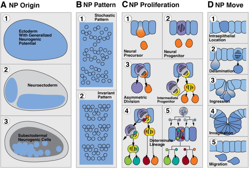
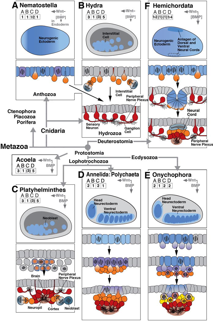
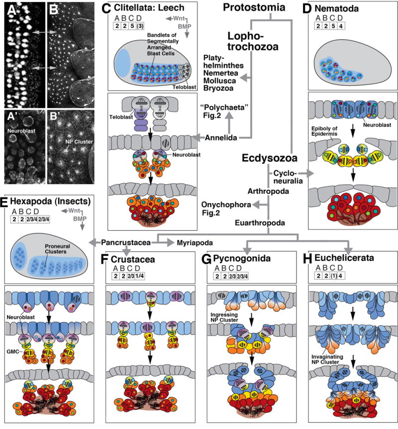
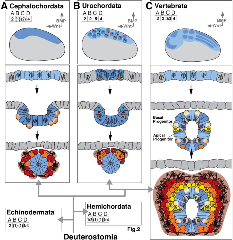
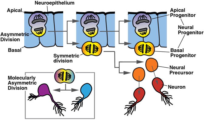
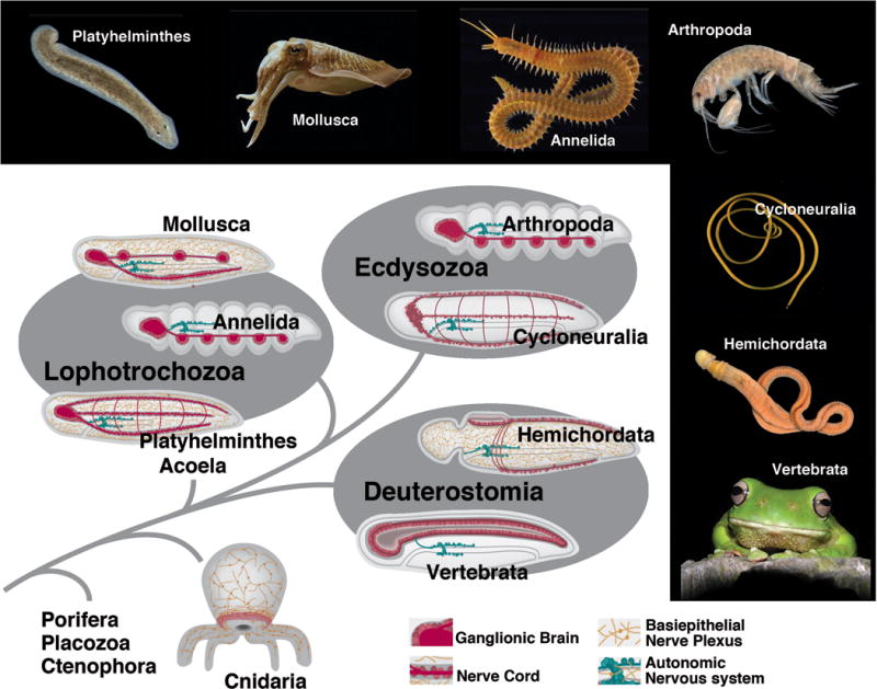
The foundation of the diverse metazoan nervous systems is laid by embryonic patterning mechanisms, involving the generation and movement of neural progenitors and their progeny. Here we divide early neurogenesis into discrete elements, including origin, pattern, proliferation, and movement of neuronal progenitors, which are controlled by conserved gene cassettes. We review these neurogenetic mechanisms in representatives of the different metazoan clades, with the goal to build a conceptual framework in which one can ask specific questions, such as which of these mechanisms potentially formed part of the developmental "toolkit" of the bilaterian ancestor and which evolved later.


Figures

Figure 1
Figure 1
Modular representation of early neurogenesis, focusing on the origin (A), pattern (B), proliferation (C), and movement (D) of neural progenitor (NP) cells. The color coding that is applied in column (C), and maintained throughout the following figures 2–4, reflects the neurogenic potential and proliferative status of a cell: neuroepithelium: blue; actively dividing progenitor: purple; intermediate (basal) progenitors: yellow; undifferentiated precursor that is postmitotic or that has an undetermined, yet limited mitotic potential: orange. Small colored circles in cells C5 and C6 symbolize hypothetical intrinsic determinants that are channeled through asymmetric divisions into distinct neural cells (fixed lineages).
Figure 2
Figure 2
Early neurogenesis in cnidarians and basal bilaterians. In boxes (A)-(F), key events of early neurogenesis are depicted in a schematic, uniform manner for six different animal clades. Represented in the upper panel of each box is a “thumbnail” of an embryo of the corresponding clade at the onset of neurogenesis (side view; anterior to the left, dorsal up). Territories with neurogenic potential are outlined in blue. Role of BMP/BMP antagonist and Wnt signaling pathway, if present, is indicated by vertical and horizontal arrows at top right corner of upper panel. Arrows point in direction of decreasing activity (i.e., away from source) of these morphogens. Numbers in grids (ABCD) at top left capture the elements of early neurogenesis, as shown and explained in text section 1 and Figure 1. Drawings in lower panels of each box show schematic cross sections of embryos at sequential stages of development, capturing spatial characteristics and proliferatory behavior of neural progenitors. The drawings use the color code introduced in Box 1 and Figure 1. As an example, box (A) represents early neurogenesis in the cnidarian Nematostella. The Nematostella embryo shows generalized neurogenic potential all over the ectoderm (# 1 in grid A; colored blue in upper section). Neural cells are scattered stochastically over ectoderm (# 1 in grid B). Ectodermal cells form neural precursors (orange in upper section; #1 in grid C) and differentiate as epithelial, sensory neurons or delaminate to become ganglion cells (both red in bottom section). Ectoderm also contains dividing neural progenitors (# 2 in grid C; purple color in upper section). Based on published reports (Richards and Rentzsch, 2014) progenitors appear to divide in ectoderm (bracketed # 1 in grid D). Bracketing of numbers generally indicates that the implied aspect of neurogenesis is the most likely scenario, based on published data, but needs further confirmation. Bracketing of “BMP” indicates that morphogen is present but excerts no effect on neural organization. The sign “1>2” in grid A and “1>4” in grid D of box (F) signify that during an early embryonic phase of hemichordate neurogenesis, a generalized neurogenic ectoderm gives rise to neural precursors forming a nerve net; this is followed in the later embryo by a phase where the dorsal ectoderm invaginates as the dorsal neural cord, and the ventral ectoderm also gives rise to a ventral cord of higher neuronal density. Phylogenetic relationships between clades, in this and the following figures, are indicated by thick grey lines/arrows connecting the corresponding boxes. The remainder of the clades shown in this figure [(B)-(F)] and the following figures are composed in the manner explained for (A)
Figure 3
Figure 3
Early neurogenesis in derived protostome clades; composition of figure as explained in legend of Figure 2. Lophotrochozoa represented by leeches [Hirudinea, (C)]), a derived branch of the annelids. Within the Ecdysozoa, the branch Cycloneuralia is represented by the nematodes (D). The Arthropoda (E-H) comprise Pancrustacea, including Hexapoda (insects; E) and crustaceans, Myriapoda (centipedes; F), and Checlicerata, including Pycnogonida (sea spiders, G) and Euchelicerata (H). Thumbnail of embryo shown for Hexapoda (E) applies to all other arthropod clades. Early neurogenesis in leech (C) and nematode (D) is characterized by fixed lineages, generated by asymmetric cell division. Positional fate (antero-posterior and dorsoventral) is controlled by intrinsic determinants and local cell-cell interactions, rather than by long range BMP/Wnt gradients. Panels at upper left show horizontal confocal sections of the ventral neuroectoderm of an insect (A: D. melanogaster; A’: T. castaneum) and a spider (B/B’: C. salei), illustrating the similarity between the pattern of individual neuroblasts (insect) and invaginating NEURAL PROGENITOR clusters (spider). Grey arrows in A/B indicate neuromere boundaries.
Figure 4
Figure 4
Early neurogenesis in deuterostomes; composition of figure as explained in legend of Figure 2. Deuterostomes include the basally branching echinoderms, hemichordates (represented in panel (F) of Figure 2), and cephalochordates [(A); lancelets], as well as more derived urochordates [(B); sea squirts] and vertebrates (C). Urochordates show fixed lineages with intrinsically specified neural fates.
Figure Box 1
Figure Box 1
Figure Box 2
Figure Box 2

References

    1. Aguinaldo AM, Turbeville JM, Linford LS, Rivera MC, Garey JR, Raff RA, Lake JA. Evidence for a clade of nematodes, arthropods and other moulting animals. Nature. 1997;387:489–493. - PubMed
    1. Altmann CR, Brivanlou AH. Neural patterning in the vertebrate embryo. Int Rev Cytol. 2001;203:447–482. - PubMed
    1. Angerer LM, Yaguchi S, Angerer RC, Burke RD. The evolution of nervous system patterning: insights from sea urchin development. Development. 2011;138:3613–3623. - PMC - PubMed
    1. Arendt D, Denes AS, Jékely G, Tessmar-Raible K. The evolution of nervous system centralization. Philos Trans R Soc Lond B Biol Sci. 2008;363:1523–1528. - PMC - PubMed
    1. Ashraf SI, Ip YT. The Snail protein family regulates neuroblast expression of inscuteable and string, genes involved in asymmetry and cell division in Drosophila. Development. 2001;128:4757–4767. - PubMed

MeSH terms