

Review
doi: 10.1016/j.devcel.2015.01.016.
Dynamics and mechanisms of CNS myelination
Affiliations
- PMID: 25710531
- PMCID: PMC6715306
- DOI: 10.1016/j.devcel.2015.01.016
Item in Clipboard
Review
Dynamics and mechanisms of CNS myelination
Dev Cell.
.
Abstract
Vertebrate myelination is an evolutionary advancement essential for motor, sensory, and higher-order cognitive function. CNS myelin, a multilamellar differentiation of the oligodendrocyte plasma membrane, ensheaths axons to facilitate electrical conduction. Myelination is one of the most pivotal cell-cell interactions for normal brain development, involving extensive information exchange between differentiating oligodendrocytes and axons. The molecular mechanisms of myelination are discussed, along with new perspectives on oligodendrocyte plasticity and myelin remodeling of the developing and adult CNS.
Copyright © 2015 Elsevier Inc. All rights reserved.
Figures
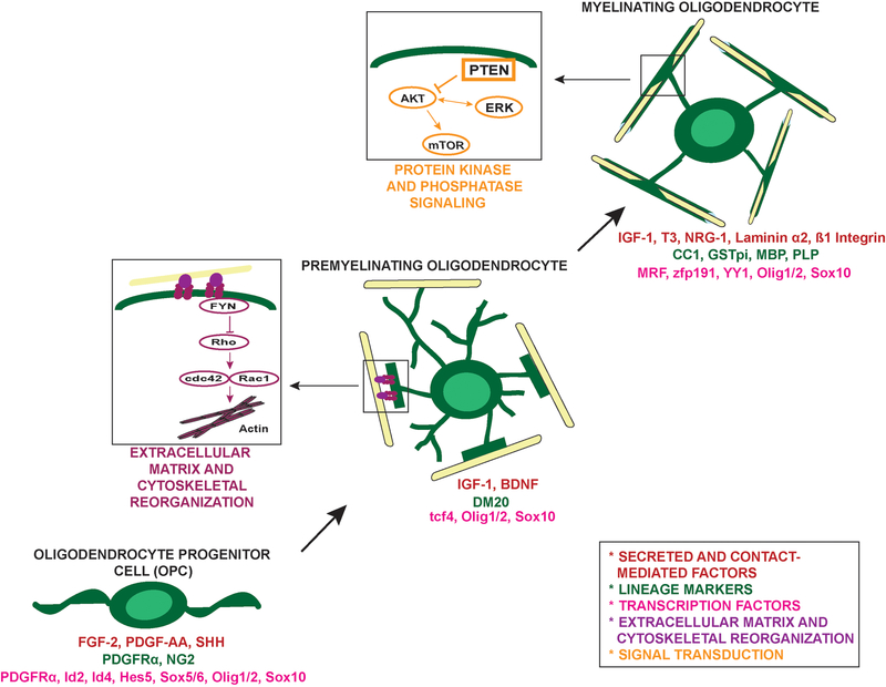
Progression through the oligodendrocyte lineage is mediated by numerous factors. As highlighted, signaling from the extracellular matrix (sub-figure adapted from Bauer et al., 2009) is critical in modulating the dynamic cytoskeletal reorganization of the premyelinating-myelinating oligodendrocyte transition. Additionally, signal transduction through the Akt/mTOR pathway, modified by PTEN, has been shown to be a regulator of myelin biogenesis. It should be noted that the ERK1/2 signaling pathway is also involved in regulating CNS myelination (Ishii et al., 2012), but this figure focuses on the Akt/mTOR pathway investigated in Snaidero et al. (2014).
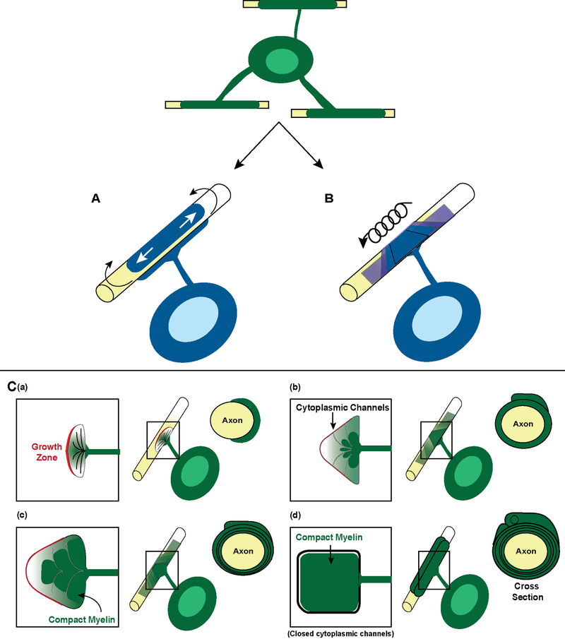
In the past, two models of myelination (blue) have been proposed to ensheath an axon. (A) Schematic of the proposed “jelly roll” model of myelination in which myelin concentrically wraps around the axon repeatedly overlapping the same internode. (B) Lateral spiral movements of an oligodendrocyte process around an axon with eventual compaction. (C) Current model of myelination adapted from (Snaidero et al., 2014). (a) The grown zone (red) of an individual oligodendrocyte process contacts the axon which it will ensheath. (b) The inner tongue of the oligodendrocyte process pushes under the outer tongue to generate the compact myelin (dark green). Cytoplasmic channels (white) allow communication between the inner and outer tongue. (c) More compact myelin is generated. (d) Cytoplasmic channels close once the appropriate number of myelin wraps per axon is generated and myelination is complete.
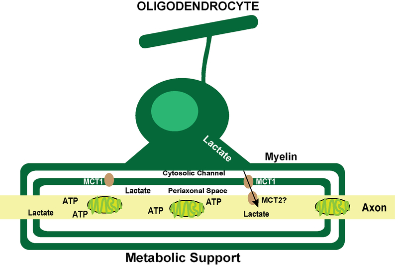
Model of oligodendrocyte-derived lactate delivered by the MCT1 transporter to the axon where it is metabolized and used as an energy source. Adapted from Funfschilling et al. (2012).
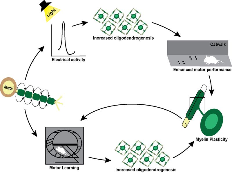
Schematic of the neural induced modification of oligodendrogenesis and the feedback loop from myelin plasticity enhancing neural-mediated behavior. In rodents, optogenetic induced electrical activity results in enhanced oligodendrogenesis resulting in enhanced swing speed on a Catwalk apparatus. Additionally, motor learning on a complex wheel requires new oligodendrogenesis. Mouse illustration provided and used with permission from Dr. Michelle Monje.
References
-
- Allen NJ, and Barres BA (2009). Neuroscience: Glia - more than just brain glue. Nature 457, 675–677. - PubMed
Publication types
MeSH terms
Grants and funding
LinkOut - more resources
Full Text Sources
Other Literature Sources
