Review
. 2015 Mar;18(3):351-9.
doi: 10.1038/nn.3950.

Microcircuits and their interactions in epilepsy: is the focus out of focus?

Affiliations
Review

Microcircuits and their interactions in epilepsy: is the focus out of focus?

Jeanne T Paz et al. Nat Neurosci. 2015 Mar.

Abstract

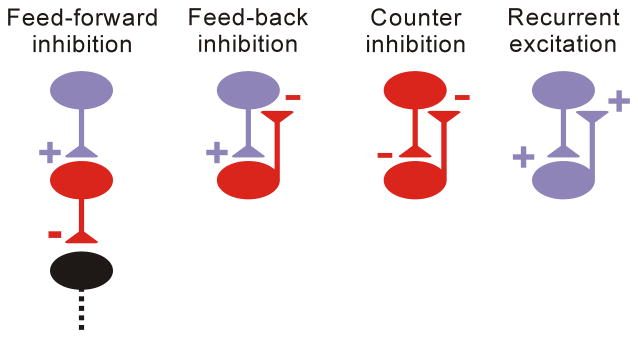
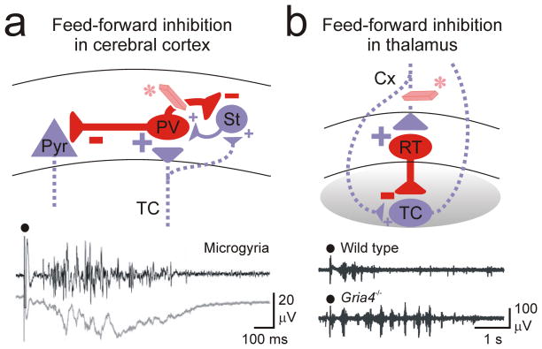
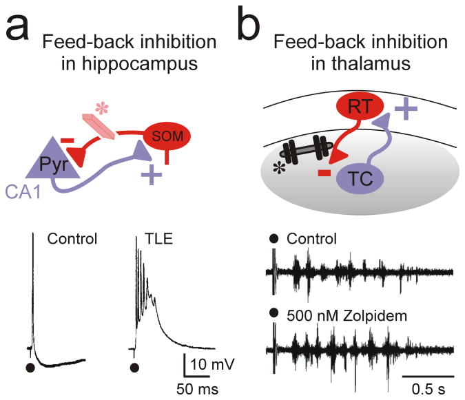
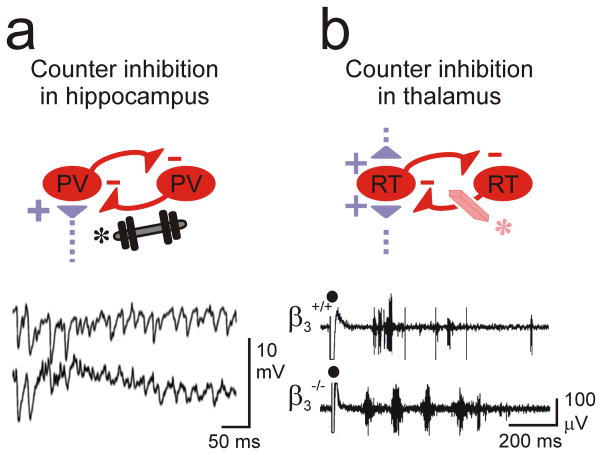
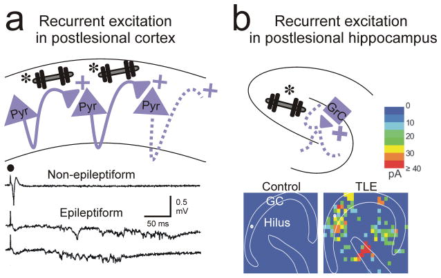
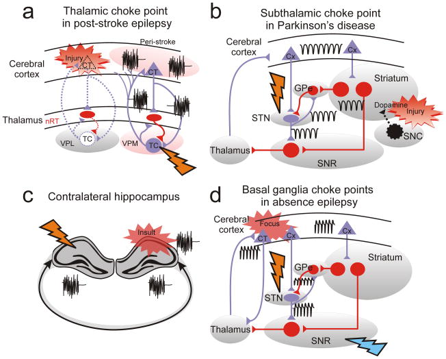
Epileptic seizures represent dysfunctional neural networks dominated by excessive and/or hypersynchronous activity. Recent progress in the field has outlined two concepts regarding mechanisms of seizure generation, or ictogenesis. First, all seizures, even those associated with what have historically been thought of as 'primary generalized' epilepsies, appear to originate in local microcircuits and then propagate from that initial ictogenic zone. Second, seizures propagate through cerebral networks and engage microcircuits in distal nodes, a process that can be weakened or even interrupted by suppressing activity in such nodes. We describe various microcircuit motifs, with a special emphasis on one that has been broadly implicated in several epilepsies: feed-forward inhibition. Furthermore, we discuss how, in the dynamic network in which seizures propagate, focusing on circuit 'choke points' remote from the initiation site might be as important as that of the initial dysfunction, the seizure 'focus'.


Figures

Figure 1
Figure 1. Microcircuit motifs whose dysfunctions have been identified in epilepsy
Feed-forward inhibition: excitatory inputs from remote brain regions recruit local inhibitory networks that control the strength of the efferent signal; Feed-back inhibition: local activation of inhibitory neurons creates local recurrent excitatory activity; Counter-inhibition: local connections between inhibitory neurons shape network-inhibitory output; Recurrent excitation: major mode of connectivity in cortical networks; Purple and red represent excitatory glutamatergic and inhibitory GABAergic neurons, respectively, in this and all following figures.
Figure 2
Figure 2. Feed-forward inhibition in cortical and thalamic microcircuits
(a) Extrinsic excitatory projections from regions outside of local cortical networks recruit feed-forward inhibition. Cortical inter-areal or thalamic inputs to the cortex result in stronger activation of FS parv cells than excitatory stellate and pyramidal cells, thus causing a robust feed-forward inhibition of excitatory cells. In case of a loss of this feed-forward inhibition (eraser*), thalamic inputs to the cortex recruit epileptiform activity in a neocortical microgyrus model of focal neocortical epilepsy (bottom multi-unit and local field recordings). (b) Excitatory inputs from the cortex to the thalamus results in stronger activation of the inhibitory interneurons, which causes a strong feed-forward inhibition of relay excitatory neurons. Loss of feed-forward inhibition (eraser*) has been implicated in the gria4−/− mouse model of absence epilepsy (multi-unit recordings) Black circle: electrical stimulation of excitatory afferents. Cx, cortex; parv, parvalbumin-positive interneuron; Pyr, pyramidal neuron; RT, reticular thalamic neuron; St, stellate; TC, thalamocortical neuron.
Figure 3
Figure 3. Feed-back inhibition in cortical and thalamic microcircuits
(a) In the cortex, inhibitory SOM interneurons provide a feed-back inhibition to pyramidal neurons that excite them. Loss of this inhibition (eraser*) has been implicated in temporal lobe epilepsy (TLE). (b) In the somatosensory thalamus, inhibitory interneurons provide a robust feed-back inhibition to TC neurons that excite them. Increasing this feed-back inhibition (dumbbell weight *) by Zolpidem, or by clonazepam in α3H126R mice (not shown), which specifically affects RT-TC but not RT-RT connections, enhances epileptiform oscillations. Pyr, pyramidal; SOM, somatostatin-positive; RT, reticular thalamic neuron; TC, thalamocortical neuron.
Figure 4
Figure 4. Counter-inhibition in hippocampal and thalamic microcircuits
(a) Inhibition between FS parv cells in the hippocampus can enhance gamma rhythmicity. Increasing this inhibition (weight*) has been suggested to enhance network synchrony associated with epilepsy. (b) Inhibition between RT neurons in the thalamus desynchronizes the thalamic network oscillations between TC and RT cells. Loss of RT-RT counter-inhibition (eraser*) in a β3−/− mouse enhances intra-thalamic network synchrony and has been implicated in epilepsy. RT, reticular thalamic neuron; TC, thalamocortical neuron.
Figure 5
Figure 5. Recurrent excitation in cortex and hippocampus
(a) Recurrent excitation between pyramidal excitatory cells (weights*) develops after neocortical lesions and has been implicated in epileptiform activities in the undercut model of focal neocortical epilepsy . Bottom traces: local recordings of epileptiform field potentials from the injured neocortex evoked by electrical stimulation (black circle). (b) Ectopic recurrent excitation (weight*) between presynaptic excitatory neurons in dentate, hilus, and CA3 and post-synaptic granule cells in the hippocampus develops in the pilocarpine model of temporal lobe epilepsy. Bottom: Connectivity maps based on glutamate photo-uncaging evoked excitatory postsynaptic currents in slices from control and epileptic (TLE) mice.
Figure 6
Figure 6. Circuit therapy: focus on choke points
(a) The thalamus is a choke point for epileptic seizures in post-stroke epilepsy. Note that the choke point (flash: thalamus) is remote from the initial dysfunction (red flash), which is a stroke in the cerebral cortex. (b) The subthalamus (STN) is an efficient choke point for pathological circuit oscillations in Parkinson’s disease. Note that the choke point (black flash: STN) is remote from the initial dysfunction (yellow flash), which results from degeneration of dopaminergic cells (Dopamine) projecting from the substantia nigra compacta (SNC) to striatum. (c) Contralateral hippocampus is a choke point for controlling ipsilateral hippocampal epileptic activity. (d) STN and SNR are choke points for spike-and-wave discharges associated with absence epilepsy and generated in somatosensory cortex . Black oscillations: pathological oscillations; Red flash: initial injury or insult; Orange flash: choke point for pathological network oscillation. Other abbreviations: GPe: External globus pallidus; SNR: substantia nigra pars reticulata. Purple cells/projections: excitatory glutamatergic; Red cells/projections: inhibitory GABAergic.

References

    1. Avoli M. A brief history on the oscillating roles of thalamus and cortex in absence seizures. Epilepsia. 2012;53:779–789. - PMC - PubMed
    1. Noebels JL, Avoli M, Rogawski MA, Olsen RW, Delgado-Escueta AV. Jasper’s Basic Mechanisms of the Epilepsies. National Center for Biotechnology Information; Bethesda: 2012. - PubMed
    1. Jones EG. Viewpoint: The core and matrix of thalamic organization. Neuroscience. 1998;85:331–345. - PubMed
    1. Douglas RJ, Martin KA. A functional microcircuit for cat visual cortex. J Physiol. 1991;440:735–769. - PMC - PubMed
    1. Douglas RJ, Koch C, Mahowald M, Martin KA, Suarez HH. Recurrent excitation in neocortical circuits. Science. 1995;269:981–985. - PubMed

Publication types