. 2015 Apr 1;194(7):3267-74.
doi: 10.4049/jimmunol.1402581. Epub 2015 Feb 20.

Blimp-1, an intrinsic factor that represses HIV-1 proviral transcription in memory CD4+ T cells

Affiliations

Blimp-1, an intrinsic factor that represses HIV-1 proviral transcription in memory CD4+ T cells

Katarzyna Kaczmarek Michaels et al. J Immunol. .

Abstract

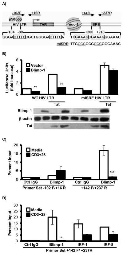
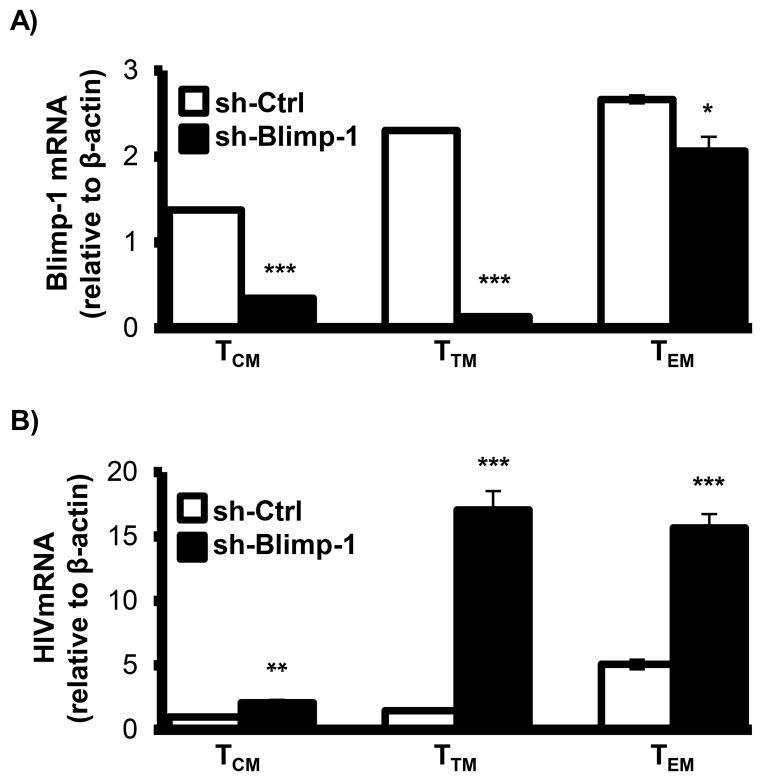
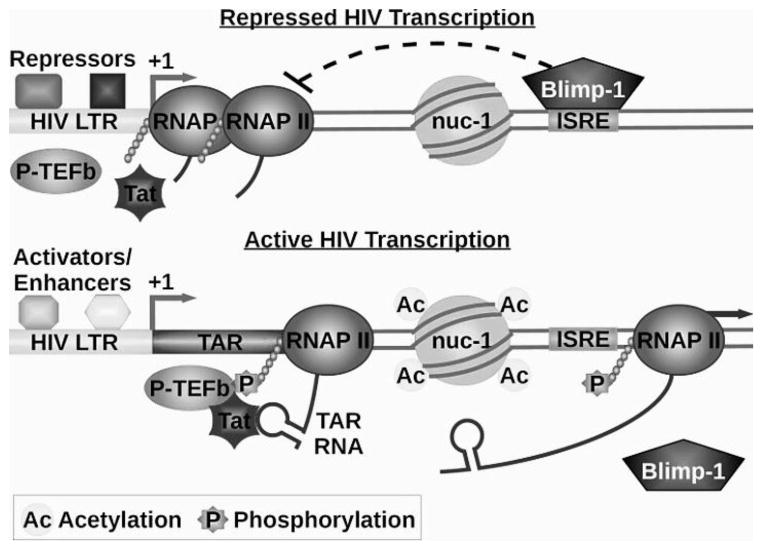
CD4(+) T cell subsets differentially support HIV-1 replication. For example, quiescent CD4(+) memory T cells are susceptible to HIV-1 infection but do not support robust HIV-1 transcription and have been implicated as the primary reservoir of latent HIV-1. T cell transcription factors that regulate maturation potentially limit HIV-1 transcription and mediate the establishment and maintenance of HIV-1 latency. We report that B lymphocyte-induced maturation protein-1 (Blimp-1), a critical regulator of B and T cell differentiation, is highly expressed in memory CD4(+) T cells compared with naive CD4(+) T cells and represses basal and Tat-mediated HIV-1 transcription. Blimp-1 binds an IFN-stimulated response element within HIV-1 provirus, and it is displaced following T cell activation. Reduction of Blimp-1 in infected primary T cells including CD4(+) memory T cells increases RNA polymerase II processivity, histone acetylation, and baseline HIV-1 transcription. Therefore, the transcriptional repressor, Blimp-1, is an intrinsic factor that predisposes CD4(+) memory T cells to latent HIV-1 infection.


Figures

Fig. 1
Fig. 1. Blimp-1 is expressed in CD4+ memory T cells
A) CD4+ cells were isolated from peripheral blood and tonsils and Blimp-1 was measured by qRT-PCR. B) Blimp-1 levels were measured by qRT-PCR in unstinmulated or CD3 + CD28 stimulated primary CD4+ T cells enriched from peripheral blood. β-actin was used as a reference gene to normalize values. C) Immnoblots of lysates from resting and CD3 + CD28 activated primary CD4+ T cells obtained from whole blood for Blimp-1, Sp1 and β-actin. Sp1, a transcription factor, and β-acting served as loading controls. D) Sorting strategy and staining profiles for T cell subsets. Peripheral blood T cells were stained with Pacific Blue–labeled antibody to CD3, APC–labeled antibody to CD4, PE-Cy7–labeled antibody to CCR7, PE-Cy5.5-labeled antibody to CD45RA and PE-labeled antibody to CD27. T cell subsets were separated by FACSAria into naïve T cells (TN; CD4+CD3+CD45RA+), central memory T cells (TCM; CD4+CD3+CD45RACCR7+CD27+), transitional memory T cells (TTM; CD4+CD3+CD45RACCR7CD27+) and effector memory T cells (TEM; CD4+CD3+CD45RACCR7CD27). E) and F) Expression of Blimp-1 in sorted memory CD4+ T cell populations measured by E) qRT-PCR using β-actin as a reference gene and by F) immunoblots. These experiments represent CD4+ cells obtained from at least three healthy individuals. Bars show average values ±SD, n=3. *p < 0.05 and ***p < 0.001 (Student’s t test).
Fig. 2
Fig. 2. Blimp-1 represses HIV-1 transcription in the presence of Tat
A) and B) HEK293T cells were transfected with viral clones HIV-1 LTR-LUC containing the TAR element (A) or ΔTat- HIV-1-LUC (B) and vector control or Blimp-1 in the absence or presence of Tat. Luciferase assays and western blot analyses were performed 48 h post-transfection. (C) HEK293T cells were transfected with HIV-1 LTR-LUC, RSV LTR-LUC, FIP-LUC and vector control or Blimp-1. Luciferase assays and western blot analyses were performed 48 h post-transfection. (D) Jurkat T cells stably transduced with Blimp-1 lentivirus were infected with HIV-1. 48 h post-infection cells were lysed and luciferase activity was measured. (E) 16 h post-HIV-1 infection primary CD4+ T cells purified from peripheral blood were transduced with empty lentiviral vector or with vector expressing Blimp-1. 72 h post-transduction expression of Blimp-1 and HIV-1 were measured by qRT-PCR. HIV-1 released into supernatants was assessed by measuring p24 by an ELISA. These experiments were performed in triplicates and the data represent at least three independent experiments. Experiments with primary cells were performed with cells from at least three different people. White bars indicate vector control; black bars indicate Blimp-1 lentiviral vector. Bars show average values ±SD, n=3. *p < 0.05, **p < 0.01 and ***p < 0.001 (Student’s t test).
Fig. 3
Fig. 3. Blimp-1 binds HIV-1 provirus
(A) The location of the HIV-1 LTR, four putative Blimp-1-binding sites and ISRE mutations in provirus. (B) HEK293T cells were transfected with HIV-1 LTR-LUC or mISRE-HIV-1 LTR-LUC and control vector or Blimp-1 in the absence or presence of Tat. Luciferase assays and western blot analyses were performed 48 h post-transfection. (C, D) 96 h post-HIV-1 infection primary human CD4+ T cells from whole blood were activated with anti-CD3 and anti-CD28 antibodies for 24 h and ChIPs were performed using anti-Blimp-1, anti-IRF-1, anti-IRF-8 or anti-rabbit antibody. Binding was detected with -102F/+16R and +142F/+237R HIV-1 primer sets. These data are performed in triplicate and represent at three independent experiments. Bars show average values ±SD, n=3. *p < 0.05, **p < 0.01 and ***p < 0.001 (Student’s t test).
Fig. 4
Fig. 4. Blimp-1 represses HIV-1 transcription in resting CD4+ T cells
HIV-1 -infected primary CD4+ T cells enriched from peripheral blood were transduced with sh-Ctrl and sh-Blimp-1. 72 h post-knockdown cells were activated with anti-CD3 and anti-CD28 antibodies for 24 h. Expression of Blimp-1 was measure by A) qRT-PCR and B) immunoblots. HIV-1 expression was assayed by qRT-PCR using primers for (C) elongated HIV-1 mRNA and (G) initiated short transcripts, D) luciferase assay and E) HIV-1 p24 ELISA. ChIP analysis used anti-rabbit and anti-RNAP II (F) oranti-AcH3 (H) antibody and +30F/+134R and +2415F/+2522R HIV-1 primer sets. These experiments were performed in triplicate and the data are representative of at least three independent experiments. White bars indicate sh-control; black bars indicate sh-Blimp-1. Bars show average values ±SD, n=3. *p < 0.05, **p < 0.01 and ***p < 0.001 (Student’s t test).
Fig. 5
Fig. 5. Blimp-1 represses basal HIV-1 transcription in primary memory CD4+ T cells
TCM, TTM and TEM sorted as described above were infected with NL4-3 HIV-1 by spinoculation. 16 h post infection cells were transduced with sh-Ctrl and sh-Blimp-1. 72 h post-knockdown mRNA was collected. Expression of (A) Blimp-1 and (B) HIV-1 was measured by qRT-PCR using β-actin as a reference gene. These experiments were performed in triplicate and are representative of three separate infections from T cells obtained from three patients. Bars show average values ±SD, n=3. *p < 0.05, **p < 0.01 and ***p < 0.001 (Student’s t test).
Fig. 6
Fig. 6. Model for the role of Blimp-1 in HIV-1 transcription
Blimp-1 is highly expressed in memory CD4+ T cells, binds the HIV-1 ISRE and inhibits HIV-1 transcription in the presence of Tat. Following T cell activation Blimp-1 is released from HIV-1 provirus which correlates with increased RNAP II processivity, histone H3 acetylation and enhanced HIV-1 transcription.

References

    1. Siliciano RF, Greene WC. HIV latency. Cold Spring Harbor Perspective Medicine. 2011;1:a007096. - PMC - PubMed
    1. Richman DD, Margolis DM, Delaney M, Greene WC, Hazuda D, Pomerantz RJ. The challenge of finding a cure for HIV infection. Science. 2009;323:1304–1307. - PubMed
    1. Kaczmarek K, Morales A, Henderson AJ. T cell transcription factors and their impact on HIV expression. Virology: Res Treat. 2013;4:41–47. - PMC - PubMed
    1. Turner CA, Mack DH, Davis MM. Blimp-1, a novel zinc finger-containing protein that can drive the maturation of B lymphocytes into immunoglobulin-secreting cells. Cell. 1994;77:297–306. - PubMed
    1. Lin Y, Wong K, Calame K. Repression of c-myc transcription by Blimp-1, an inducer of terminal B cell differentiation. Science. 1997;276:596–599. - PubMed

Publication types

MeSH terms

Substances