. 2015 Apr 1;194(7):3487-500.
doi: 10.4049/jimmunol.1401717. Epub 2015 Feb 20.

Specific roles of each TCR hemichain in generating functional chain-centric TCR

Affiliations

Specific roles of each TCR hemichain in generating functional chain-centric TCR

Munehide Nakatsugawa et al. J Immunol. .

Abstract

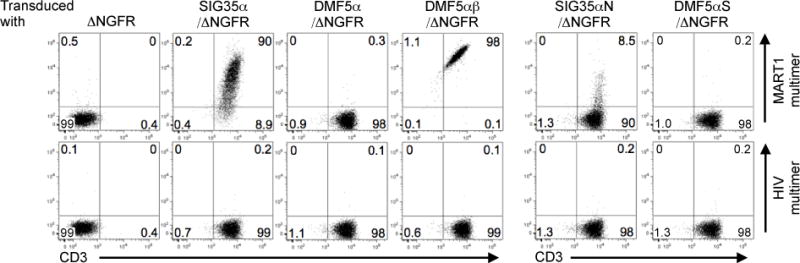
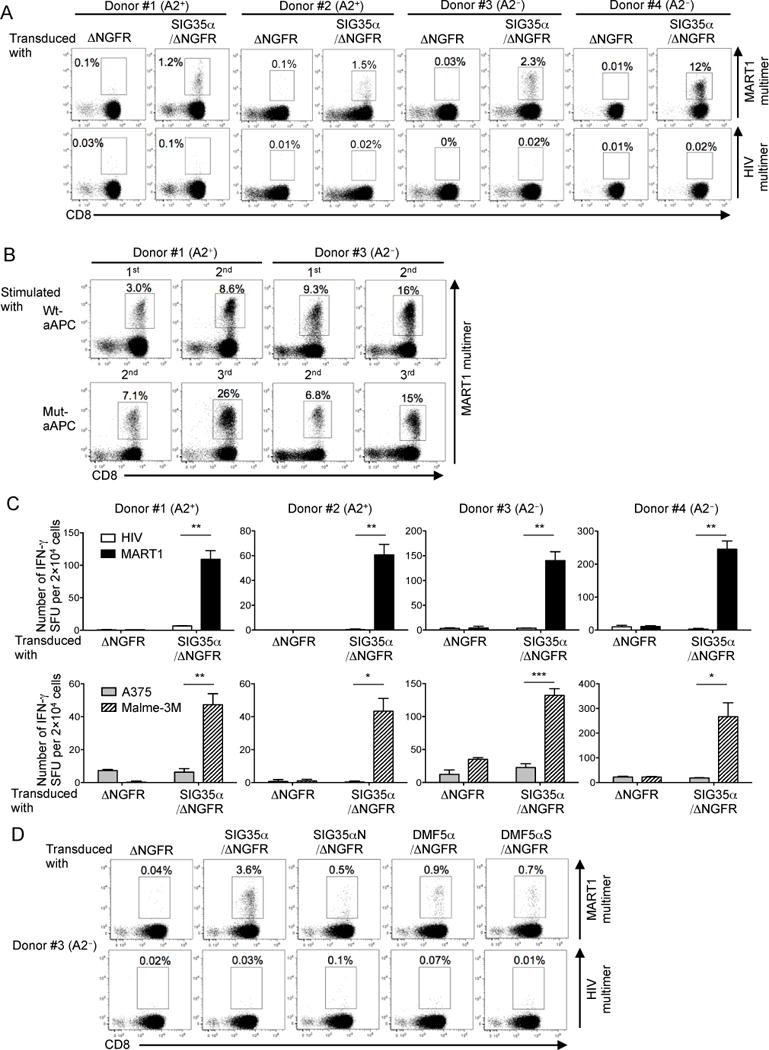
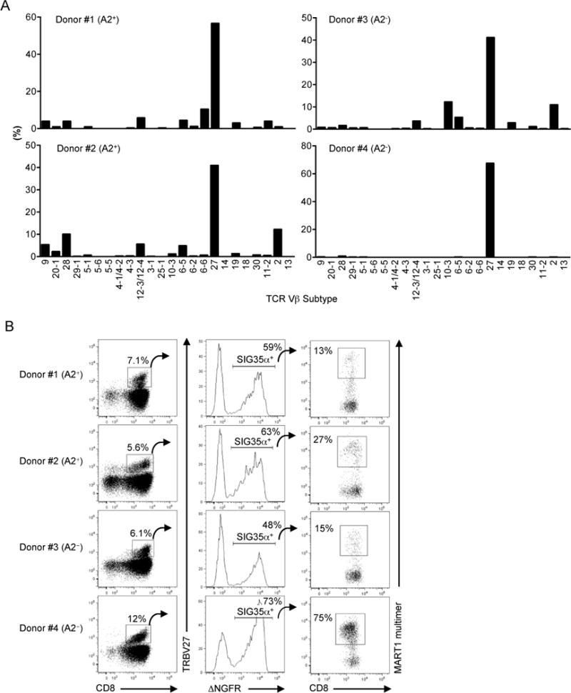
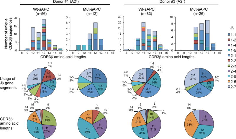
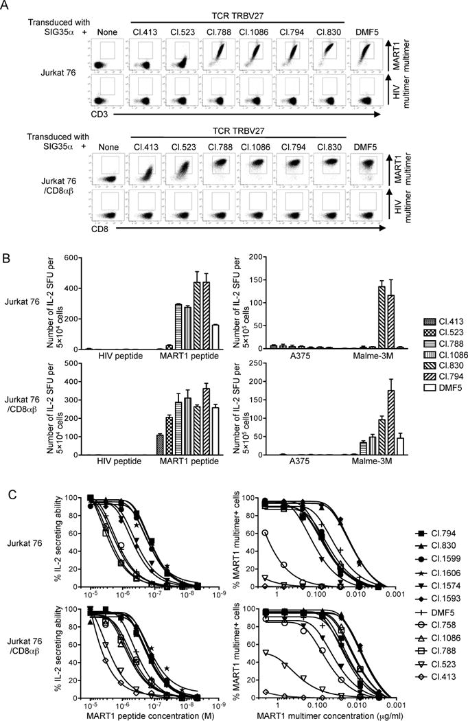
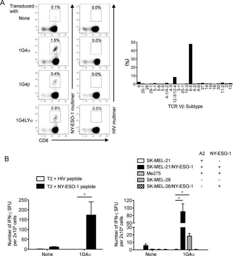
TCRα- and β-chains cooperatively recognize peptide-MHC complexes. It has been shown that a "chain-centric" TCR hemichain can, by itself, dictate MHC-restricted Ag specificity without requiring major contributions from the paired TCR counterchain. Little is known, however, regarding the relative contributions and roles of chain-centric and its counter, non-chain-centric, hemichains in determining T cell avidity. We comprehensively analyzed a thymically unselected T cell repertoire generated by transducing the α-chain-centric HLA-A*02:01(A2)/MART127-35 TCRα, clone SIG35α, into A2-matched and unmatched postthymic T cells. Regardless of their HLA-A2 positivity, a substantial subset of peripheral T cells transduced with SIG35α gained reactivity for A2/MART127-35. Although the generated A2/MART127-35-specific T cells used various TRBV genes, TRBV27 predominated with >10(2) highly diverse and unique clonotypic CDR3β sequences. T cells individually reconstituted with various A2/MART127-35 TRBV27 TCRβ genes along with SIG35α possessed a wide range (>2 log orders) of avidity. Approximately half possessed avidity higher than T cells expressing clone DMF5, a naturally occurring A2/MART127-35 TCR with one of the highest affinities. Importantly, similar findings were recapitulated with other self-Ags. Our results indicate that, although a chain-centric TCR hemichain determines Ag specificity, the paired counterchain can regulate avidity over a broad range (>2 log orders) without compromising Ag specificity. TCR chain centricity can be exploited to generate a thymically unselected Ag-specific T cell repertoire, which can be used to isolate high-avidity antitumor T cells and their uniquely encoded TCRs rarely found in the periphery because of tolerance.


Conflict of interest statement

Competing financial interests

S.T. is an employee of Takara Bio, Inc. This study was partly sponsored by Takara Bio, Inc. The University Health Network has filed a provisional patent application related to this study on which NH, MN, and TO are named as inventors.

Figures

Fig. 1
Fig. 1. SIG35α but not DMF5α expressing SupT1 cells are stained by A2/MART1 multimers when paired with the endogenous irrelevant TCRβ chain of SupT1 cells
The human TCRα+ pre-T cell leukemia cell line, SupT1 was transduced with 5 different clonotypic A2/MART1-specific TCRα chains, SIG35α/ΔNGFR, DMF5α/ΔNGFR, SIG35αN/ΔNGFR, or DMF5αS/ΔNGFR, or TCRαβ chains, DMF5αβ/ΔNGFR. SIG35αN is a SIG35α-derived mutant encoding Asn instead of Ser at the V-J junction. DMF5 is a high affinity A2/MART1 TCR (48). DMF5αS is a DMF5α-derived mutant coding for Ser instead of Asn at the V-J junction. All TCRα genes were fused with ΔNGFR gene via an optimized intervening sequence consisting of a furin cleavage site, an SGSG spacer sequence, and an F2A sequence (35). ΔNGFR alone was employed as a control. The transduction efficiency of SupT1 transfectants was approximately 90% as determined by the percentage of ΔNGFR+ cells (data not shown). All SupT1 transfectants were stained with A2/MART1 or A2/HIV multimer along with anti-CD3 mAb. Data shown are gated on ΔNGFR+ cells and a representative of two independent experiments.
Fig. 2
Fig. 2. Both HLA-A2+ and A2 peripheral T cells can recognize A2/MART1 when transduced with chain-centric SIG35α
(A), Both HLA-A2+ and A2 peripheral T cells become A2/MART1-reactive upon transduction of chain-centric SIG35α. Peripheral CD8+ T cells freshly isolated from two HLA-A2+ donors (#1 and #2) and two A2 donors (#3 and #4) were retrovirally transduced with ΔNGFR or SIG35α/ΔNGFR and stained with A2/MART1 multimer or A2/HIV multimer in conjunction with anti-CD8 mAb and anti-NGFR mAb. Data shown are gated on ΔNGFR+ cells. Data of donors #1 and #3 are representative of three independent experiments and data of donors #2 and #4 are representative of two independent experiments. (B), SIG35α-transduced A2/MART1 CD8+ T cells expand in an A2/MART1-specific manner. A2+ and A2 CD8+ T cells transduced with SIG35α/ΔNGFR were stimulated with wt-aAPC or mut-aAPC pulsed with wild-type A2/MART1 peptide once a week. Between stimulations, the T cells were supplemented with IL-2 (10 IU/ml) and IL-15 (10 ng/ml) every 3 days. Data depict A2/MART1 multimer staining performed following the first and second stimulations using wt-aAPC and the second and third stimulations utilizing mut-aAPC. Data shown are gated on ΔNGFR+ cells. Representative multimer-staining data from one of two HLA-A2+ donors and one of two A2 donors are shown. (C), Peripheral T cells transduced with SIG35α are highly avid for A2/MART1 recognition. CD8+ T cells following stimulation with wt-aAPC pulsed with wild-type A2/MART1 peptide were used as responder cells in IFN-γ ELISPOT analysis. T2 cells pulsed with 10 μg/ml A2/HIV control peptide or wild-type A2/MART1 peptide were used as stimulator cells (top). The A2+ MART1 melanoma line, A375, and the A2+ MART1+ melanoma line, Malme-3M, were used as stimulator cells (bottom). Data shown are representative of two independent experiments. All experiments were carried out in triplicate and error bars depict SD. *P < 0.05, **P < 0.01, ***P < 0.001. (D), Peripheral CD8+ T cells isolated from an A2 donor #3 were transduced with ΔNGFR, SIG35α/ΔNGFR, SIG35αN/ΔNGFR, DMF5α/ΔNGFR, or DMF5αS/ΔNGFR and stained by A2/MART1 multimer or A2/HIV multimer in conjunction with anti-CD8 mAb and anti-NGFR mAb as described in Fig. 1. Data shown are gated on ΔNGFR+ cells.
Fig. 3
Fig. 3. SIG35α predominantly pairs with TRBV27 TCRβ chains to recognize A2/MART1
(A), SIG35α/ΔNGFR-transduced peripheral CD8+ T cells from two HLA-A2+ and two A2 donors were stained with A2/MART1 multimer, mAbs for TCR Vβ subtypes, and anti-CD8 mAb. The percentage of A2/MART1 multimer+ CD8+ T cells expressing each subtype is shown. Data shown are gated on ΔNGFR+ cells. (B), A significant proportion of TRBV27 TCRβ chains in the periphery can recognize A2/MART1 when paired with SIG35α. The percentage of A2/MART1 multimer+ cells in CD8+ TRBV27+ T cells transduced with SIG35α/ΔNGFR gene is shown.
Fig. 4
Fig. 4. TRBV27 TCRβ chains which recognize A2/MART1 when paired with SIG35α are highly heterogeneous and unique
SIG35α/ΔNGFR-transduced CD8+ T cells from the HLA-A2+ donor #1 and the A2 donor #3 were stimulated with wt-aAPC or mut-aAPC pulsed with wild-type A2/MART1 peptide. A2/MART1 multimer+ CD8+ T cells were collected by fluorescence activated cell sorting (>99% purity) and their TRBV27 CDR3β regions were amplified by PCR and sequenced after cloning. The number of unique CDR3β sequences (top), the relative usage of Jβ gene segments (middle), and the CDR3β amino acid lengths (bottom) are depicted separately for the A2+ donor #1 (left) and A2 donor #3 (right). Data were analyzed by the aAPC used for stimulations, wt-aAPC vs. mut-aAPC, in each donor.
Fig. 5
Fig. 5. The structural and functional avidity range of A2/MART1 TCRs consisting of SIG35α is very broad and further enhanced by the presence of CD8
Jurkat 76 cells, which lack the expression of CD8αβ and endogenous TCRs, were retrovirally transduced with CD8αβ to produce Jurkat 76/CD8αβ. Jurkat 76 or Jurkat 76/CD8αβ cells were individually transduced with eleven distinct TRBV27 TCRβ chains along with SIG35α or with DMF5αβ chains. (A), A2/MART1 TCRs reconstituted on Jurkat 76 or Jurkat 76/CD8αβ cells were differentially stained by A2/MART1 multimer. All Jurkat 76 or Jurkat 76/CD8αβ transfectants were stained with 2 μg/ml A2/MART1 or A2/HIV multimer along with anti-CD3 mAb (top) or anti-CD8 mAb (bottom). Data for multimer staining of seven representative Jurkat 76 or Jurkat 76/CD8αβ transfectants are shown. Data for multimer staining of the remaining 5 transfectants are shown in Supplementary Fig. 3. (B), Reconstituted A2/MART1 TCRs are highly avid for A2/MART1 recognition. IL-2 ELISPOT assays were performed using seven representative Jurkat 76 or Jurkat 76/CD8αβ transfectants as responder cells. T2 cells pulsed with 10 μg/ml wild-type A2/MART1 or A2/HIV control peptide were used as stimulator cells (left). The A2+ MART1 melanoma line, A375, and the A2+ MART1+ melanoma line, Malme-3M were used as stimulator cells (right). All experiments were conducted in triplicate and error bars show SD. Data shown are representative of two independent experiments. (C), Reconstituted A2/MART1 TCRs possess a broad range of functional and structural avidities. Functional avidities of Jurkat 76 or Jurkat 76/CD8αβ cells expressing 11 different A2/MART1 TCRβ chains paired with SIG35α and DMF5 are depicted as % IL-2 secreting abilities determined by IL-2 ELISPOT assays using T2 cells pulsed with graded concentrations of wild-type A2/MART1 peptide as stimulator cells (left). Structural avidities of the same transfectants are shown as multimer staining percentages determined by staining with graded concentrations of A2/MART1 multimer (right). Data shown are representative of two independent experiments.
Fig. 6
Fig. 6. TCR chain centricity is observed with other HLA-restricted antitumor TCRs
(A), Peripheral CD8+ T cells transduced with 1G4α, 1G4β, or 1G4LYα recognize A2/NY-ESO-1. Peripheral CD8+ T cells transduced with 1G4α, 1G4β, or 1G4LYα were stimulated with IL-21-secreting wt-aAPC pulsed with heteroclitic A2/NY-ESO-1 peptide once a week. Between stimulations, IL-2 (10 IU/ml) and IL-15 (10 ng/ml) were added every 3 days. Data for A2/NY-ESO-1 multimer staining conducted after second stimulation are shown (left). Data are representative of two donors. 1G4α-transduced CD8+ T cells were costained with A2/NY-ESO-1 multimer, mAbs for TCR TRBV subtypes, and anti-CD8 mAb. The percentage of A2/NY-ESO-1 multimer+ CD8+ T cells expressing each subtype after second stimulation is shown (right). (B), Peripheral T cells transduced with 1G4α hemichain are highly avid for A2/NY-ESO-1 recognition. 1G4α-transduced or non-transduced CD8+ T cells were stimulated with mut-aAPC pulsed with heteroclitic NY-ESO-1 peptide and used as responder cells in IFN-γ ELISPOT analysis. T2 cells pulsed with 10 μg/ml A2/HIV control or A2/NY-ESO-1 peptide were used as stimulator cells (left). Various target cells, which did or did not express HLA-A2 and/or NY-ESO-1, were used as stimulator cells (right). Experiments were carried out in triplicate and error bars depict SD. *P < 0.05.

Similar articles

Cited by

References

    1. Turner SJ, Doherty PC, McCluskey J, Rossjohn J. Structural determinants of T-cell receptor bias in immunity. Nature reviews Immunology. 2006;6:883–894. - PubMed
    1. Venturi V, Price DA, Douek DC, Davenport MP. The molecular basis for public T-cell responses? Nature reviews Immunology. 2008;8:231–238. - PubMed
    1. Miles JJ, Douek DC, Price DA. Bias in the alphabeta T-cell repertoire: implications for disease pathogenesis and vaccination. Immunology and cell biology. 2011;89:375–387. - PubMed
    1. Dong L, Li P, Oenema T, McClurkan CL, Koelle DM. Public TCR use by herpes simplex virus-2-specific human CD8 CTLs. Journal of immunology. 2010;184:3063–3071. - PMC - PubMed
    1. Brennan RM, Petersen J, Neller MA, Miles JJ, Burrows JM, Smith C, McCluskey J, Khanna R, Rossjohn J, Burrows SR. The impact of a large and frequent deletion in the human TCR beta locus on antiviral immunity. Journal of immunology. 2012;188:2742–2748. - PubMed

Publication types

MeSH terms