Review
. 2015 Mar 12;34(6):710-38.
doi: 10.15252/embj.201490563. Epub 2015 Feb 20.

Programming and reprogramming a human heart cell

Affiliations
Review

Programming and reprogramming a human heart cell

Makoto Sahara et al. EMBO J. .

Abstract

The latest discoveries and advanced knowledge in the fields of stem cell biology and developmental cardiology hold great promise for cardiac regenerative medicine, enabling researchers to design novel therapeutic tools and approaches to regenerate cardiac muscle for diseased hearts. However, progress in this arena has been hampered by a lack of reproducible and convincing evidence, which at best has yielded modest outcomes and is still far from clinical practice. To address current controversies and move cardiac regenerative therapeutics forward, it is crucial to gain a deeper understanding of the key cellular and molecular programs involved in human cardiogenesis and cardiac regeneration. In this review, we consider the fundamental principles that govern the "programming" and "reprogramming" of a human heart cell and discuss updated therapeutic strategies to regenerate a damaged heart.

Keywords: cardiac progenitor cell; cardiac regeneration; cardiomyocyte proliferation; embryonic heart field; reprogramming.


Figures

Figure 1
Figure 1
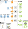
Fate map of cardiac cell lineages during development (A) A cellular flow chart shows the stepwise commitment of pluripotent stem cells via various cardiac progenitor cells, including the first and second heart field (FHF and SHF) progenitors, epicardium-derived progenitor cells (EPDCs) and so-called tertiary heart field (HF) progenitors, and putative intermediates toward mature cardiac cell types during heart development. Mature cardiac cells include cardiomyocytes (CMs), vascular smooth muscle cells (SMCs), arterial and venous endothelial cells (ECs), fibroblasts (FBs), and conductive cells of the cardiac conduction system (CS), which include pacemaker (sino-atrial [SA] nodal) cells, atrio-ventricular (AV) nodal cells, and the ventricular CS cells (ex. Purkinje fibers). The gray boxes in the middle indicate the major mature cell types that each cardiac progenitor differentiates into. Cardiac neural crest cells (cNCCs) originating from the ectoderm also contribute to vascular SMCs of the outflow tract (OFT) and thereby to OFT separation and patterning. (B) Cardiac development at the early stage of the mouse embryo. Developing hearts at murine embryonic day (E) 7.5 and 9.0 are shown. A, atria; EMT, epithelial-to-mesenchymal transition; LV, left ventricle; ML, midline; PEO, proepicardial organ; PM, pharyngeal mesoderm; and RV, right ventricle.
Figure 2
Figure 2
Signaling pathways programming a heart cell during embryogenesis The flow chart shows the representative signaling pathways and/or modulators working in a stepwise manner at various stages of cardiac development/cardiac cell differentiation, including mesodermal formation, cardiac specification, maintenance of heart field (HF) progenitors, and cardiac maturation and patterning (see text for details). A circular arrow denotes self-renewal. First heart field (FHF) marker gene is in blue, and second heart field (SHF) marker genes in red. CM, cardiomyocyte; CS, conduction system; and SA, sino-atrial.
Figure 3
Figure 3
Heart regeneration in various model organisms (Top) Schematic representation indicates cardiac regenerative capabilities of various model organisms from lower vertebrates (zebrafish and amphibians) to human. (Bottom) Heart regenerative/replicative processes following injury in zebrafish and amphibians, neonatal/adult mice, and humans are shown. In zebrafish, various injury models (genetic ablation, apical resection (AR), and cryoinjury) are reported and exhibit different recovery processes, especially in terms of transient fibrotic scar formation. All ultimately lead to full regeneration of the ablated myocardium. Similar post-injury regenerative processes are seen in hearts of 1-day-old mice, but not after 7 days nor in humans, where fibrotic scar formation and pathological remodeling occur. Regeneration enhancers are indicated in green, inhibitors in red (see text for details). A-V, atrial-to-ventricular; CM, cardiomyocyte; EMT, epithelial-to-mesenchymal transition; MI, myocardial infarction.
Figure 4
Figure 4
Therapeutic strategies for cardiac regeneration (Left) Cell-based therapies involve transplantation into the damaged heart of in vitro-derived cardiomyocytes (CMs) or somatic stem cells. CM differentiation can be induced from embryonic stem cells (ESCs) or induced pluripotent stem cells (iPSCs) obtained by reprogramming differentiated somatic cells, such as fibroblasts. Alternatively, by forced expression of cardiac-specific factors, the pluripotent state can be bypassed and fibroblasts directly converted into CMs or cardiac progenitor cells (CPCs). Different types of cardiac and non-cardiac somatic stem cells can also be transplanted. Cell-based therapies can produce beneficial effects on heart function directly, by engrafting into the host tissue and differentiating/proliferating in vivo, or indirectly via paracrine effects that act on host cells. (Right) Cell-free therapies involve administration of chemical compounds or genes (via viral vectors, non-viral DNA vectors, or modified mRNAs) that act on host cells to stimulate cardiac regeneration. Mechanisms of action include the stimulation of proliferation of preexisting CMs, direct conversion of cardiac fibroblasts (CFs) into CMs, trans-differentiation of CMs or other cardiac cells from one cellular subtype to another, and activation and differentiation of endogenous CPCs. Cardiac delivery routes for both cell-based and cell-free therapeutic agents are also indicated.

References

    1. Ai D, Fu X, Wang J, Lu MF, Chen L, Baldini A, Klein WH, Martin JF. Canonical Wnt signaling functions in second heart field to promote right ventricular growth. Proc Natl Acad Sci USA. 2007;104:9319–9324. - PMC - PubMed
    1. Andersen DC, Ganesalingam S, Jensen CH, Sheikh SP. Do neonatal mouse hearts regenerate following heart apex resection? Stem Cell Reports. 2014;2:406–413. - PMC - PubMed
    1. Anderson ME, Goldhaber J, Houser SR, Puceat M, Sussman MA. Embryonic stem cell-derived cardiac myocytes are not ready for human trials. Circ Res. 2014;115:335–338. - PMC - PubMed
    1. Andersson ER, Sandberg R, Lendahl U. Notch signaling: simplicity in design, versatility in function. Development. 2011;138:3593–3612. - PubMed
    1. Ang KL, Chin D, Leyva F, Foley P, Kubal C, Chalil S, Srinivasan L, Bernhardt L, Stevens S, Shenje LT, Galinanes M. Randomized, controlled trial of intramuscular or intracoronary injection of autologous bone marrow cells into scarred myocardium during CABG versus CABG alone. Nat Clin Pract Cardiovasc Med. 2008;5:663–670. - PubMed

Publication types