. 2015 Jun;35(6):1015-23.
doi: 10.1038/jcbfm.2015.14. Epub 2015 Feb 25.

Calibrated MRI to evaluate cerebral hemodynamics in patients with an internal carotid artery occlusion

Affiliations

Calibrated MRI to evaluate cerebral hemodynamics in patients with an internal carotid artery occlusion

Jill B De Vis et al. J Cereb Blood Flow Metab. 2015 Jun.

Abstract

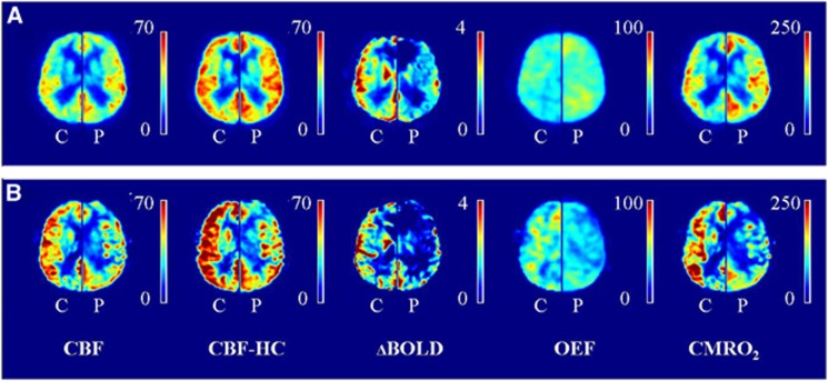
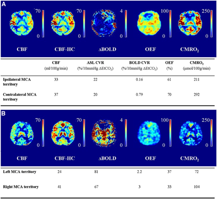
The purpose of this study was to assess whether calibrated magnetic resonance imaging (MRI) can identify regional variances in cerebral hemodynamics caused by vascular disease. For this, arterial spin labeling (ASL)/blood oxygen level-dependent (BOLD) MRI was performed in 11 patients (65±7 years) and 14 controls (66±4 years). Cerebral blood flow (CBF), ASL cerebrovascular reactivity (CVR), BOLD CVR, oxygen extraction fraction (OEF), and cerebral metabolic rate of oxygen (CMRO2) were evaluated. The CBF was 34±5 and 36±11 mL/100 g per minute in the ipsilateral middle cerebral artery (MCA) territory of the patients and the controls. Arterial spin labeling CVR was 44±20 and 53±10% per 10 mm Hg ▵EtCO2 in patients and controls. The BOLD CVR was lower in the patients compared with the controls (1.3±0.8 versus 2.2±0.4% per 10 mm Hg ▵EtCO2, P<0.01). The OEF was 41±8% and 38±6%, and the CMRO2 was 116±39 and 111±40 μmol/100 g per minute in the patients and the controls. The BOLD CVR was lower in the ipsilateral than in the contralateral MCA territory of the patients (1.2±0.6 versus 1.6±0.5% per 10 mmHg ▵EtCO2, P<0.01). Analysis was hampered in three patients due to delayed arrival time. Thus, regional hemodynamic impairment was identified with calibrated MRI. Delayed arrival artifacts limited the interpretation of the images in some patients.


Figures

Figure 1
Figure 1
Hemodynamic maps. (A) Mean maps of all patients (P) without delayed arrival artifacts (DAAs) are shown (left hemisphere), mean maps of their corresponding controls (C, right hemisphere). (B) Mean maps of the patients with DAA (P, left hemisphere) and their corresponding controls (C, right hemisphere). Delayed arrival artifacts are visible on the ASL images of the patients with DAA and this propagates to the OEF and CMRO2 maps. Note that the images of the control subjects of the patients with DAA (B, right hemispheres) seem to have higher values compared with the control subjects of the patients without DAA (A, right hemispheres). We hypothesize this to be caused by the fact that the right hemisphere in (B) shows the mean image of three controls while the right hemisphere in (A) shows the mean image of 11 controls which decreases the amount of variation on which the mean perfusion images are based. BOLD CVR, blood oxygen level-dependent cerebrovascular reactivity (% per 10 mm Hg ▵EtCO2); CBF, cerebral blood flow (in mL/100 g per minute); CBF-HC, cerebral blood flow during hypercapnia (in mL/100 g per minute); CMRO2, cerebral metabolic rate of oxygen (in μmol/100 g per minute); OEF, oxygen extraction fraction (in %).
Figure 2
Figure 2
Patient example. (A) Example images and quantitative data of a 54-year-old male patient with a right-sided internal carotid artery (ICA) occlusion. The oxygen extraction fraction is increased in the gray matter of both MCA territories and the ability of the vasculature to modulate its tone in response to a hypercapnic stimulus (ASL CVR) is exhausted in both hemispheres. (B) Example images and quantitative data of a 70-year-old male patient with a bilateral ICA occlusion. The OEF is higher and the CMRO2 are lower in the gray matter of the left compared with the right MCA territory. ASL CVR, arterial spin labeling cerebrovascular reactivity; BOLD, blood oxygenation level-dependent; CBF, cerebral blood flow; CMRO2, cerebral metabolic rate of oxygen; MCA, middle cerebral artery; OEF, oxygen extraction fraction.
Figure 3
Figure 3
(A) Demonstration of the error in the OEF estimate introduced by errors in the CBF and ΔBOLD measurements performed at hypercapnia and hyperoxia breathing level. (B) Demonstration of the OEF error introduced by errors in the α, β, ΔCMRO2, and the ΔCaCv. CBFCO2, cerebral blood flow measured at hypercapnia breathing; CBFO2, CBF measured at hyperoxia breathing; ΔBOLDCO2, change in blood oxygenation level-dependent signal change from baseline to hypercapnia breathing; ΔBOLDO2, change in BOLD signal change from baseline to hyperoxia breathing; OEF, oxygen extraction fraction; ΔCMRO2, the relative change in the CMRO2 as a result of hypercapnia breathing; ΔCaCv, the ratio between the arterial to venous concentration of oxygen before and during hyperoxia breathing.

References

    1. 1Derdeyn CP, Videen TO, Yundt KD, Fritsch SM, Carpenter DA, Grubb RL et al. Variability of cerebral blood volume and oxygen extraction: stages of cerebral haemodynamic impairment revisited. Brain 2002; 125: 595–607. - PubMed
    1. 2MacKenzie ET, Farrar JK, Fitch W, Graham DI, Gregory PC, Harper AM. Effects of hemorrhagic hypotension on the cerebral circulation. I. Cerebral blood flow and pial arteriolar caliber. Stroke 1979; 10: 711–718. - PubMed
    1. 3Powers WJ, Grubb RL, Darriet D, Raichle ME. Cerebral blood flow and cerebral metabolic rate of oxygen requirements for cerebral function and viability in humans. J Cereb Blood Flow Metab 1985; 5: 600–608. - PubMed
    1. 4Vernieri F, Pasqualetti P, Passarelli F, Rossini PM, Silvestrini M. Outcome of carotid artery occlusion is predicted by cerebrovascular reactivity. Stroke 1999; 30: 593–598. - PubMed
    1. 5Yonas H, Smith HA, Durham SR, Pentheny SL, Johnson DW. Increased stroke risk predicted by compromised cerebral blood flow reactivity. J Neurosurg 1993; 79: 483–489. - PubMed

Publication types

LinkOut - more resources