Review
. 2015 Jun;282(11):2076-88.
doi: 10.1111/febs.13249. Epub 2015 Mar 16.

Parkin structure and function

Affiliations
Review

Parkin structure and function

Marjan Seirafi et al. FEBS J. 2015 Jun.

Abstract

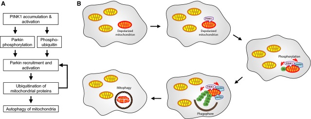
Mutations in the parkin or PINK1 genes are the leading cause of the autosomal recessive form of Parkinson's disease. The gene products, the E3 ubiquitin ligase parkin and the serine/threonine kinase PINK1, are neuroprotective proteins, which act together in a mitochondrial quality control pathway. Here, we review the structure of parkin and mechanisms of its autoinhibition and function as a ubiquitin ligase. We present a model for the recruitment and activation of parkin as a key regulatory step in the clearance of depolarized or damaged mitochondria by autophagy (mitophagy). We conclude with a brief overview of other functions of parkin and considerations for drug discovery in the mitochondrial quality control pathway.

Keywords: PINK1; mitophagy; parkin; ubiquitin; ubiquitination.


Figures

Figure 1
Figure 1
Ubiquitination pathway. (A) Cascade of ubiquitination enzymes. The ubiquitin-activating enzyme E1 uses ATP to conjugate the C-terminal carboxylic acid group of ubiquitin to an active site cysteine. This is then transferred to a cysteine on one of a number of E2 enzymes that work in concert with E3 enzymes to ubiquitinate substrate proteins on amino groups of lysine residues or protein N termini. The formations of mono-ubiquitin or polyubiquitin chains on substrates are signals for different downstream pathways. (B) Classes of E3 ubiquitin ligases. E3 ligases can be distinguished based on the presence of a RING domain, a catalytic cysteine or both. RING and U-box domain ligases act as scaffolds to bring the substrate and ubiquitin-conjugated E2 together. HECT ligases have a catalytic cysteine in their C-lobe that is transiently conjugated to ubiquitin as an intermediate in a two-step process of substrate ubiquitination. Parkin is a RING/HECT hybrid ligase that contains a RING domain that binds the E2 enzyme and a catalytic cysteine that transfers ubiquitin to the substrate.
Figure 2
Figure 2
The structure of parkin reveals the mechanism of its autoinhibition. (A) Domain architecture of the five parkin domains and identification of selected PD mutations. (B) Cartoon representation of parkin (PDB: 4K95) . Parkin activity depends on two functional sites: a binding site for ubiquitin-conjugated E2 enzyme on the RING1 of parkin and a catalytic site with a cysteine that forms a transient covalent linkage with ubiquitin on the RING2 domain. Both sites are occluded in the autoinhibited structure. The Ubl domain and REP linker between the IBR and RING2 domains prevent the E2 from binding to RING1. The RING0 domain partially covers the catalytic cysteine on the RING2 domain. Mutations at interdomain interfaces as well as phosphorylation of Ser65 in the Ubl domain increase parkin ubiquitin ligase activity. The eight structural zinc ions in parkin are shown as gray spheres. Dashed lines indicate portions of the IBR and RING2 linker that were not observed in the crystal structure. (C) Model of an E2 enzyme bound to parkin with Ubl and REP linker removed. An additional structural rearrangement must occur to allow ubiquitin on the E2 enzyme to transfer to the catalytic cysteine of parkin. The E2 and parkin catalytic cysteines are ∼ 50 Å apart in the model.
Figure 3
Figure 3
Mechanism of parkin ubiquitin ligase activity. (A) Sequence alignment of RBR E3 ubiquitin ligases shows conservation in the catalytic RING2 domains. The catalytic cysteine (highlighted in yellow) is invariant across RBR proteins, whereas histidine (green) and glutamate residues (red) that play secondary roles in catalysis are less conserved. Gray indicates zinc-coordinating residues. (B) Structural comparison of catalytic domains of parkin, HHARI and HOIP (PDB: 4K7D ; 4KBL ; 4LJO 44). The alignment of the catalytic cysteine, histidine and glutamate/glutamine is suggestive of a catalytic triad where the histidine acts as a general base to promote transesterification of ubiquitin onto the substrate. (C) Model of the positions of the donor and acceptor ubiquitin molecules in the catalytic site of parkin based on the crystal structure of HOIP . The C terminus of the donor ubiquitin lies in a groove of the RING2 domain and terminates next to the catalytic cysteine, mimicking the thioester intermediate. The amino group of the acceptor ubiquitin approaches Cys431 from the opposite side but would be sterically blocked by the parkin RING0 domain in the absence of a conformational change.
Figure 4
Figure 4
Pathway of PINK1 activation of parkin leading to autophagy of depolarized mitochondria. (A) Flowchart of feedforward and feedback activation of parkin. (B) Schematic of the quality control pathway. PINK1 accumulates on depolarized or damaged mitochondria and phosphorylates ubiquitin and parkin present on the surface. Activated parkin produces additional ubiquitin chains on the mitochondria, which in turn are phosphorylated by PINK1 to promote the recruitment of more parkin. The autophagosome forms around the heavily ubiquitinated mitochondria, which are then eliminated by autophagy.

References

    1. Venderova K. Park DS. Programmed cell death in Parkinson’s disease. Cold Spring Harb Perspect Med. 2012;2:pii: a009365. - PMC - PubMed
    1. Valente EM, Abou-Sleiman PM, Caputo V, Muqit MM, Harvey K, Gispert S, Ali Z, Del Turco D, Bentivoglio AR, Healy DG, et al. Hereditary early-onset Parkinson’s disease caused by mutations in PINK1. Science. 2004;304:1158–1160. - PubMed
    1. Narendra D, Walker JE. Youle R. Mitochondrial quality control mediated by PINK1 and parkin: links to parkinsonism. Cold Spring Harb Perspect Biol. 2012;4:pii: a011338. - PMC - PubMed
    1. Lazarou M, Jin SM, Kane LA. Youle RJ. Role of PINK1 binding to the TOM complex and alternate intracellular membranes in recruitment and activation of the E3 ligase parkin. Dev Cell. 2012;22:320–333. - PMC - PubMed
    1. Deas E, Plun-Favreau H, Gandhi S, Desmond H, Kjaer S, Loh SHY, Renton AEM, Harvey RJ, Whitworth AJ, Martins LM, et al. PINK1 cleavage at position A103 by the mitochondrial protease PARL. Hum Mol Genet. 2011;20:867–879. - PMC - PubMed

Publication types