. 2015 Jun;22(3):353-67.
doi: 10.1530/ERC-15-0038. Epub 2015 Feb 24.

X-linked acrogigantism syndrome: clinical profile and therapeutic responses

Affiliations

X-linked acrogigantism syndrome: clinical profile and therapeutic responses

Albert Beckers et al. Endocr Relat Cancer. 2015 Jun.

Abstract

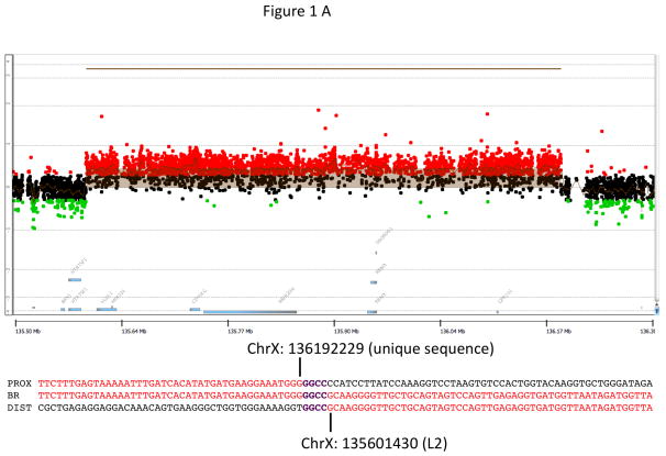
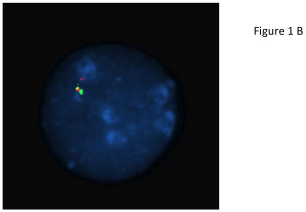
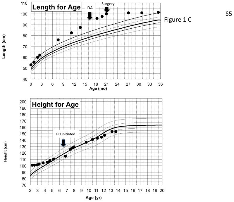
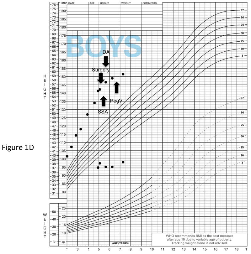
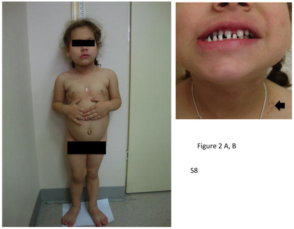
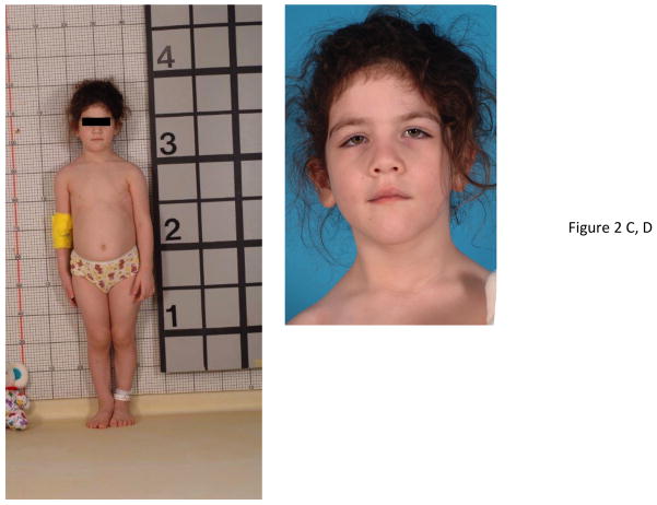
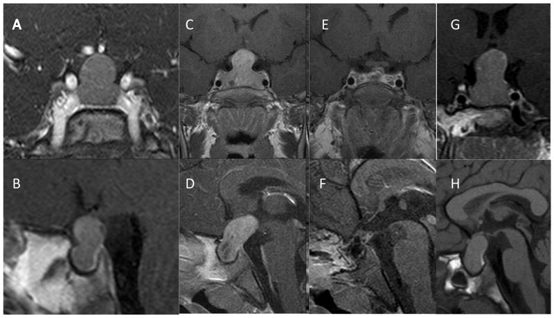
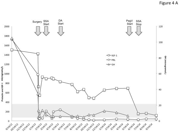
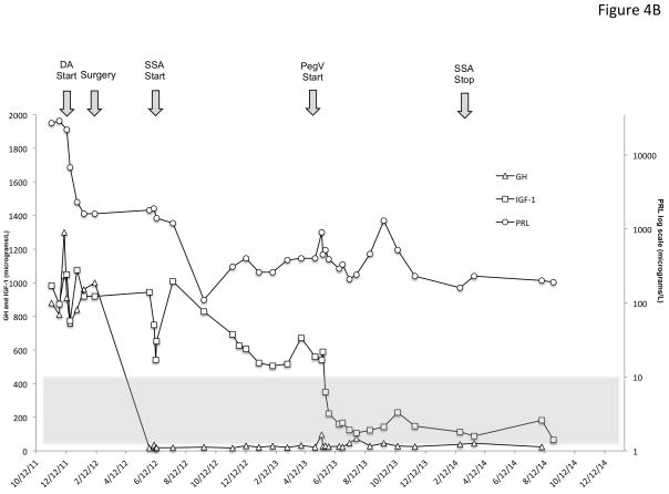
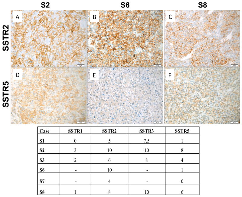
X-linked acrogigantism (X-LAG) is a new syndrome of pituitary gigantism, caused by microduplications on chromosome Xq26.3, encompassing the gene GPR101, which is highly upregulated in pituitary tumors. We conducted this study to explore the clinical, radiological, and hormonal phenotype and responses to therapy in patients with X-LAG syndrome. The study included 18 patients (13 sporadic) with X-LAG and microduplication of chromosome Xq26.3. All sporadic cases had unique duplications and the inheritance pattern in two families was dominant, with all Xq26.3 duplication carriers being affected. Patients began to grow rapidly as early as 2-3 months of age (median 12 months). At diagnosis (median delay 27 months), patients had a median height and weight standard deviation scores (SDS) of >+3.9 SDS. Apart from the increased overall body size, the children had acromegalic symptoms including acral enlargement and facial coarsening. More than a third of cases had increased appetite. Patients had marked hypersecretion of GH/IGF1 and usually prolactin, due to a pituitary macroadenoma or hyperplasia. Primary neurosurgical control was achieved with extensive anterior pituitary resection, but postoperative hypopituitarism was frequent. Control with somatostatin analogs was not readily achieved despite moderate to high levels of expression of somatostatin receptor subtype-2 in tumor tissue. Postoperative use of adjuvant pegvisomant resulted in control of IGF1 in all five cases where it was employed. X-LAG is a new infant-onset gigantism syndrome that has a severe clinical phenotype leading to challenging disease management.

Keywords: FIPA; GPR101; X chromosome; X-LAG syndrome; duplication; gigantism; pediatric; pituitary adenoma.


Figures

Figure 1
Figure 1
Genetic and growth features in patients with X-LAG syndrome. Panel A Representative chromosome Xq26.3 microduplication in female sporadic patient S10 on HD-CGH array showing the duplicated region (red) and details of the breakpoint regions of microhomology leading to the duplication (below in purple GGCC). FISH results for sporadic patient S1 (male) showing a duplication of the proximal and distal probes (red/green) in a representative leukocyte. Growth patterns during early life in sporadic patients S5 (female) and S1 (male). Panel C shows the growth in patient S5 that exceeded the 97 centile for length as early as 6 months of age; she was treated with cabergoline DA and then surgery, after which time the total resection of tumor was associated with flattening of her growth curve. She eventually was diagnosed with panhypopituitarism and diabetes insipidus and required GH supplementation as indicated. Panel D shows the early growth of patient S1, a sporadic male case that began between the ages of 9 and 12 months and continued despite subtotal surgical resection, SSA and DA; the introduction of pegvisomant (PegV) at the age of 6 led to a decrease in gain in height.
Figure 1
Figure 1
Genetic and growth features in patients with X-LAG syndrome. Panel A Representative chromosome Xq26.3 microduplication in female sporadic patient S10 on HD-CGH array showing the duplicated region (red) and details of the breakpoint regions of microhomology leading to the duplication (below in purple GGCC). FISH results for sporadic patient S1 (male) showing a duplication of the proximal and distal probes (red/green) in a representative leukocyte. Growth patterns during early life in sporadic patients S5 (female) and S1 (male). Panel C shows the growth in patient S5 that exceeded the 97 centile for length as early as 6 months of age; she was treated with cabergoline DA and then surgery, after which time the total resection of tumor was associated with flattening of her growth curve. She eventually was diagnosed with panhypopituitarism and diabetes insipidus and required GH supplementation as indicated. Panel D shows the early growth of patient S1, a sporadic male case that began between the ages of 9 and 12 months and continued despite subtotal surgical resection, SSA and DA; the introduction of pegvisomant (PegV) at the age of 6 led to a decrease in gain in height.
Figure 2
Figure 2
Physical changes in representative cases of X-LAG syndrome with Xq26.3 microduplications. Patient S8 is shown at the age of 32 months at which time her height was >4 SDS (she had been growing at an abnormal rate since the age of 11 months). Of note are her large hands and feet and facial features that are moderately coarsened but well proportioned. Panel B shows widely spaced teeth and a patch of acanthosis nigricans on the left side of her neck. In Panel C, patient S9 who began to grow abnormally at the age of 3 months is pictured shortly after diagnosis (aged 33 months), when her height was >5 SDS and hands and feet are enlarged. Panel D shows her facial features, which include a large nose and prominent mandible and the impression of hypertelorism.
Figure 2
Figure 2
Physical changes in representative cases of X-LAG syndrome with Xq26.3 microduplications. Patient S8 is shown at the age of 32 months at which time her height was >4 SDS (she had been growing at an abnormal rate since the age of 11 months). Of note are her large hands and feet and facial features that are moderately coarsened but well proportioned. Panel B shows widely spaced teeth and a patch of acanthosis nigricans on the left side of her neck. In Panel C, patient S9 who began to grow abnormally at the age of 3 months is pictured shortly after diagnosis (aged 33 months), when her height was >5 SDS and hands and feet are enlarged. Panel D shows her facial features, which include a large nose and prominent mandible and the impression of hypertelorism.
Figure 3
Figure 3
T1 weighted gadolinium enhanced MRI images of female patient S9 at diagnosis (age 2 yr 11 mo) revealed a large, hypoattenuated hourglass (“peanut”) shaped mass within the sella with expansion of the diaphragma sella (A, B). Panels B and C show coronal and saggital T1 weighted post contrast images at diagnosis in female patient S6 (age 3 yr) showing a large pituitary mass with marked upward and posterior extension and areas of degenerative changes. Post operative images (E, F) from the same patient reveal that the adenoma has been visibly resected (hormonal and growth control, however, required SSA and pegvisomant). Panels G and H show coronal and saggital T1-weighted MRI images of female patient S10 at diagnosis (aged 3 yr) showing a large homogeneous pituitary mass extending superiorly and posteriorly (“bean shaped”); the posterior pituitary is clearly seen as a hyperattenuated posterior bright spot in Panel H.
Figure 4
Figure 4
Time course of the effects of treatment modalities on GH, IGF-1 and PRL in two representative patients with X-LAG syndrome. Panel A shows the evolution of sporadic patient S1, a male who was diagnosed at the age of 56 months and underwent primary neurosurgical treatment to grossly resect the visible tumor. This surgical intervention lead to marked decreases in GH and PRL, but IGF-1 remained very high (normal IGF-1 range shown as a grey shaded zone). Addition of a SSA (octreotide LAR 30 mg/month) and later a DA (cabergoline 0.25 mg 5× week) reduced the IGF-1 by about 50% from the postoperative level but as the IGF-1 remained elevated and overgrowth continued, pegvisomant (10mg/day) was eventually added and lead to a rapid decrease in IGF-1, which allowed the SSA to be withdrawn. In Panel B, a female sporadic case S6 first received a dopamine agonist and later underwent a neurosurgical intervention, which removed the majority of the visible tumor. The post-operative GH levels were decreased greatly but remained elevated to an extent that IGF-1 secretion was still greatly increased (normal range for IGF-1 shown as a grey shaded zone)and growth continued. Addition of a depot somatostatin analog (octreotide LAR 30mg/month) did not control IGF-1 alone or in combination with cabergoline, although IGF-1 levels showed approximately a 40% decrease from levels before SSA were started. Due to the lack of control and continuing somatic overgrowth, pegvisomant was added and rapidly brought IGF-1 levels and growth under control. As IGF-1 levels fell to near the lower limit of normal for the age of the patient (5 yr), the SSA was withdrawn and subsequent IGF-1 was in the middle of the normal range on pegvisomant 10mg/day.
Figure 4
Figure 4
Time course of the effects of treatment modalities on GH, IGF-1 and PRL in two representative patients with X-LAG syndrome. Panel A shows the evolution of sporadic patient S1, a male who was diagnosed at the age of 56 months and underwent primary neurosurgical treatment to grossly resect the visible tumor. This surgical intervention lead to marked decreases in GH and PRL, but IGF-1 remained very high (normal IGF-1 range shown as a grey shaded zone). Addition of a SSA (octreotide LAR 30 mg/month) and later a DA (cabergoline 0.25 mg 5× week) reduced the IGF-1 by about 50% from the postoperative level but as the IGF-1 remained elevated and overgrowth continued, pegvisomant (10mg/day) was eventually added and lead to a rapid decrease in IGF-1, which allowed the SSA to be withdrawn. In Panel B, a female sporadic case S6 first received a dopamine agonist and later underwent a neurosurgical intervention, which removed the majority of the visible tumor. The post-operative GH levels were decreased greatly but remained elevated to an extent that IGF-1 secretion was still greatly increased (normal range for IGF-1 shown as a grey shaded zone)and growth continued. Addition of a depot somatostatin analog (octreotide LAR 30mg/month) did not control IGF-1 alone or in combination with cabergoline, although IGF-1 levels showed approximately a 40% decrease from levels before SSA were started. Due to the lack of control and continuing somatic overgrowth, pegvisomant was added and rapidly brought IGF-1 levels and growth under control. As IGF-1 levels fell to near the lower limit of normal for the age of the patient (5 yr), the SSA was withdrawn and subsequent IGF-1 was in the middle of the normal range on pegvisomant 10mg/day.
Figure 5
Figure 5
Immunohistochemistry of pituitary adenomas from Xq26.3 microduplication cases. In case S1 (sporadic male) Panel A demonstrates strong GHRH-R staining (brown) in the pituitary adenoma using a C-terminal GHRH-R peptide (20× magnification). The tumor was a mixed GH and PRL adenoma and staining of consecutive slices demonstrated largely different cell populations that stained for GH (brown; Panel B) and PRL (brown; Panel C). SSTR immunohistochemistry was also performed on tumors from patients with the Xq26.3 microduplication. AIP staining (brown) shown in 3 pituitary tumors from X-LAG syndrome cases (Panels D–F)). In all 6 cases tested AIP staining was preserved at moderate to high levels and was predominantly cytoplasmic (20× magnification).
Figure 6
Figure 6
SSTR immunohistochemistry was performed on tumors from patients with the Xq26.3 microduplication and results are shown in the tabulation (lower Panel). Expression of SSTR2 in representative tumor samples of three X-LAG patients are shown in Panels D-F and SSTR5 in Panels G-I. Immunoreactive scores for SSTR2 for all three samples illustrated were high, whereas SSTR5 scores varied from low (S6) to high (S2). Original magnification: ×400. Scale bar: 20 μm.

References

    1. Asa SL, Kovacs K, Stefaneanu L, Horvath E, Billestrup N, Gonzalez-Manchon C, Vale W. Pituitary mammosomatotroph adenomas develop in old mice transgenic for growth hormone-releasing hormone. Proceedings of the Society for Experimental Biology and Medicine Society for Experimental Biology and Medicine. 1990;193:232–235. - PubMed
    1. Asa SL, Kovacs K, Stefaneanu L, Horvath E, Billestrup N, Gonzalez-Manchon C, Vale W. Pituitary adenomas in mice transgenic for growth hormone-releasing hormone. Endocrinology. 1992;131:2083–2089. doi: 10.1210/endo.131.5.1425411. - DOI - PubMed
    1. AvRuskin TW, Sau K, Tang S, Juan C. Childhood acromegaly: successful therapy with conventional radiation and effects of chlorpromazine on growth hormone and prolactin secretion. The Journal of Clinical Endocrinology & Metabolism. 1973;37:380–388. - PubMed
    1. Beckers A, Aaltonen LA, Daly AF, Karhu A. Familial isolated pituitary adenomas (FIPA) and the pituitary adenoma predisposition due to mutations in the aryl hydrocarbon receptor interacting protein (AIP) gene. Endocrine Reviews. 2013;34:239–277. doi: 10.1210/er.2012-1013. - DOI - PMC - PubMed
    1. Borson-Chazot F, Garby L, Raverot G, Claustrat F, Raverot V, Sassolas G group GTE. Acromegaly induced by ectopic secretion of GHRH: a review 30 years after GHRH discovery. Annales d’Endocrinologie. 2012;73:497–502. doi: 10.1016/j.ando.2012.09.004. - DOI - PubMed

Publication types

MeSH terms