. 2015 Aug 15;212(4):645-53.
doi: 10.1093/infdis/jiv103. Epub 2015 Feb 23.

Selection and Counterselection of Hia Expression Reveals a Key Role for Phase-Variable Expression of Hia in Infection Caused by Nontypeable Haemophilus influenzae

Affiliations

Selection and Counterselection of Hia Expression Reveals a Key Role for Phase-Variable Expression of Hia in Infection Caused by Nontypeable Haemophilus influenzae

John M Atack et al. J Infect Dis. .

Abstract

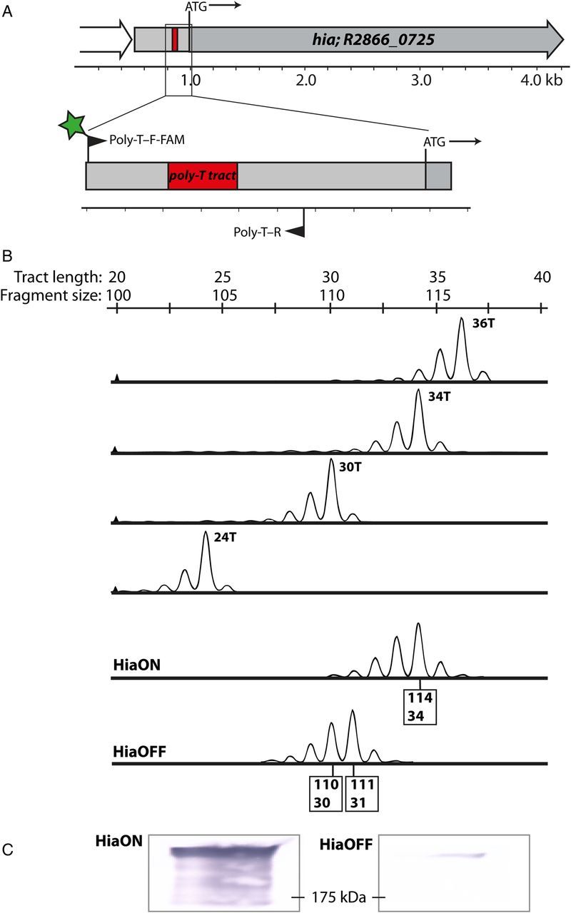
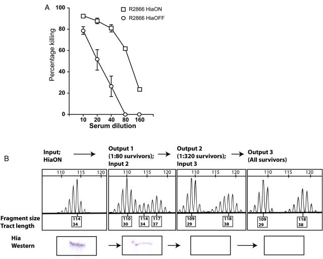
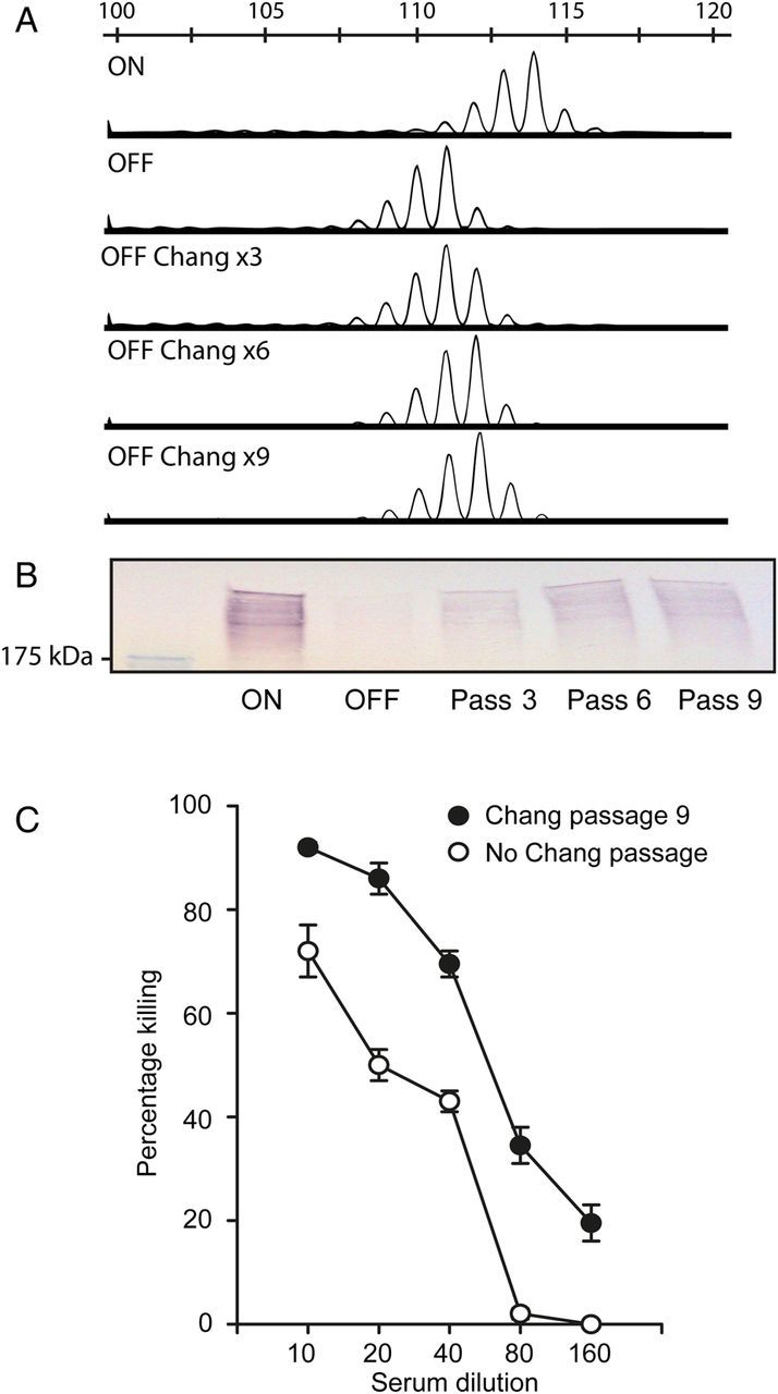
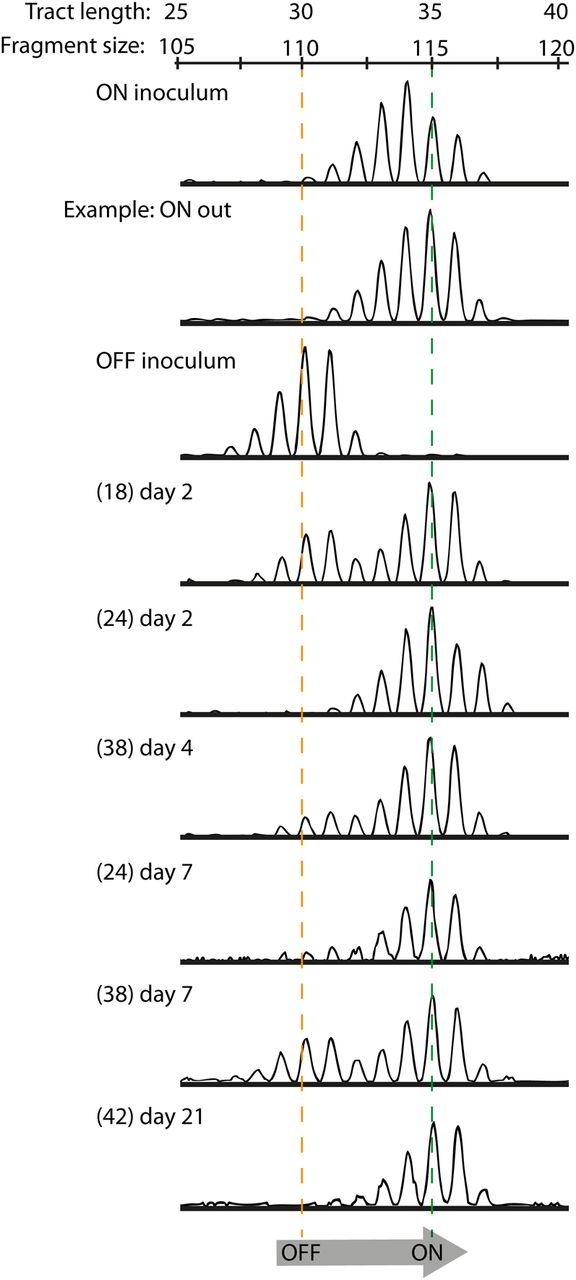
Hia is a major adhesin of nontypeable Haemophilus influenzae (NTHi) and has long been investigated as a vaccine candidate. Here we show that Hia phase variation is controlled by changes in the length of a polythymidine tract located in the hia promoter. Studies of an invasive clinical isolate (strain R2866) show that strains expressing high Hia levels are more efficiently killed by opsonophagocytosis. An opsonophagocytic assay was used to select for a subpopulation of variants that expressed a low level of Hia, which facilitated their escape from killing by anti-Hia antisera. Conversely, a subpopulation of variants expressing a high level of Hia was selected for during passaging through Chang cells. In both cases, phase variation of Hia expression corresponded directly with discrete modal changes in polythymidine tract length. In the chinchilla model of NTHi infection, we observed consistent selection for high Hia expression upon nasopharyngeal colonization, confirming the key role of phase-variable expression of Hia within a specific niche in vivo.

Keywords: Haemophilus; adhesion; colonization; phase variation.


Figures

Figure 1.
Figure 1.
Identification of poly-T tract in the hia promoter and design and validation of a fluorescent polymerase chain reaction (PCR) technique to monitor changes. A, Shown are a large (34 T in HiaON) poly-T tract in the promoter region of the hia gene and the location of primers designed to map this tract (6 carboxyfluorescein [FAM]–labeled poly-T–F and poly-T–R). B, Results of FAM-labeled PCR and fragment analysis, using purified standards containing a known number of T residues in the tract to validate this PCR method. The sequence of the standards matches the sequence of the promoter containing the poly-T tract amplified by primer pair poly-T–F-FAM and poly-T–R, with each containing the specified number of T residues. The bottom 2 panels show results of mapping the poly-T tract in nontypeable Haemophilus influenzae strains R2866 HiaON and HiaOFF, with sizes of major fragments corresponding to the number of T residues. C, Western blots from samples used to make the genomic DNA for HiaON and HiaOFF samples in panel B, confirming that changes in tract length match differences in Hia protein levels. The molecular weight size marker is shown at 175 kDa.
Figure 2.
Figure 2.
Opsonophagocytic killing assays. A, Opsonophagocytic killing of nontypeable Haemophilus influenzae strains R2866 HiaON and HiaOFF. At all dilutions of sera, P ≤ .0001 (by the Student t test). B, Results of multiple rounds of opsonophagocytic killing using R2866 HiaON as the initial input. Survivors from the 1:80 dilution of anti-3248 Hia sera in round 1 were selected as the input for round 2, using anti-11 Hia antisera. The 1:320 survivors from round 2 were then selected as the input for round 3, also using anti-11 Hia antisera. Poly-T tract polymerase chain reactions and Western blots of inputs and survivors were performed as described in “Materials and Methods” section. Full bacterial counts from each round can be located in Supplementary Table 2.
Figure 3.
Figure 3.
Chang passaging of nontypeable Haemophilus influenzae (NTHi) strain R2866 HiaOFF. A, Poly-T polymerase chain reaction (PCR) fragment analyses of multiple rounds of Chang adherence assays were performed using NTHi strain R2866 HiaOFF, and the size of the poly-T tract and Hia protein levels were probed as described in “Materials and Methods” section. B, Western blots using the 3, 6, and 9 passaged outputs. C, R2866 HiaOFF Chang × 9 passed strain (HiaOFF-Chang) was then used in an opsonophagocytic killing assay with R2866 HiaOFF as a reference. P = .0001, by the Student t test, at all titrations.
Figure 4.
Figure 4.
In vivo assay for hia expression levels in a chinchilla model. Nontypeable Haemophilus influenzae (NTHi) strain R2866 HiaON and HiaOFF were used to infect chinchillas at a colony-forming unit dose of approximately 1 × 10⁸, with samples collected from the nasopharynx on days 2, 4, 7, 10, 14, 18, and 21 after infection. Plate counts were enumerated by plating 10 µL of each sample (full plate counts for the 3- and 5-animal cohorts are shown in Supplementary Table 2), with the remaining sample snap frozen so that bacterial cultures represented the state at the time and site of sampling. Polymerase chain reaction analysis of poly-T fragments was performed to characterize phase variation in the poly-T tract length relative to the input strains. All HiaON-infected animals were colonized by strains containing the same number of T residues (34 or 35) that would give high levels of Hia protein (ON inoculum; only a single example [ie, ON out] of an output from an animal infected with HiaON is shown; the full data set is in Supplementary Figure 2). All animals infected with HiaOFF (OFF input) showed a clear switch from 31 T residues in the input to 35 T residues in all output samples, which would correlate with a switch from low to high Hia protein levels. Numbers of animals from which samples could be analyzed from HiaOFF input are shown in brackets, with the day of sample collection shown on each individual trace (the nasopharyngeal lavage label was only used in the Supplementary Figure 3).

Similar articles

Cited by

References

    1. Haggard M. Otitis media: prospects for prevention. Vaccine 2008; 26(suppl 7):G20–4. - PubMed
    1. Johnson RH. Community-acquired pneumonia: etiology, diagnosis, and treatment. Clin Ther 1988; 10:568–73. - PubMed
    1. Sethi S, Murphy TF. Infection in the pathogenesis and course of chronic obstructive pulmonary disease. N Engl J Med 2008; 359:2355–65. - PubMed
    1. A.A.P. Diagnosis and management of acute otitis media. Pediatrics 2004; 113:1451–65. - PubMed
    1. Alsarraf R, Jung CJ, Perkins J, Crowley C, Alsarraf NW, Gates GA. Measuring the indirect and direct costs of acute otitis media. Arch Otolaryngol Head Neck Surg 1999; 125:12–8. - PubMed

Publication types