. 2015 Apr 10;290(15):9465-77.
doi: 10.1074/jbc.M114.620724. Epub 2015 Feb 24.

Mechanisms of membrane binding of small GTPase K-Ras4B farnesylated hypervariable region

Affiliations

Mechanisms of membrane binding of small GTPase K-Ras4B farnesylated hypervariable region

Hyunbum Jang et al. J Biol Chem. .

Abstract

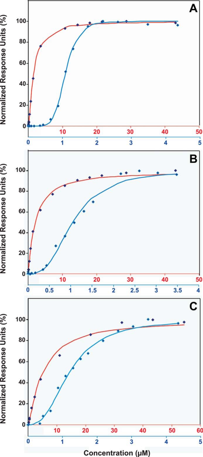
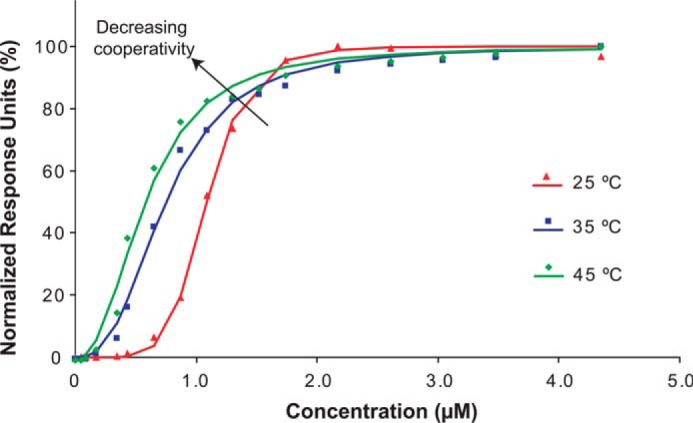
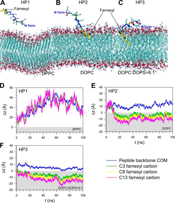
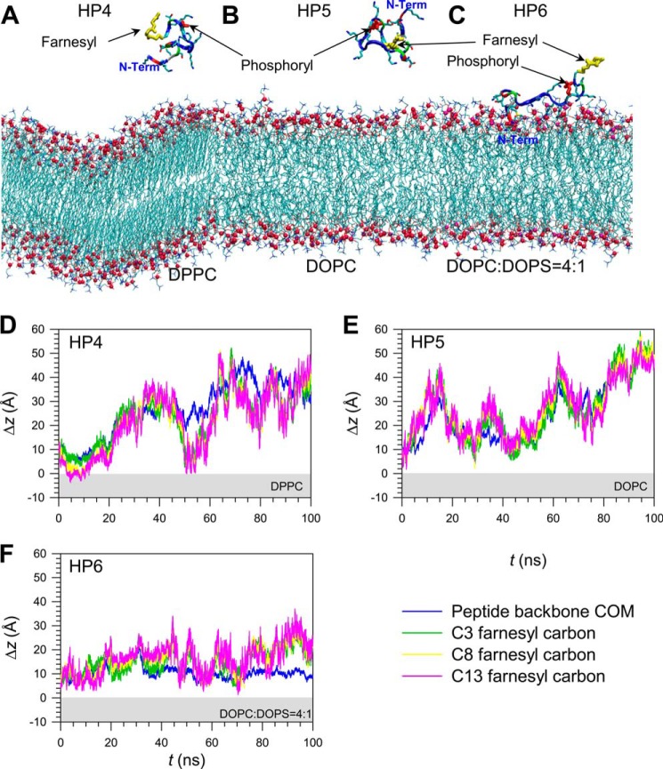
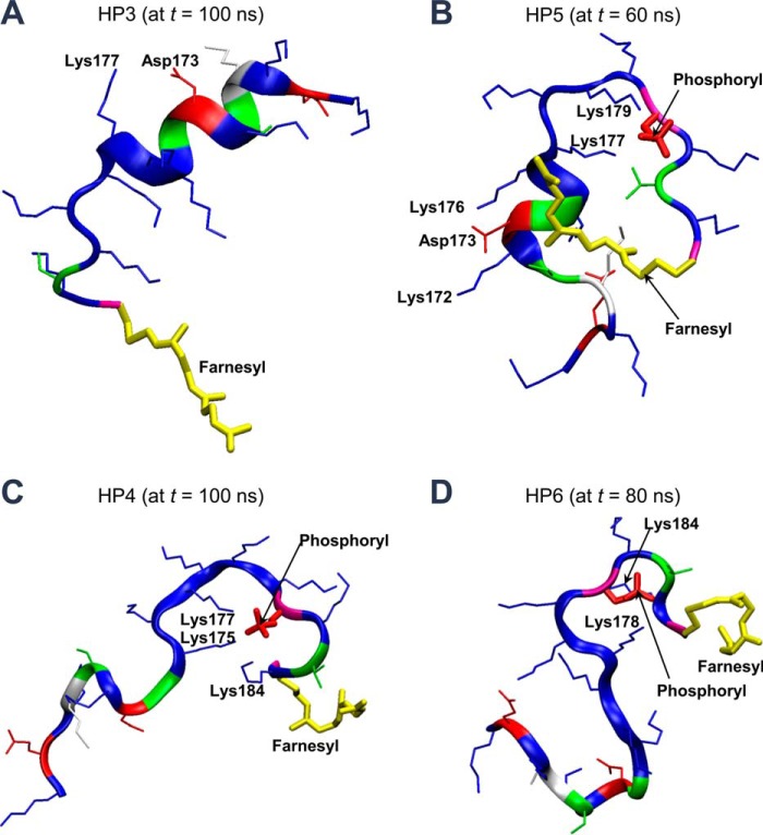
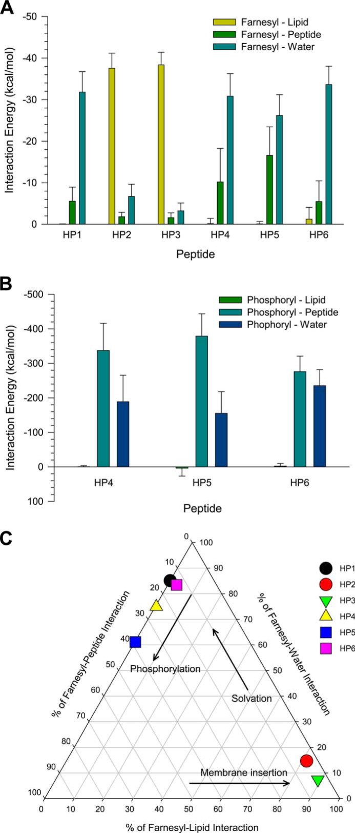
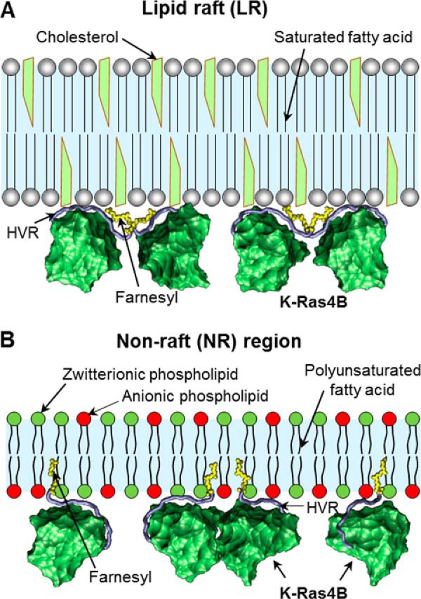
K-Ras4B belongs to a family of small GTPases that regulates cell growth, differentiation and survival. K-ras is frequently mutated in cancer. K-Ras4B association with the plasma membrane through its farnesylated and positively charged C-terminal hypervariable region (HVR) is critical to its oncogenic function. However, the structural mechanisms of membrane association are not fully understood. Here, using confocal microscopy, surface plasmon resonance, and molecular dynamics simulations, we observed that K-Ras4B can be distributed in rigid and loosely packed membrane domains. Its membrane binding domain interaction with phospholipids is driven by membrane fluidity. The farnesyl group spontaneously inserts into the disordered lipid microdomains, whereas the rigid microdomains restrict the farnesyl group penetration. We speculate that the resulting farnesyl protrusion toward the cell interior allows oligomerization of the K-Ras4B membrane binding domain in rigid microdomains. Unlike other Ras isoforms, K-Ras4B HVR contains a single farnesyl modification and positively charged polylysine sequence. The high positive charge not only modulates specific HVR binding to anionic phospholipids but farnesyl membrane orientation. Phosphorylation of Ser-181 prohibits spontaneous farnesyl membrane insertion. The mechanism illuminates the roles of HVR modifications in K-Ras4B targeting microdomains of the plasma membrane and suggests an additional function for HVR in regulation of Ras signaling.

Keywords: Cooperativity; HVR; Membrane Microdomains; Phospholipid; Phosphorylation; Post-translational Modification (PTM); Protein Isoprenylation.


Figures

FIGURE 1.
FIGURE 1.
Induction of HA-tagged K-Ras4B-G12V expression in mouse type II alveolar epithelial cells. A, immunoblot analysis of whole cell lysates of E10 cells induced to express HA-tagged K-Ras4B-G12V using increasing concentrations of doxycycline was performed using HRP-conjugated anti-HA antibody (GenScript), with anti-β-Actin (Santa Cruz) used as a loading control. Cells were lysed in ice-cold radioimmune precipitation assay buffer before sonication. Confocal microscopy analysis of HA-KRas4B expression was performed as described, without GM1 staining.
FIGURE 2.
FIGURE 2.
Confocal microscopy images of HA-K-Ras4B co-localization with the GM1 LR marker in E10 cells incubated in culture media at 18 °C (upper panels), 37 °C (middle panels), and 42 °C (lower panels) for 15 min before fixing. Shown are slides stained with DAPI (blue), anti-HA antibody conjugated with Alexa-Fluor 488 (green), and cholera toxin subunit B labeled with Vybrant Alexa Fluor 594 (red) for GM1 recognition. The merged images show co-localization of K-Ras4B and GM1 at 18 °C (Mander's coefficient = 0.764, 0.887), 37 °C (Mander's coefficient = 0.794, 0.839), and 42 °C (Mander's coefficient = 0.891, 0.940). The images were generated using Zeiss LSM 700 confocal microscope, and the data were analyzed using LSM Browser Software (Zeiss).
FIGURE 3.
FIGURE 3.
SPR titration of DPPC (A), DOPC (B), and DOPS (C) nanodiscs with non-farnesylated (red) and farnesylated HVR (blue) of K-Ras4B. The normalized response units at equilibrium for various injection points were plotted versus concentration, and the Hill equation was used to fit the curves. Red and blue horizontal labels of concentration correspond to non-farnesylated and farnesylated HVRs, respectively.
FIGURE 4.
FIGURE 4.
Binding cooperativity of farnesylated HVR of K-Ras4B to DPPC phospholipids decreases with an increase in temperature. At 25 °C, the peptide binds DPPC nanodiscs with high cooperativity (HC = 6.17 ± 0.28). When the temperature is increased to 35 °C, the cooperativity decreases (HC = 2.69 ± 0.04). At the temperature of 45 °C, the cooperativity is further reduced (HC = 2.39 ± 0.02).
FIGURE 5.
FIGURE 5.
Snapshots of farnesylated HVR peptides of K-Ras4B, HP1, HP2, and HP3, at a simulation time of t = 100 ns, representing the peptide interaction with the zwitterionic DPPC bilayer in the gel phase (A), the zwitterionic DOPC bilayer in the liquid phase (B), and the anionic DOPC:DOPS (mole ratio 4:1) bilayer in the liquid phase (C). In the peptides, hydrophobic residues are shown in white, polar and Gly residues are shown in green, negatively charged residues are shown in red, and positively charged residues are in blue. The thick yellow stick represents the farnesyl group. Shown is the time series of the deviation, Δz, from the upper bilayer leaflet for the center of mass of the HVR peptide and selected carbon atoms in the farnesyl group for the HP1 (D), HP2 (E), and HP3 peptides (F). The light gray area denotes the interior of the lipid bilayers.
FIGURE 6.
FIGURE 6.
Snapshots of farnesylated and phosphorylated HVR peptides of K-Ras4B, HP4, HP5, and HP6, at a simulation time of t = 100 ns, representing the peptide interaction with the zwitterionic DPPC bilayer in the gel phase (A), the zwitterionic DOPC bilayer in the liquid phase (B), and the anionic DOPC:DOPS (mole ratio 4:1) bilayer in the liquid phase (C). In the peptides, hydrophobic residues are shown in white, polar and Gly residues are shown in green, negatively charged residues are in red, and positively charged residues are in blue. Thick yellow and red sticks represent the farnesyl and phosphoryl groups, respectively. Shown is the time series of the deviation, Δz, from the upper bilayer leaflet for the center of mass of HVR peptide and selected carbon atoms in the farnesyl group for the HP4 (D), HP5 (E), and HP6 peptides (F). The light gray area denotes the interior of the lipid bilayers.
FIGURE 7.
FIGURE 7.
Snapshots depicted at t = 100 ns for the HP3 peptide on the anionic DOPC:DOPS (mole ratio 4:1) bilayer in the liquid phase (A), at t = 60 ns for the HP5 peptide on the zwitterionic DOPC bilayer in the liquid phase (B), at t = 100 ns for the HP4 peptide on the zwitterionic DPPC bilayer in the gel phase (C), and at t = 80 ns for the HP6 peptide on the anionic DOPC:DOPS (mole ratio 4:1) bilayer in the liquid phase (D). In the peptide ribbon, hydrophobic residues are shown in white, polar and Gly residues are shown in green, negatively charged residues in red, and positively charged residues are in blue. Thick yellow and red sticks represent the farnesyl and phosphoryl groups, respectively.
FIGURE 8.
FIGURE 8.
Shown are the averaged total interaction energies of the farnesyl (A) and phosphoryl groups (B) in the HVR peptides of K-Ras4B with the surrounding environments including lipid, its own peptide, and water. C, interaction map representing the percentage of farnesyl interactions with surrounding environments including lipid, its own peptide, and water. The interaction energies of each farnesyl with lipid, peptide, and water are calculated and then averaged over the time.
FIGURE 9.
FIGURE 9.
Model for membrane association of K-Ras4B at different plasma membrane microdomains. A, LR is rich in cholesterol and contains saturated lipids such as sphingolipids. It is highly ordered and tightly packed into an ordered gel phase. The K-Ras4B farnesyls are not able to insert into the LR microdomain. Instead, they bind together via farnesyl-farnesyl interactions leading to high cooperativity. B, NR microdomain primarily contains phosphatidylserine and phosphatidic acid with acidic headgroup. These lipids are polyunsaturated and pack into a less ordered fluid-like phase. The farnesyl groups can insert spontaneously into the NR microdomain.

Similar articles

Cited by

References

    1. Friday B. B., Adjei A. A. (2005) K-ras as a target for cancer therapy. Biochim. Biophys. Acta 1756, 127–144 - PubMed
    1. Ellis C. A., Clark G. (2000) The importance of being K-Ras. Cell. Signal. 12, 425–434 - PubMed
    1. Esteban L. M., Vicario-Abejón C., Fernández-Salguero P., Fernández-Medarde A., Swaminathan N., Yienger K., Lopez E., Malumbres M., McKay R., Ward J. M., Pellicer A., Santos E. (2001) Targeted genomic disruption of H-ras and N-ras, individually or in combination, reveals the dispensability of both loci for mouse growth and development. Mol. Cell. Biol. 21, 1444–1452 - PMC - PubMed
    1. Koera K., Nakamura K., Nakao K., Miyoshi J., Toyoshima K., Hatta T., Otani H., Aiba A., Katsuki M. (1997) K-ras is essential for the development of the mouse embryo. Oncogene 15, 1151–1159 - PubMed
    1. Plowman S. J., Williamson D. J., O'Sullivan M. J., Doig J., Ritchie A. M., Harrison D. J., Melton D. W., Arends M. J., Hooper M. L., Patek C. E. (2003) While K-ras is essential for mouse development, expression of the K-ras 4A splice variant is dispensable. Mol. Cell. Biol. 23, 9245–9250 - PMC - PubMed

Publication types

MeSH terms