. 2015 Apr 24;290(17):10717-28.
doi: 10.1074/jbc.M115.646430. Epub 2015 Feb 24.

Fo-driven Rotation in the ATP Synthase Direction against the Force of F1 ATPase in the FoF1 ATP Synthase

Affiliations

Fo-driven Rotation in the ATP Synthase Direction against the Force of F1 ATPase in the FoF1 ATP Synthase

James Martin et al. J Biol Chem. .

Abstract

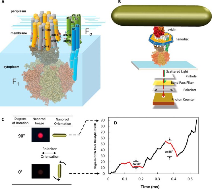
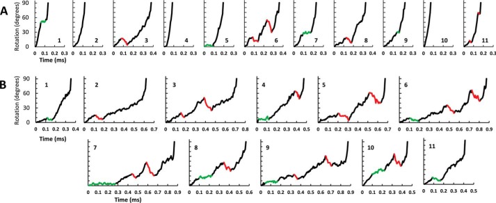
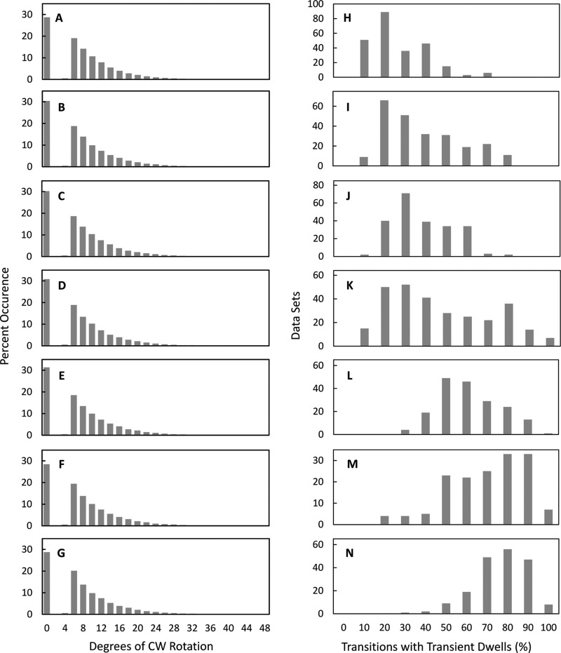
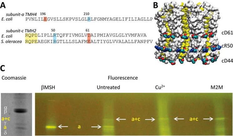
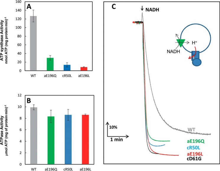
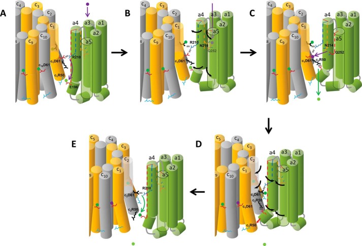
Living organisms rely on the FoF1 ATP synthase to maintain the non-equilibrium chemical gradient of ATP to ADP and phosphate that provides the primary energy source for cellular processes. How the Fo motor uses a transmembrane electrochemical ion gradient to create clockwise torque that overcomes F1 ATPase-driven counterclockwise torque at high ATP is a major unresolved question. Using single FoF1 molecules embedded in lipid bilayer nanodiscs, we now report the observation of Fo-dependent rotation of the c10 ring in the ATP synthase (clockwise) direction against the counterclockwise force of ATPase-driven rotation that occurs upon formation of a leash with Fo stator subunit a. Mutational studies indicate that the leash is important for ATP synthase activity and support a mechanism in which residues aGlu-196 and cArg-50 participate in the cytoplasmic proton half-channel to promote leash formation.

Keywords: ATP Synthase; F1Fo ATPase; Molecular Motor; Nanodiscs; Proton Transport; Single-molecule Biophysics.


Figures

FIGURE 1.
FIGURE 1.
A, subunit components of the FoF1 ATP synthase. The E. coli Fo motor shown as a schematic includes subunits a (gray) and b1 and b2 (green) and the c10-ring, where individual c subunits are distinguished in yellow and white. The F1 ATPase motor (PDB code 3OAA) subunits include α (orange), β (tan), γ (light brown), and ϵ (dark brown). B, single molecule measurements of rotation using gold nanorods as a probe. The FoF1 complexes were incorporated into lipid bilayer nanodiscs (n-FoF1) that each contained a bilayer of phospholipid molecules surrounded by the membrane scaffold protein to stabilize Fo (27). The n-FoF1 molecules were attached to the microscope slide via His6 tags on the N termini of the F1 β subunits. The c2▿C mutation to E. coli FoF1 was biotinylated to attach the avidin-coated gold nanorod (78 × 34 nm). The intensity of red light scattered from a single nanorod was measured as a function of time through a polarizing filter that was aligned for minimum intensity at one of the three ATPase-dependent catalytic dwells. C, scattered light intensity when the long axis of a nanorod is perpendicular and parallel to the direction of polarization. D, transient dwells that rotate the c-ring CW (red data) against the force of ATPase-driven CCW rotation (black data) observed during a single F1ATPase power stroke. The rotational positions for the first 90° of single power strokes as a function of time were determined from the arcsine (31) where minimum and maximum intensity values of light scattered from a nanorod in the presence of 1 mm Mg2+ATP represent 0°, the end of the F1 catalytic dwell, and 90°.
FIGURE 2.
FIGURE 2.
A and B, examples of successive F1 ATPase transitions observed during single-molecule data acquisition data sets of n-FoF1 at a viscosity of (A) 0. 9 cP (aqueous buffer) or (B) 3.0 cP (25% PEG-400). Examples of transient dwells where rotation was halted or contained CW rotation are indicated in green and red, respectively. Transient dwells were not observed in transitions A2, A4, and A10.
FIGURE 3.
FIGURE 3.
A–G, the extent of Fo-dependent CW rotation against the force of F1 ATPase-driven rotation. Shown is the distribution of transient dwells as a function of the degrees of CW rotation observed against the force of ATPase-driven CCW rotation. H–N, distribution of single molecule n-FoF1 transition data sets as a function of the percentage of occurrence of transient dwells during ATPase-dependent power strokes. Viscosities of 0.9 cP (A and H), 1.2 cP (B and I), 1.5 cP (C and J), 1.8 cP (D and K), 2.3 cP (E and L), 3.0 cP (F and M), and 4.3 cP (G and N) were obtained by the presence of 0%, 5%, 10%, 15%, 20%, 25%, and 30% PEG-400 in the buffer, respectively.
FIGURE 4.
FIGURE 4.
A, sequence alignments of subunit a TMH4 with subunit c TMH2, where subunit c residues in the F1 docking loop, are shown in yellow. B, the c14-ring from Pisum sativum Fo (PDB code 3V3C). C, verification that residues aGlu-196 and cArg-50 are juxtaposed in Fo at a distance capable of forming an electrostatic interaction. Following addition of either βMSH, 1,2-ethanediyl bismethanethiosulfonate, or CuCl2, FoF1-aE196C/cR50C/c2▿C, available sulfhydryl groups were fluorescently labeled with fluorescein maleimide. The subunits were separated by SDS-PAGE and visualized by fluorescence and Coomassie stain.
FIGURE 5.
FIGURE 5.
A–D, effects of site-directed mutations on the distribution of single-molecule n-FoF1 transition data sets as a function of the percentage of occurrence of transient dwells during F1 ATPase-dependent power strokes. E–H, the effects of mutations on the extent of Fo-dependent CW rotation. Shown is the distribution of transient dwells as a function of the degrees of CW rotation observed against the force of ATPase-driven CCW rotation. Data were collected at a viscosity of 4.3 cP for WT (A and E), aR210G (B and F), cD61G (C and G), and aE196Q (D and H) mutants of FoF1.
FIGURE 6.
FIGURE 6.
A–C, effects of site-directed mutations on the distribution of single-molecule n-FoF1 transition data sets as a function of the percentage of occurrence of transient dwells during F1 ATPase-dependent power strokes. D–F, the effects of mutations on the extent of Fo-dependent CW rotation. Shown is the distribution of transient dwells as a function of the degrees of CW rotation observed against the force of ATPase-driven CCW rotation. Data were collected at a viscosity of 3.0 cP for WT (A and D), cR50L (B and E), and cD44A/cR50L/cD61G (C and F) mutants of FoF1.
FIGURE 7.
FIGURE 7.
A–C, effects of the FoF1 mutations cR50L, aE196L, and aE196Q using inverted membrane vesicles on ATP synthesis (A) and ATP hydrolysis (B) activity measured by ensemble coupled enzyme assays and on NADH-driven ACMA quenching (C). The green triangle represents electron transfer complexes that pump protons in response to NADH, and the red arrow indicates the mutation-dependent restriction of Fo-dependent proton flow that results in an increase in ACMA quenching.
FIGURE 8.
FIGURE 8.
Model for the mechanism of Fo-dependent CW rotation in the ATP synthase direction. A, the deprotonated periplasmic channel conformation of subunit a, where the cytoplasmic end of aTMH4 is positioned between c-ring subunits c10 and c1. The position of aArg-210 on aTMH4 has displaced the proton (green dot) from c1Asp-61 to protonate aGlu-196. Vertical dotted lines in subunit a TMHs indicate the rotational position of residues. B, protonation of the periplasmic half-channel (purple dot) induces the subunit a conformational change where aTMH4 rotates relative to the c-ring. C, the PPC subunit a conformation. The cytoplasmic end of aTMH4 containing aGlu-196 is positioned between c-ring subunits c1 and c2 to form the leash. Leash formation is facilitated by a salt bridge between c2Arg-50 and aGlu-196, which moves the proton to the cytoplasm. The PPG conformation caused aR210 to be rotated from subunit c1 to c2 in the c-ring. This conformation also facilitates proton transfer from the periplasmic half channel to c1Asp-61. D, the reversion of subunit a from the PPG to the DPG conformation by van der Waals repulsion when the leash is engaged is responsible for CW c-ring rotation (E). Release of the leash occurs after CW rotation by 36°. As subunit a forms the DPG conformation, aArg-210 rotates into position to transfer the proton from c2Asp-61 to aGlu-196, which facilitates the release of the leash.
FIGURE 9.
FIGURE 9.
Sequence alignments of subunit a TMH4 and subunit c TMH2 aligned by organism. Sequence alignments of subunit c TMH2 are sorted by positively charged residues exposed to the outer surface of the c-ring near the cytoplasmic side of the membrane. Organisms for which a crystal structure of the c-ring is available are highlighted in yellow, and residues in which the c subunit side chains are not exposed to the outer surface are highlighted in gray.

Similar articles

Cited by

References

    1. Spetzler D., Ishmukhametov R., Hornung T., Martin J., York J., Jin-Day L., Frasch W. D. (eds) (2012) Photosynthesis, in Advances in Photosynthesis and Respiration (Eaton-Rye J., Tripathy B., Sharkey T., eds.), Vol. 34, pp. 561–590, Springer, Dordrecht, The Netherlands
    1. Girvin M. E., Fillingame R. H. (1994) Hairpin folding of subunit c of F1Fo ATP synthase: 1H distance measurements to nitroxide-derivatized aspartyl-61. Biochemistry 33, 665–674 - PubMed
    1. Watt I. N., Montgomery M. G., Runswick M. J., Leslie A. G., Walker J. E. (2010) Bioenergetic cost of making an adenosine triphosphate molecule in animal mitochondria. Proc. Natl. Acad. Sci. U.S.A. 107, 16823–16827 - PMC - PubMed
    1. Pogoryelov D., Yildiz O., Faraldo-Gómez J. D., Meier T. (2009) High-resolution structure of the rotor ring of a proton-dependent ATP synthase. Nat. Struct. Mol. Biol. 16, 1068–1073 - PubMed
    1. Pogoryelov D., Krah A., Langer J. D., Yildiz Ö., Faraldo-Gómez J. D., Meier T. (2010) Microscopic rotary mechanism of ion translocation in the F-0 complex of ATP synthases. Nat. Chem. Biol. 6, 891–899 - PubMed

Publication types

MeSH terms

Substances

LinkOut - more resources