. 2015 Apr 3;290(14):8913-24.
doi: 10.1074/jbc.M114.621003. Epub 2015 Feb 24.

Recruitment of β-catenin to N-cadherin is necessary for smooth muscle contraction

Affiliations

Recruitment of β-catenin to N-cadherin is necessary for smooth muscle contraction

Tao Wang et al. J Biol Chem. .

Abstract

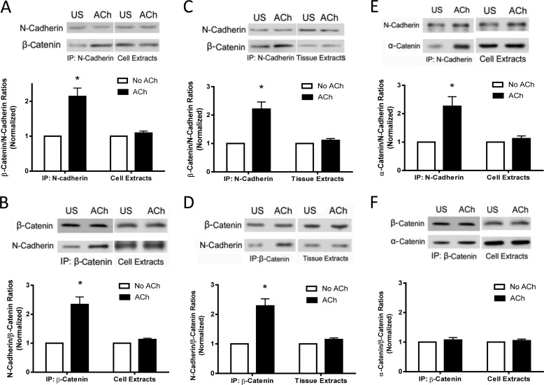
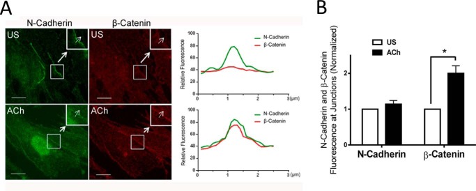
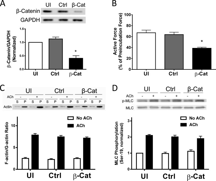
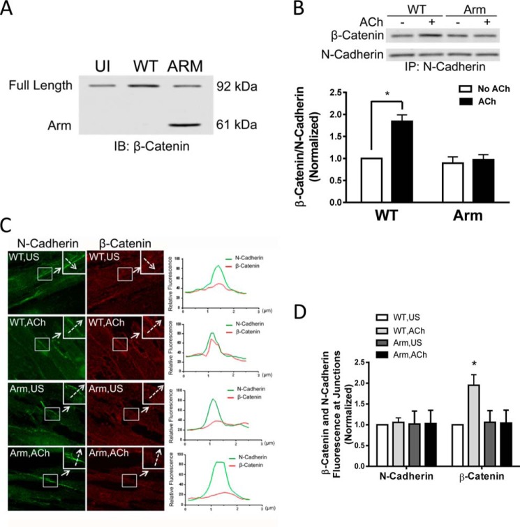
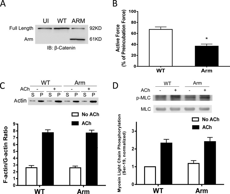
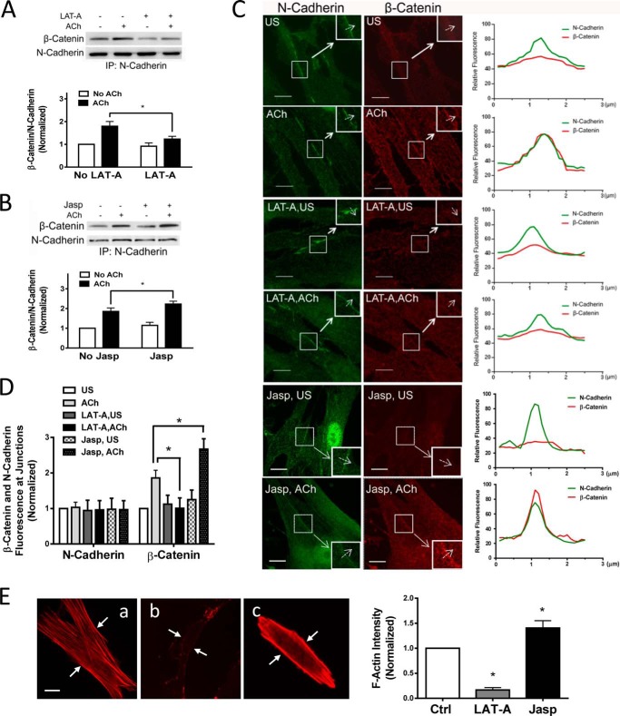
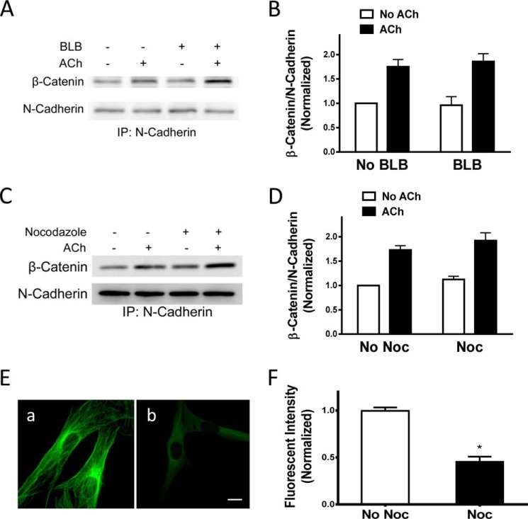
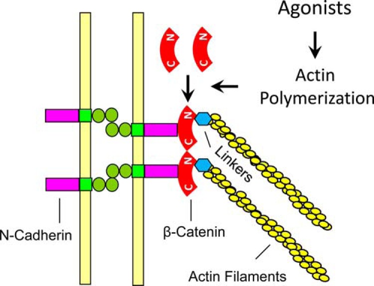
β-Catenin is a key component that connects transmembrane cadherin with the actin cytoskeleton at the cell-cell interface. However, the role of the β-catenin/cadherin interaction in smooth muscle has not been well characterized. Here stimulation with acetylcholine promoted the recruitment of β-catenin to N-cadherin in smooth muscle cells/tissues. Knockdown of β-catenin by lentivirus-mediated shRNA attenuated smooth muscle contraction. Nevertheless, myosin light chain phosphorylation at Ser-19 and actin polymerization in response to contractile activation were not reduced by β-catenin knockdown. In addition, the expression of the β-catenin armadillo domain disrupted the recruitment of β-catenin to N-cadherin. Force development, but not myosin light chain phosphorylation and actin polymerization, was reduced by the expression of the β-catenin armadillo domain. Furthermore, actin polymerization and microtubules have been implicated in intracellular trafficking. In this study, the treatment with the inhibitor latrunculin A diminished the interaction of β-catenin with N-cadherin in smooth muscle. In contrast, the exposure of smooth muscle to the microtubule depolymerizer nocodazole did not affect the protein-protein interaction. Together, these findings suggest that smooth muscle contraction is mediated by the recruitment of β-catenin to N-cadherin, which may facilitate intercellular mechanotransduction. The association of β-catenin with N-cadherin is regulated by actin polymerization during contractile activation.

Keywords: Adherens Junction; Cytoskeleton; Excitation-Contraction Coupling (E-C Coupling); Signal Transduction; Smooth Muscle.


Figures

FIGURE 1.
FIGURE 1.
Contractile activation increases the association of β-catenin with N-cadherin in smooth muscle cells/tissues. A, N-cadherin immunoprecipitates (IP) or extracts of HASM cells stimulated with ACh (10−4 m, 5 min) or left unstimulated (US) were separated by SDS-PAGE and blotted with antibodies against N-cadherin and β-catenin. Ratios of β-catenin/N-cadherin in stimulated cells were normalized to corresponding unstimulated cells. The ratios of β-catenin/N-cadherin in immunoprecipitates were significantly higher in stimulated cells than in unstimulated cells (*, p < 0.05). However, the protein ratios in cell extracts are similar in stimulated and unstimulated cells. Data are mean ± S.E. of four independent experiments. B, blots of β-catenin immunoprecipitates or extracts of cells treated with ACh or left untreated were probed with antibodies against N-cadherin and β-catenin. The ratios of N-cadherin/β-catenin in stimulated cells were normalized to corresponding unstimulated cells. Data are mean ± S.E. of four independent experiments. *, p < 0.05. C, N-Cadherin immunoprecipitates or extracts of human bronchial rings treated with ACh or left untreated were analyzed by immunoblotting. The ratios of β-catenin/N-cadherin in stimulated tissues were normalized to corresponding unstimulated tissues. Data are mean ± S.E. of three independent experiments. *, p < 0.05. D, β-catenin precipitates or extracts of human bronchial rings treated with ACh or untreated were evaluated by immunoblot analysis. The ratios of N-cadherin/β-catenin in stimulated tissues were normalized to corresponding unstimulated tissues. Data are mean ± S.E. of three independent experiments. *, p < 0.05. E, N-cadherin immunoprecipitates or extracts of HASM cells treated with ACh or left untreated were analyzed by immunoblotting. The ratios of α-catenin/N-cadherin in stimulated cells were normalized to corresponding unstimulated cells. Data are mean ± S.E. of four independent experiments. *, p < 0.05. F, β-catenin precipitates or extracts of HASM cells treated with ACh or left untreated were evaluated by immunoblot analysis. The ratios of α-catenin/β-catenin in stimulated cells were normalized to corresponding unstimulated cells. Data are mean ± S.E. of four independent experiments.
FIGURE 2.
FIGURE 2.
Activation with ACh induces the translocation of β-catenin to the intercellular junctions. A, representative micrographs illustrating the effects of ACh (10−4 m, 5 min) on the spatial localization of β-catenin and N-cadherin in HASM cells. The insets are ×1.5 magnifications of the selected areas. Dashed arrows indicate a single line scan to quantify the fluorescence signals at cell-cell junctions. The right panel shows relative fluorescence intensity. Scale bars = 10 μm. US, unstimulated. B, the fluorescence intensity of β-catenin or N-cadherin in stimulated cells was normalized to corresponding unstimulated cells. Data are mean ± S.E. of 10–12 independent experiments. *, p < 0.05.
FIGURE 3.
FIGURE 3.
β-Catenin is required for smooth muscle contraction. A, human bronchial rings were transduced with lentiviruses encoding control shRNA or β-catenin shRNA. These tissues were then incubated in serum-free medium for 3 days. Immunoblot analysis was used to assess protein expression in tissues. UI, uninfected; Ctrl, control shRNA; β-Cat, β-catenin shRNA. *, p < 0.05 (significantly lower protein ratios of β-catenin/GAPDH in tissues transduced with virus encoding β-catenin shRNA than in uninfected tissues and tissues expressing control shRNA). Data are mean ± S.E. of three independent experiments. B, the contraction of human bronchial rings was evaluated, after which they were transduced with lentiviruses as described above. Contractile responses were compared before and after incubation. *, p < 0.05 (significantly lower contractile force in bronchial rings treated with β-catenin shRNA compared with uninfected tissues or tissues infected with viruses encoding control shRNA). Data are mean ± S.E. of three independent experiments. C, uninfected cells and cells expressing control shRNA or β-catenin shRNA were stimulated with ACh (10−4 m, 5 min) or left unstimulated. F/G-actin ratios in the cells were evaluated using a fractionation assay. Data are mean ± S.E. of four independent experiments. (p > 0.05). S, supernatant; P, pellet. D, myosin light chain (MLC) phosphorylation at Ser-19 in uninfected cells and cells transduced with lentivirus encoding control or β-catenin shRNA was assessed by immunoblot analysis. Myosin phosphorylation was similar in uninfected cells, cells expressing control shRNA, or β-catenin shRNA (p > 0.05). Data are mean ± S.E. of four to five independent experiments.
FIGURE 4.
FIGURE 4.
Expression of the Arm domain of β-catenin attenuates the recruitment of β-catenin to N-cadherin. A, representative immunoblots illustrating the expression of WT β-catenin or the Arm domain of β-catenin in cells. Extracts of cells transfected with plasmids encoding WT β-catenin or the Arm domain of β-catenin were immunoblotted (IB) with β-catenin full-length antibody. The Arm domain with a molecular mass of 60 kDa was detected in the extracts of cells transfected with the plasmid for Arm domain but not in untransfected cells (UI) or in cells transfected with WT β-catenin plasmid, indicating the effective expression of the Arm domain of β-catenin in these cells. The blots are representative of four identical experiments. B, cells expressing WT β-catenin or the Arm domain of β-catenin were stimulated with 10−4 m ACh for 5 min or left unstimulated. The interaction of N-cadherin with β-catenin was evaluated by coimmunoprecipitation (IP) analysis using antibody against the β-catenin C terminus and antibody against N-cadherin. The ratios of β-catenin/N-cadherin in stimulated cells were normalized to corresponding unstimulated cells. *, p < 0.05 (significantly lower ACh-induced β-catenin/N-cadherin ratios in cells expressing the Arm domain of β-catenin compared with cells expressing WT β-catenin). Data are mean ± S.E. of four independent experiments. C, representative images illustrating the effects of the expression of the Arm domain of β-catenin on the localization of β-catenin and N-cadherin at the adherens junctions in response to ACh activation. Cells were immunostained with antibodies against the β-catenin C terminus and N-cadherin. The insets are ×1.5 magnifications of the selected areas. Dashed arrows indicate a single line scan to quantify the signals at cell-cell junctions. The right panel shows the relative fluorescence intensity. Scale bars = 10 μm. D, the fluorescence intensity of β-catenin or N-cadherin in stimulated cells was normalized to corresponding unstimulated (US) WT cells. Data are mean ± S.E. of 10–12 independent experiments. *, p < 0.05.
FIGURE 5.
FIGURE 5.
Expression of the Arm domain of β-Catenin inhibits smooth muscle contraction. A, representative immunoblots showing the expression of β-catenin and its mutant in tissues. Extracts of human bronchial rings transduced with plasmids encoding WT β-catenin or the Arm domain of β-catenin were immunoblotted (IB) with antibodies against full-length β-catenin. The blots are representative of three identical experiments. UI, uninfected. B, the contraction of human bronchial rings was evaluated, after which they were transduced with plasmids as described under “Experimental Procedures.” Contractile responses were compared before and after incubation. *, p < 0.05 (significantly lower contractile force in bronchial rings expressing the Arm domain of β-catenin compared with tissues transfected with WT β-catenin). Data are mean ± S.E. of three independent experiments. C, cells expressing WT or mutant β-catenin were stimulated with ACh or left unstimulated. F/G-actin ratios in the cells were evaluated using a fractionation assay. Data are mean ± S.E. of four independent experiments (p > 0.05). S, supernatant; P, pellet. D, myosin light chain (MLC) phosphorylation at Ser-19 in uninfected cells and cells transduced with lentivirus encoding control or β-catenin shRNA was assessed by immunoblot analysis. Myosin phosphorylation was similar in uninfected cells and cells expressing control shRNA or β-catenin shRNA (p > 0.05). Data are mean ± S.E. of four to five independent experiments.
FIGURE 6.
FIGURE 6.
Actin polymerization regulates the recruitment of β-catenin to N-cadherin upon contractile activation. A, cells were pretreated with 1 μm latrunculin A (LAT-A) for 15 min. They were then stimulated with 10−4 m ACh for 5 min or left unstimulated. The protein-protein interaction was evaluated by coimmunoprecipitation (IP). Ratios of β-catenin/N-cadherin under various treatments were normalized to unstimulated cells not treated with latrunculin A. Data are mean ± S.E. of four independent experiments. *, p < 0.05. B, cells were pretreated with 3 μm jasplakinolide (Jasp) for 30 min. The protein-protein interaction in unstimulated and stimulated cells was evaluated by coimmunoprecipitation. The ratios of β-catenin/N-cadherin under various treatments were normalized to unstimulated cells not treated with jasplakinolide. Error bars indicate S.E. *, p < 0.05; n = 4. C, cells pretreated with latrunculin A or jasplakinolide were stimulated with ACh or left unstimulated (US). Cells were then immunostained with antibodies against the β-catenin C terminus and N-cadherin. The insets are ×1.5 magnifications of the selected areas. Dashed arrows indicate a single line scan to quantify the fluorescence signals at cell-cell junctions. The right panel shows relative fluorescence intensity. Scale bar = 10 μm. D, the fluorescence intensity of β-catenin or N-cadherin in stimulated cells was normalized to corresponding unstimulated and untreated cells. Data are mean ± S.E. of 10–12 independent experiments. *, p < 0.05. E, untreated HASM cells (a) or cells treated with latrunculin A (1 μm, 15 min) (b) or jasplakinolide (3 μm, 30 min) (c) were stained with rhodamine-phalloidin to visualize F-actin. National Institutes of Health ImageJ software was used to quantify the fluorescence intensity of the total cell areas. Arrows point to the cell edges. Data are mean ± S.E. *, p < 0.05 versus control; n = 38–40 cells. Scale bars = 10 μm.
FIGURE 7.
FIGURE 7.
Tension and microtubules do not regulate the interaction of β-catenin with N-cadherin upon contractile activation. A, representative immunoblots illustrating the effects of blebbistatin (BLB) on the interaction of β-catenin with N-cadherin. Cells were pretreated with 30 μm blebbistatin for 15 min. They were then stimulated with 10−4 m ACh for 5 min or left unstimulated. β-Catenin/N-cadherin coupling was evaluated by coimmunoprecipitation (IP). B, β-catenin/N-cadherin ratios were normalized to the ratios in unstimulated cells not treated with blebbistatin. Data are mean ± S.E. of four independent experiments. C, representative immunoblots illustrating the effects of nocodazole on the interaction of β-catenin/N-cadherin. Cells were pretreated with 1 μm nocodazole for 15 min. They were then stimulated with 10−4 m ACh for 5 min or left unstimulated. β-Catenin/N-cadherin coupling was evaluated by coimmunoprecipitation. D, β-catenin/N-cadherin ratios were normalized to the ratios in unstimulated cells not treated with nocodazole (Noc). Data are mean ± S.E. of four independent experiments. E, untreated cells and HASM cells treated with nocodazole (1 μm, 15 min) were stained with α-tubulin antibody. a, untreated cells display a well defined microtubule structure. b, treated cells have a less defined filamentous structure. F, the fluorescence intensity in treated cells was normalized to untreated cells. Data are mean ± S.E. of 37–41 independent experiments. *, p < 0.05.
FIGURE 8.
FIGURE 8.
Proposed mechanism for intercellular force transmission. In addition to myosin activation, contractile agonists induce actin polymerization, which promotes the recruitment of β-catenin to N-cadherin. The increase in protein-protein interaction may enhance linkage of actin filaments to adherens junctions and promote intercellular force transmission and smooth muscle contraction. C, C terminus; N, N terminus; Linkers, linker proteins such as α-catenin, vinculin, and VASP.

Similar articles

Cited by

References

    1. Kamm K. E., Stull J. T. (1989) Regulation of smooth muscle contractile elements by second messengers. Annu. Rev. Physiol 51, 299–313 - PubMed
    1. Morgan K. G. (2008) Contractility in health and disease. J. Cell Mol. Med. 12, 2157. - PMC - PubMed
    1. Amrani Y., Panettieri R. A. (2003) Airway smooth muscle: contraction and beyond. Int. J. Biochem. Cell Biol. 35, 272–276 - PubMed
    1. Somlyo A. V., Khromov A. S., Webb M. R., Ferenczi M. A., Trentham D. R., He Z. H., Sheng S., Shao Z., Somlyo A. P. (2004) Smooth muscle myosin: regulation and properties. Philos. Trans. R. Soc. Lond. B. Biol. Sci. 359, 1921–1930 - PMC - PubMed
    1. Gunst S. J., Zhang W. (2008) Actin cytoskeletal dynamics in smooth muscle: a new paradigm for the regulation of smooth muscle contraction. Am. J. Physiol. Cell Physiol. 295, C576–C587 - PMC - PubMed

Publication types