. 2015 Apr 10;290(15):9604-13.
doi: 10.1074/jbc.M114.633198. Epub 2015 Feb 24.

Cyclic AMP signaling reduces sirtuin 6 expression in non-small cell lung cancer cells by promoting ubiquitin-proteasomal degradation via inhibition of the Raf-MEK-ERK (Raf/mitogen-activated extracellular signal-regulated kinase/extracellular signal-regulated kinase) pathway

Affiliations

Cyclic AMP signaling reduces sirtuin 6 expression in non-small cell lung cancer cells by promoting ubiquitin-proteasomal degradation via inhibition of the Raf-MEK-ERK (Raf/mitogen-activated extracellular signal-regulated kinase/extracellular signal-regulated kinase) pathway

Eui-Jun Kim et al. J Biol Chem. .

Abstract

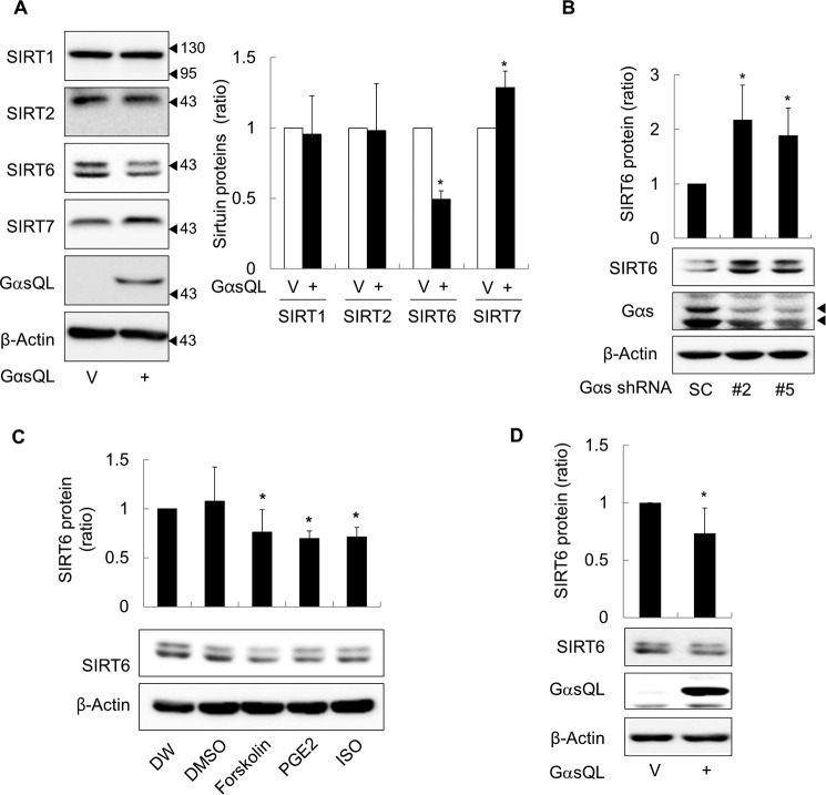
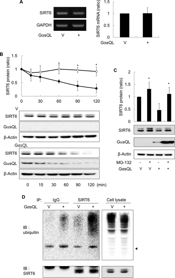
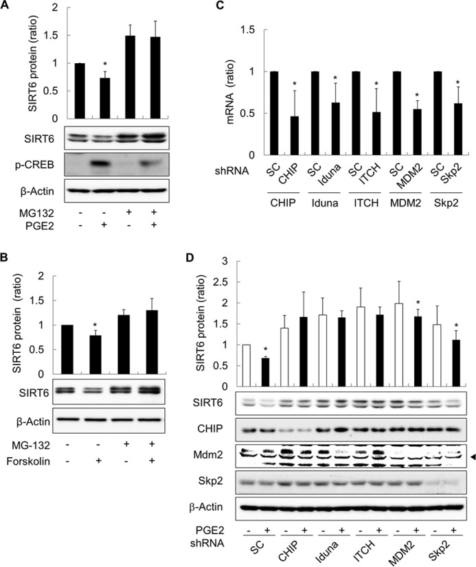
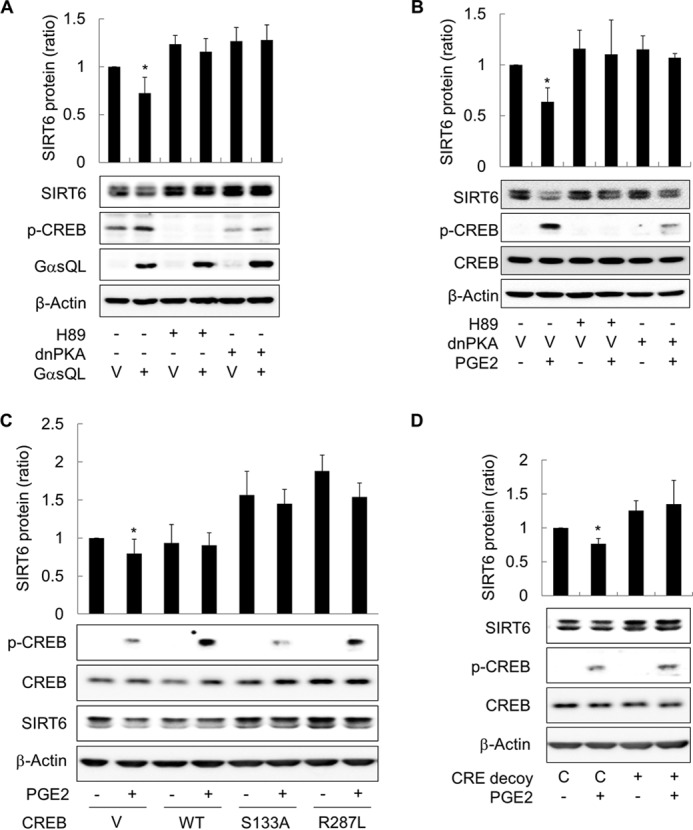
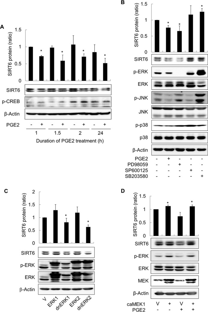
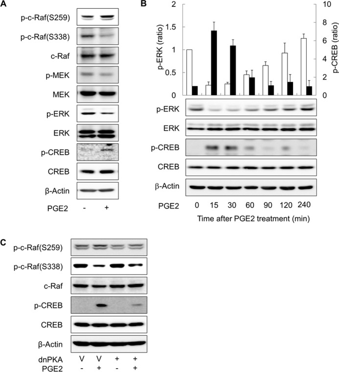
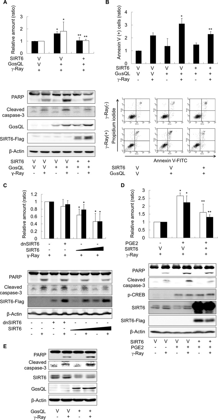
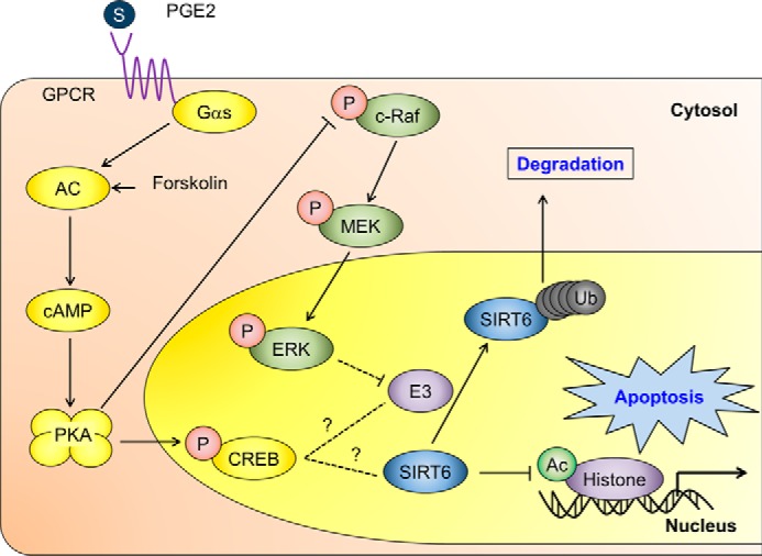
The cAMP signaling system regulates various cellular functions, including metabolism, gene expression, and death. Sirtuin 6 (SIRT6) removes acetyl groups from histones and regulates genomic stability and cell viability. We hypothesized that cAMP modulates SIRT6 activity to regulate apoptosis. Therefore, we examined the effects of cAMP signaling on SIRT6 expression and radiation-induced apoptosis in lung cancer cells. cAMP signaling in H1299 and A549 human non-small cell lung cancer cells was activated via the expression of constitutively active Gαs plus treatment with prostaglandin E2 (PGE2), isoproterenol, or forskolin. The expression of sirtuins and signaling molecules were analyzed by Western blotting. Activation of cAMP signaling reduced SIRT6 protein expression in lung cancer cells. cAMP signaling increased the ubiquitination of SIRT6 protein and promoted its degradation. Treatment with MG132 and inhibiting PKA with H89 or with a dominant-negative PKA abolished the cAMP-mediated reduction in SIRT6 levels. Treatment with PGE2 inhibited c-Raf activation by increasing inhibitory phosphorylation at Ser-259 in a PKA-dependent manner, thereby inhibiting downstream MEK-ERK signaling. Inhibiting ERK with inhibitors or with dominant-negative ERKs reduced SIRT6 expression, whereas activation of ERK by constitutively active MEK abolished the SIRT6-depleting effects of PGE2. cAMP signaling also augmented radiation-induced apoptosis in lung cancer cells. This effect was abolished by exogenous expression of SIRT6. It is concluded that cAMP signaling reduces SIRT6 expression by promoting its ubiquitin-proteasome-dependent degradation, a process mediated by the PKA-dependent inhibition of the Raf-MEK-ERK pathway. Reduced SIRT6 expression mediates the augmentation of radiation-induced apoptosis by cAMP signaling in lung cancer cells.

Keywords: Apoptosis; Cyclic AMP (cAMP); Extracellular Signal-regulated Kinase (ERK); Lung Cancer; Protein Kinase A (PKA); Sirtuin.


Figures

FIGURE 1.
FIGURE 1.
s inhibits SIRT6 expression in lung cancer cells. A, effects of Gαs on the expression of sirtuin isoforms in H1299 human lung cancer cells. B, effects of Gαs knockdown on the expression of SIRT6. C, effects of cAMP signaling on the expression of SIRT6. D, effects of Gαs on the expression of SIRT6 in A549 human lung cancer cells. Lung cancer cells (H1299 and A549 cells) were transfected with EE-tagged GαsQL (GαsQL) or vector pcDNA3.1 (V) using the calcium phosphate method and then incubated for 24 h. The cells were then harvested, homogenized, and analyzed by Western blotting. Gαs expression was knocked down by transfection of 10 μg of shRNA. Scrambled shRNA was used as a control (SC). The two arrowheads indicate long- and short-forms of Gαs proteins (B). H1299 cells were treated with 40 μm forskolin, 10 μm PGE2, 1 μm isoproterenol (ISO), distilled water (DW), or DMSO for 24 h before Western blotting analysis. An asterisk indicates a statistically significant difference compared with the respective control or vector-transfected cells (*, p < 0.05; Mann-Whitney U test).
FIGURE 2.
FIGURE 2.
s promotes the proteasome-dependent degradation of SIRT6 in H1299 cells. A, effects of Gαs on the expression of SIRT6 mRNA. H1299 cells were transfected with GαsQL or vector (V), and the expression of SIRT6 mRNA was analyzed by RT-PCR and real-time quantitative PCR 24 h later. B, effects of Gαs on the degradation of SIRT6 protein. H1299 cells were transfected with GαsQL or vector (V) and then treated with 50 μg/ml cycloheximide (CHX) 24 h later. Cells were harvested at the indicated times. C, effect of MG-132 on the SIRT6 expression. H1299 cells were transfected with GαsQL or vector (V) and incubated for 6 h before exposure to 10 μm MG132 for 18 h. Cells were then harvested for analysis. D, effects of Gαs on the ubiquitination of SIRT6. H1299 cells were transfected with GαsQL or vector (V) and incubated 24 h before harvesting. Whole cell lysates (800 μg) were precleared by incubation with protein A-agarose for 1 h followed by centrifugation. IP, immunoprecipitation. The precleared lysate was then incubated with an anti-SIRT6 antibody (1 μg) or with control IgG at 4 °C for 16 h followed by protein A-agarose for 2 h. Finally, the samples were washed three times and analyzed by Western blotting (IB) with antibodies against SIRT6 or ubiquitin. An arrowhead indicates the molecular weight of SIRT6 (D). Asterisks (*) on the histograms indicate a statistically significant difference from the respective control or vector-transfected control cells (p < 0.05, Mann-Whitney U test). One-way analysis of variance analysis was also performed to compare the amount of HDAC6 protein remained following cycloheximide treatment (B).
FIGURE 3.
FIGURE 3.
Prostaglandin E2 promotes the proteasome-dependent degradation of SIRT6 in H1299 cells. A and B, effect of PGE2 or forskolin on the degradation of SIRT6 protein. H1299 cells were treated with 10 μm PGE2, 40 μm forskolin, or DMSO in the presence/absence of 10 μm MG132 for 24 h before Western blot analysis. C and D, effects of knocking down E3 ligases on PGE2-promoted degradation of SIRT6 protein. H1299 cells were transfected with shRNA against CHIP, iduna, ITCH, Mdm2, or Skp2 or with scrambled shRNA (SC). To confirm knock down of the target ligases, expression of mRNA for each E3 ligase was analyzed by qPCR at 48 h after transfection (C). At 24 h after transfection, cells were treated with 10 μm PGE2 for 24 h before Western blotting (D). Asterisks (*) on the histograms indicate a statistically significant difference from the respective control cells (p < 0.05, Mann-Whitney U test).
FIGURE 4.
FIGURE 4.
cAMP signaling inhibits SIRT6 expression in H1299 cells via the PKA and CREB pathways. A, inhibiting PKA abolished the effects of GαsQL on SIRT6 expression. B, inhibiting PKA blocked the effects of PGE2 on SIRT6 expression. C, inhibiting CREB activation increased SIRT6 expression. D, inhibiting CREB binding to CRE increased SIRT6 expression. H1299 cells were transfected with GαsQL, a dnPKA, wild-type CREB (WT), dominant-negative CREBs (S133A, R287L), oligonucleotides (CRE decoy, CRE control; C), or respective control vectors (V) and then incubated for 24 h. The cells were then treated with 20 μm H89, 10 μm PGE2, or DMSO for 24 h before Western blot analysis. CREB phosphorylation (pCREB) was analyzed 30 min after exposure to PGE2. Asterisks (*) on the histograms indicate a statistically significant difference from the respective control cells (p < 0.05, Mann-Whitney U test).
FIGURE 5.
FIGURE 5.
cAMP signaling reduces SIRT6 expression in H1299 cells by inhibiting the ERK pathway. A, effect of PGE2 treatment duration on SIRT6 expression. H1299 cells were treated with 10 μm PGE2 or with DMSO for the indicated times before the culture medium was aspirated. The cells were then washed with DPBS, and fresh medium was added. The cells were harvested 24 h after the start of PGE2 treatment, and SIRT6 expression was analyzed by Western blotting. B, effect of MAPK inhibition on SIRT6 expression. C, effect of ERK inhibition by dominant-negative ERKs (dnERK) on SIRT6 expression. D, effect of ERK activation on PGE2-induced inhibition of SIRT6 expression. H1299 cells were treated with 10 μm PGE2, 40 μm PD98059, 20 μm SP600125, 20 μm SB203580, or DMSO for 2 h and then harvested and analyzed by Western blotting. The expression of SIRT6 was analyzed 24 h after treatment. H1299 cells were also transfected with ERK1, ERK2, dominant-negative ERKs (dnERK1, dnERK2), constitutively active MEK1 (caMEK1), or the respective empty vectors (V) and incubated for 24 h. Transfected cells were treated with 10 μm PGE2 for 30 min (if necessary) before Western blot analysis of signaling molecules. Asterisks (*) on the histograms indicate a statistically significant difference from the respective control cells (p < 0.05, Mann-Whitney U test).
FIGURE 6.
FIGURE 6.
cAMP signaling inhibits the ERK pathway in a PKA-dependent way. A, effect of PGE2 on the activation Raf-MEK-ERK pathway. B, temporal patterns of CREB and ERK phosphorylation after PGE2 treatment. H1299 cells were treated with 10 μm PGE2 or DMSO, and CREB and ERK phosphorylation was examined at the indicated times by Western blotting. The empty bar represents p-ERK and the filled bar p-CREB. C, effect of PKA on PGE2-induced on c-Raf phosphorylation. H1299 cells were treated with 10 μm PGE2 or DMSO for 30 min and then harvested and analyzed by Western blotting. The expression of SIRT6 was analyzed 24 h after treatment. H1299 cells were also transfected with dnPKA or the empty vectors (V) and incubated for 24 h. Transfected cells were treated with 10 μm PGE2 for 30 min (if necessary) before Western blot analysis.
FIGURE 7.
FIGURE 7.
cAMP signaling increases γ-ray-induced apoptosis by reducing SIRT6 expression in lung cancer cells. A, effects of Gαs and SIRT6 on γ-ray-induced cleavage of caspase 3 and PARP in H1299 cells. B, effects of Gαs and SIRT6 on γ-ray-induced annexin V-staining of H1299 cells. C, effects of SIRT6 on γ-ray-induced apoptosis in H1299 cells. D, effect of PGE2 on γ-ray induced apoptosis in H1299 cells. E, effect of Gαs on γ-ray-induced cleavage of caspase 3 and PARP in A549 cells. H1299 or A549 cells were transfected with GαsQL, wild-type SIRT6 (SIRT6), dominant-negative SIRT6 (dnSIRT6), or empty vector (V) followed by incubation for 24 h. The cells were pretreated (or not) with PGE2 for 30 min and then irradiated with γ-rays (10 gray). Cells were incubated for 24 h before apoptosis was analyzed by Western blotting or flow cytometry after staining with annexin V and propidium iodide. The empty bar represents cleaved caspase 3, and the filled bar represents PARP (A, C, and D). The bar graph shows the proportion of annexin V-positive cells within the whole cell population (B). Asterisks (*) on the histograms indicate a statistically significant difference from the respective control or vector-transfected control cells; the double asterisks (**) represent a statistically significant difference from the GαsQL-transfected or PGE2-treated control cells (p < 0.05, Mann-Whitney U test).
FIGURE 8.
FIGURE 8.
A suggested mechanism by which cAMP signaling reduces SIRT6 expression in lung cancer cells, resulting in augmented apoptosis. The solid lines indicate proven signaling pathways, and the dotted lines indicate potential signaling pathways. GPCR, G-protein-coupled receptor; Ub, ubiquitin.

Similar articles

Cited by

References

    1. Jemal A., Bray F., Center M. M., Ferlay J., Ward E., Forman D. (2011) Global cancer statistics. CA Cancer J. Clin. 61, 69–90 - PubMed
    1. Zochbauer-Muller S., Gazdar A. F., Minna J. D. (2002) Molecular pathogenesis of lung cancer. Annu. Rev. Physiol. 64, 681–708 - PubMed
    1. Govindan R., Page N., Morgensztern D., Read W., Tierney R., Vlahiotis A., Spitznagel E. L., Piccirillo J. (2006) Changing epidemiology of small-cell lung cancer in the United States over the last 30 years: analysis of the surveillance, epidemiologic, and end results database. J. Clin. Oncol. 24, 4539–4544 - PubMed
    1. Johnson D. H., Schiller J. H., Bunn P. A., Jr. (2014) Recent clinical advances in lung cancer management. J. Clin. Oncol. 32, 973–982 - PubMed
    1. Lefkimmiatis K., Zaccolo M. (2014) cAMP signaling in subcellular compartments. Pharmacol. Ther. 143, 295–304 - PMC - PubMed

Publication types

MeSH terms

Substances