. 2015 Apr 10;290(15):9714-26.
doi: 10.1074/jbc.M114.620294. Epub 2015 Feb 24.

A metal-containing nucleoside that possesses both therapeutic and diagnostic activity against cancer

Affiliations

A metal-containing nucleoside that possesses both therapeutic and diagnostic activity against cancer

Jung-Suk Choi et al. J Biol Chem. .

Abstract

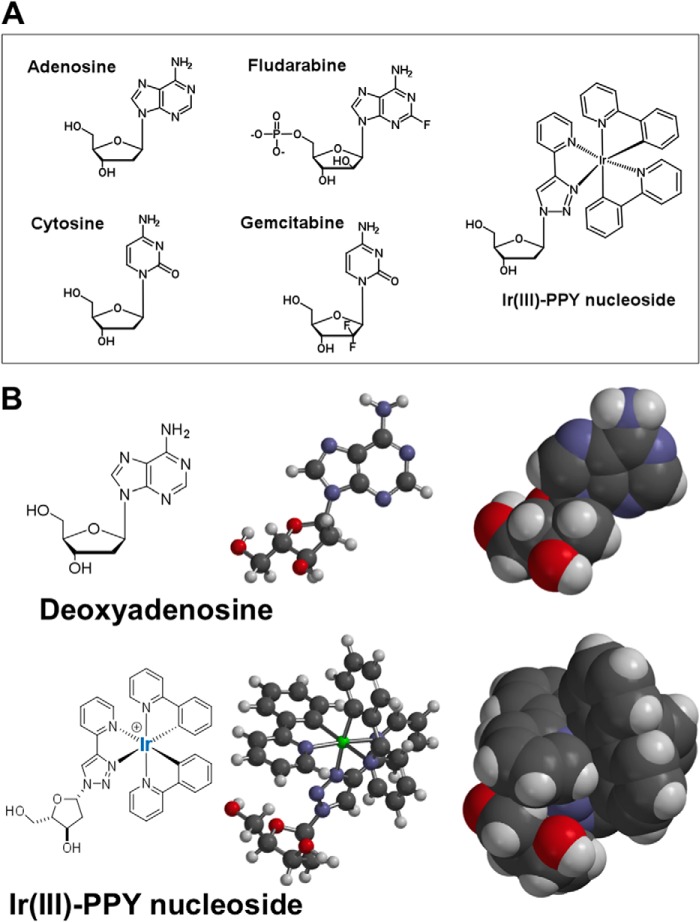
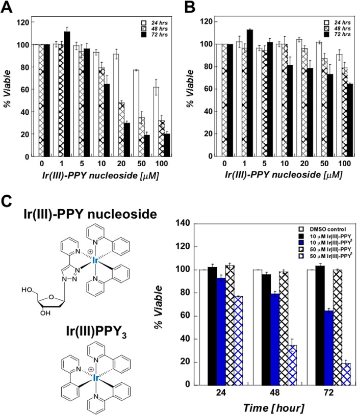
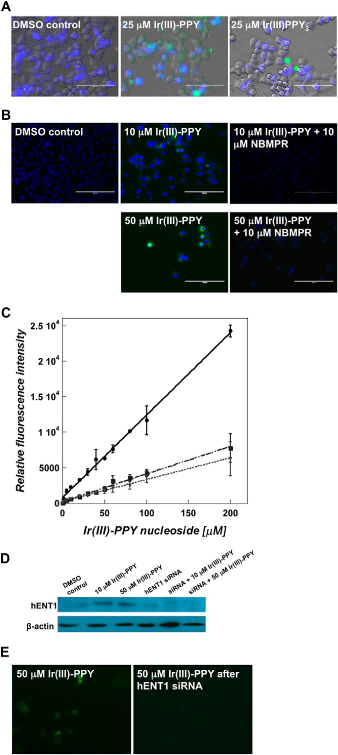
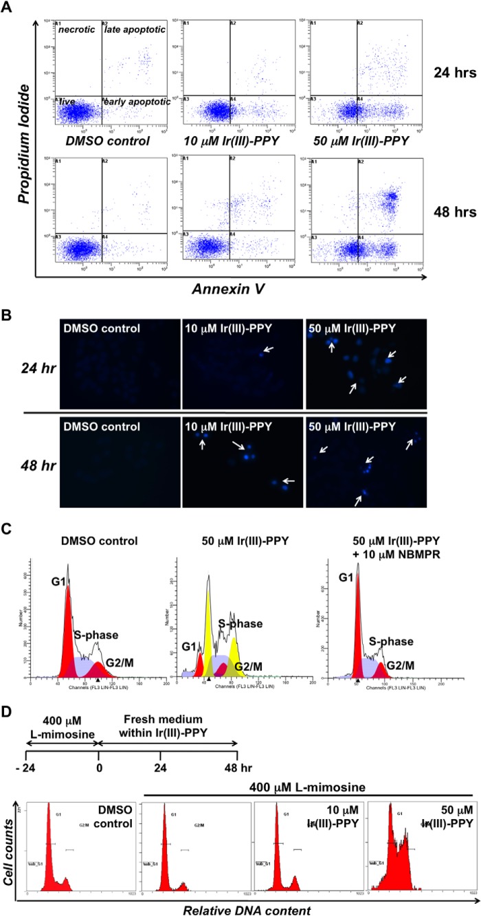
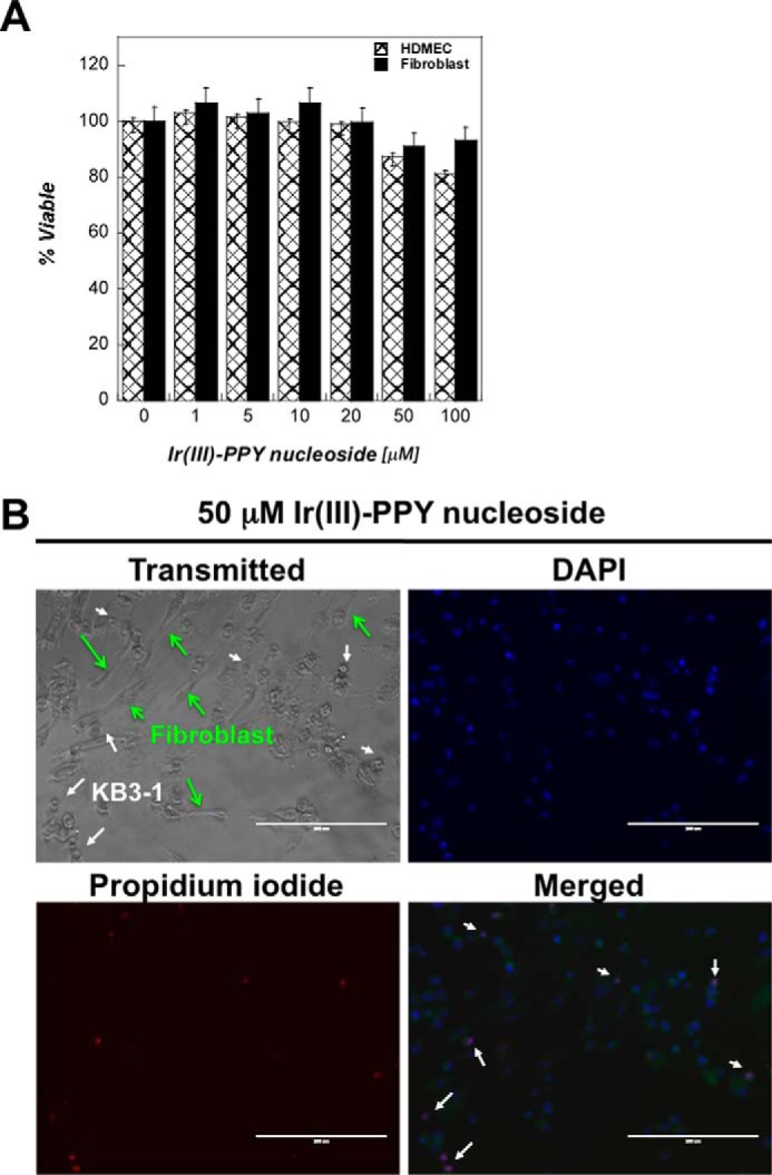
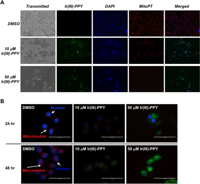
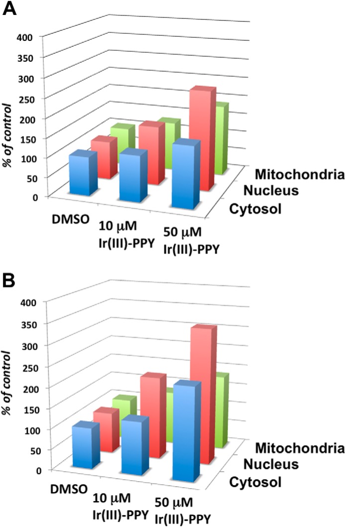
Nucleoside transport is an essential process that helps maintain the hyperproliferative state of most cancer cells. As such, it represents an important target for developing diagnostic and therapeutic agents that can effectively detect and treat cancer, respectively. This report describes the development of a metal-containing nucleoside designated Ir(III)-PPY nucleoside that displays both therapeutic and diagnostic properties against the human epidermal carcinoma cell line KB3-1. The cytotoxic effects of Ir(III)-PPY nucleoside are both time- and dose-dependent. Flow cytometry analyses validate that the nucleoside analog causes apoptosis by blocking cell cycle progression at G2/M. Fluorescent microscopy studies show rapid accumulation in the cytoplasm within 4 h. However, more significant accumulation is observed in the nucleus and mitochondria after 24 h. This localization is consistent with the ability of the metal-containing nucleoside to influence cell cycle progression at G2/M. Mitochondrial depletion is also observed after longer incubations (Δt ∼48 h), and this effect may produce additional cytotoxic effects. siRNA knockdown experiments demonstrate that the nucleoside transporter, hENT1, plays a key role in the cellular entry of Ir(III)-PPY nucleoside. Collectively, these data provide evidence for the development of a metal-containing nucleoside that functions as a combined therapeutic and diagnostic agent against cancer.

Keywords: Cancer Therapy; Molecular Imaging; Molecular Pharmacology; Nucleoside/Nucleotide Analogue; Transport.


Figures

FIGURE 1.
FIGURE 1.
A, chemical structures of natural nucleosides (adenosine and cytosine), conventional anti-cancer nucleosides (fludarabine and gemcitabine), and the metal-containing nucleoside, Ir(III)-PPY nucleoside. B, two-and three-dimensional models for adenosine and Ir(III)-PPY nucleoside, which provide a more accurate comparison of shape and size of the metal-containing nucleoside analog versus a natural nucleoside substrate. Models were developed using Spartan version 4.0 software.
FIGURE 2.
FIGURE 2.
A, dose- and time-dependent effects of Ir(III)-PPY nucleoside against the human epidermal carcinoma cell line, KB3-1. 0.1% DMSO was used as the vehicle, whereas Ir(III)-PPY nucleoside concentration was varied from 1 to 100 μm. An IC50 value of 30 ± 5 μm was obtained for the Ir(III)-PPY nucleoside after 48 h of treatment. B, dose- and time-dependent effects of Ir(III)-PPY nucleoside against the multidrug resistance-positive carcinoma cell line, KB-V1. C, structural comparison of Ir(III)-PPY nucleoside with Ir(III)PPY3, the metal complex lacking the deoxyribose moiety. Ir(III)-PPY nucleoside displays cytotoxic effects against KB3-1 cells that are time- and dose-dependent. In contrast, Ir(III)PPY3 does not produce cytostatic or cytotoxic effects even at high concentrations (50 μm) for extended time periods (3 days of treatment). The various treatments are shown as follows: 0.1% DMSO (white bars), 10 μm Ir(III)PPY3 (black bars), 10 μm Ir(III)-PPY nucleoside (blue bars), 50 μm Ir(III)PPY3 (black hashed bars), and 50 μm Ir(III)-PPY nucleoside (blue hashed bars). Error bars, S.E.
FIGURE 3.
FIGURE 3.
A, fluorescence microscope images of KB3-1 cells treated with 0.1% DMSO, 25 μm Ir(III)PPY3, or 25 μm Ir(III)-PPY nucleoside (Δt = 24 h post-treatment). Ir(III)-PPY nucleoside shows green fluorescence, whereas nuclei were stained with DAPI (blue) (magnification, ×40). B, fluorescence microscope images of 10 or 50 μm Ir(III)-PPY nucleoside uptake in the absence or presence of NBMPR in KB3-1 cells (Δt = 24 h post-treatment). Ir(III)-PPY nucleoside shows green fluorescence, whereas nuclei were stained with DAPI (blue) (magnification, ×40). C, the accumulation of Ir(III)-PPY nucleoside in KB3-1 cells was measured in the absence or presence of nucleoside transport inhibitors. Whole cell fluorescence intensities of Ir(III)-PPY nucleoside were measured using a fluorescence plate reader assay (excitation/emission = 312/510). The various treatments are shown as follows: Ir(III)-PPY nucleoside alone (solid line), Ir(III)-PPY nucleoside in the presence of 10 μm NBMPR (dashed and dotted line), and Ir(III)-PPY nucleoside in the presence of 1 μm dipyridamole (dotted line). D, Western blot analysis of cells treated with and without siRNA targeting hENT1. Cells treated with siRNA show reduced levels of the nucleoside transporter. E, cells treated with siRNA targeting hENT1 (right panel) show reduced uptake of Ir(III)-PPY nucleoside compared with cells treated with scrambled siRNA (left panel) (magnification, ×40). Error bars, S.E.
FIGURE 4.
FIGURE 4.
A, dual parameter flow cytometry of KB3-1 cells treated with 0.1% DMSO 10 μm Ir(III)-PPY nucleoside or 50 μm Ir(III)-PPY nucleoside. Treatment with 50 μm Ir(III)-PPY nucleoside for 48 h produces a robust apoptotic effect. B, DAPI staining demonstrates cell cycle-specific toxicity by Ir(III)-PPY nucleoside in a dose- and time-dependent manner. C, cell cycle analysis of KB3-1 cells treated with Ir(III)-PPY nucleoside. Treatment with 50 μm Ir(III)-PPY nucleoside for 48 h causes a significant perturbation in cell cycle progression because cells are unable to complete mitosis. The anti-cancer effects of Ir(III)-PPY nucleoside can be blocked by pretreating cells with the hENT inhibitor, NBMPR. D, KB3-1 cells were synchronized at the G1 phase of the cell cycle by treating with 400 μm l-mimosine for 24 h. After removal of medium containing l-mimosine, fresh medium was added containing 0.1% DMSO, 10 μm Ir(III)-PPY nucleoside, or 50 μm Ir(III)-PPY nucleoside. The 0 h control represents treatment with l-mimosine alone.
FIGURE 5.
FIGURE 5.
A, comparison of the anti-cancer effects of Ir(III)-PPY nucleoside measured against two non-cancerous cell lines (human dermal microvascular endothelial cells (HDMEC) and dermal fibroblast cells) versus the cancerous cell line KB3-1. All three cell lines were treated with variable concentrations of Ir(III)-PPY nucleoside (1–100 μm) for 24 h. DMSO was used as the vehicle control. B, DAPI and propidium iodide staining demonstrates that treatment with 50 μm Ir(III)-PPY nucleoside produces apoptotic effects against KB3-1 cells (cancer) but not dermal fibroblast cells (non-cancerous) after 24 h of exposure. Green arrows, position of fibroblasts; white arrows, position of KB3-1 cells (magnification, ×20). Error bars, S.E.
FIGURE 6.
FIGURE 6.
A, measuring the subcellular localization of Ir(III)-PPY nucleoside by fluorescence microscopy. KB3-1 cells were treated with DMSO, 10 μm Ir(III)-PPY nucleoside, or 50 μm Ir(III)-PPY nucleoside for 48 h. Ir(III)-PPY nucleoside shows green fluorescence. Nuclei were stained with DAPI (blue). Mitochondria were stained with MitoPT (red). Merged images show that Ir(III)-PPY nucleoside primarily accumulates in the cytoplasm and nucleus at low concentrations (10 μm). However, an increase in mitochondrial accumulation is observed at higher concentrations (50 μm). Images were obtained using an EVOSfl Advanced microscope (magnification, ×40). B, higher magnification images (magnification, ×60) show that Ir(III)-PPY nucleoside accumulates in the nucleus and mitochondria of KB3-1 cells in a time- and dose-dependent manner. Ir(III)-PPY nucleoside shows green fluorescence. Nuclei were stained with DAPI (blue). Mitochondria were stained with MitoPT (red) (magnification, ×60).
FIGURE 7.
FIGURE 7.
Results of fractionation studies to quantify the amount of Ir(III)-PPY nucleoside in the cytosol, nucleus, and mitochondria of treated KB3-1 cells. Cells were treated with 0.1% DMSO, 10 μm Ir(III)-PPY nucleoside, and 50 μm Ir(III)-PPY nucleoside. After 24 h (A) or 48 h (B) post-treatment, cells were harvested, and fractions corresponding to the cytosol, mitochondria, and nucleus were isolated. The relative amount of Ir(III)-PPY nucleoside measured using its fluorescence properties as described under “Experimental Procedures.” Error bars are omitted for clarity. In all cases, the S.E. associated with each value is less than 10%.

Similar articles

Cited by

References

    1. Parker W. B. (2009) Enzymology of purine and pyrimidine antimetabolites used in the treatment of cancer. Chem. Rev. 109, 2880–2893 - PMC - PubMed
    1. Crawford S. (2013) Is it time for a new paradigm for systemic cancer treatment? Lessons from a century of cancer chemotherapy. Front. Pharmacol. 4, 68. - PMC - PubMed
    1. Berdis A. J. (2008) DNA polymerases as therapeutic targets. Biochemistry 47, 8253–8260 - PMC - PubMed
    1. Tiwari K. N., Cappellacci L., Montgomery J. A., Secrist J. A., 3rd (2003) Synthesis and anti-cancer activity of some novel 5-azacytosine nucleosides. Nucleosides Nucleotides Nucleic Acids 22, 2161–2170 - PubMed
    1. Wang M., Liu Y., Liu S., Zheng D. (2004) 8-Chloro-adenosine sensitizes a human hepatoma cell line to TRAIL-induced apoptosis by caspase-dependent and -independent pathways. Oncol. Rep. 12, 193–199 - PubMed

Publication types

MeSH terms