. 2015 Apr 24;290(17):10572-86.
doi: 10.1074/jbc.M115.637827. Epub 2015 Feb 23.

Family 46 Carbohydrate-binding Modules Contribute to the Enzymatic Hydrolysis of Xyloglucan and β-1,3-1,4-Glucans through Distinct Mechanisms

Affiliations

Family 46 Carbohydrate-binding Modules Contribute to the Enzymatic Hydrolysis of Xyloglucan and β-1,3-1,4-Glucans through Distinct Mechanisms

Immacolata Venditto et al. J Biol Chem. .

Abstract

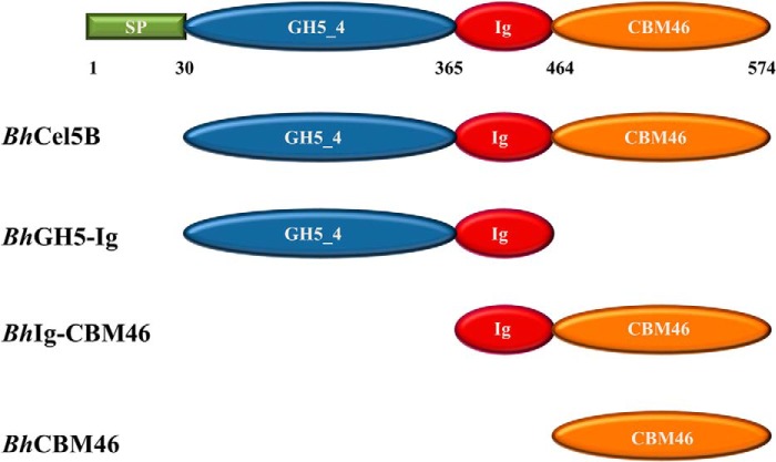
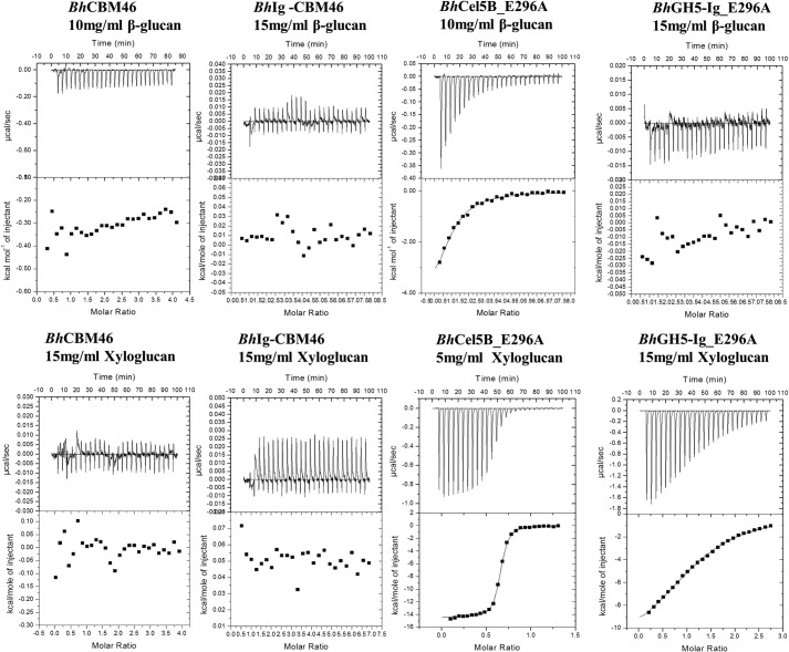
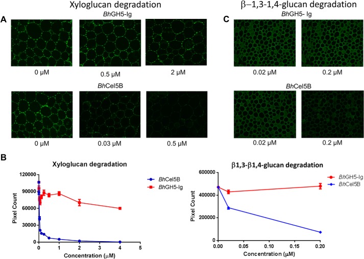
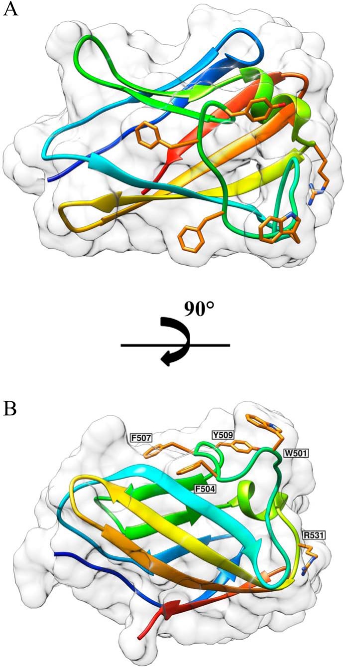
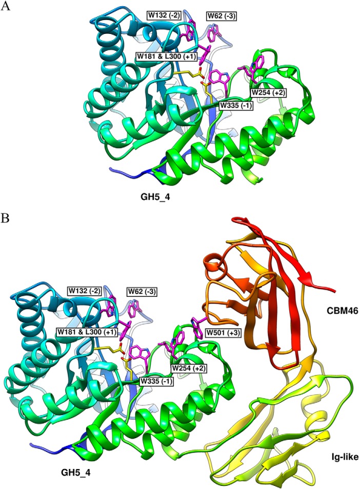
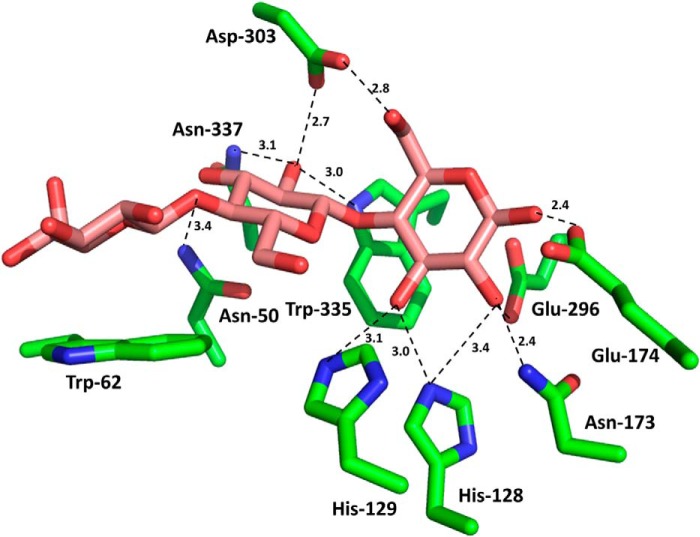
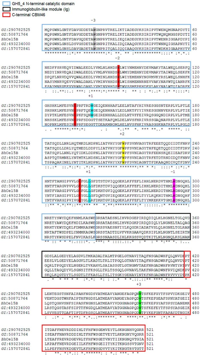
Structural carbohydrates comprise an extraordinary source of energy that remains poorly utilized by the biofuel sector as enzymes have restricted access to their substrates within the intricacy of plant cell walls. Carbohydrate active enzymes (CAZYmes) that target recalcitrant polysaccharides are modular enzymes containing noncatalytic carbohydrate-binding modules (CBMs) that direct enzymes to their cognate substrate, thus potentiating catalysis. In general, CBMs are functionally and structurally autonomous from their associated catalytic domains from which they are separated through flexible linker sequences. Here, we show that a C-terminal CBM46 derived from BhCel5B, a Bacillus halodurans endoglucanase, does not interact with β-glucans independently but, uniquely, acts cooperatively with the catalytic domain of the enzyme in substrate recognition. The structure of BhCBM46 revealed a β-sandwich fold that abuts onto the region of the substrate binding cleft upstream of the active site. BhCBM46 as a discrete entity is unable to bind to β-glucans. Removal of BhCBM46 from BhCel5B, however, abrogates binding to β-1,3-1,4-glucans while substantially decreasing the affinity for decorated β-1,4-glucan homopolymers such as xyloglucan. The CBM46 was shown to contribute to xyloglucan hydrolysis only in the context of intact plant cell walls, but it potentiates enzymatic activity against purified β-1,3-1,4-glucans in solution or within the cell wall. This report reveals the mechanism by which a CBM can promote enzyme activity through direct interaction with the substrate or by targeting regions of the plant cell wall where the target glucan is abundant.

Keywords: Carbohydrate-binding Protein; Cellulase; Cellulose; Plant Cell Wall; Protein Structure.


Figures

FIGURE 1.
FIGURE 1.
Architectural arrangement of BhCel5B and truncated derivatives produced in this work. Green, signal peptide; blue, N-terminal glycoside hydrolase family 5 catalytic module; red, immunoglobulin-like module; orange, C-terminal family 46 CBM.
FIGURE 2.
FIGURE 2.
Representative ITC data of BhCBM46, BhCel5B_E296A, and variants binding to soluble ligands. Titrations were conducted in 50 mm Na-HEPES buffer, pH 7.5, containing 200 mm NaCl at 25 °C. Top, ligand barley β-glucan in the syringe was titrated into cell contained protein (50 μm). Bottom, ligand xyloglucan in the syringe was titrated into cell contained protein (50 μm).
FIGURE 3.
FIGURE 3.
pH and temperature profile of BhCel5B (A) and thermostability of BhCel5B and BhGH5-Ig (B). A, panel 1, BhCel5B was incubated with 0.2% barley β-glucan at standard conditions in MES (●), Tris (■), and NaHCO3 (▴) buffers, and the activity was determined at 55 °C. A, panel 2, BhCel5B activity was determined with 0.2% barley β-glucan at different temperatures (●). B, panel 1, for thermostability, BhCel5B was incubated with 0.3% xyloglucan (●) or 0.3% barley β-glucan (■) for 30 min at different temperatures, and residual activity was determined at 30 °C. B, panel 2, BhGH5-Ig was incubated with 0.2% xyloglucan (●) or 0.7% barley β-glucan (■) for 30 min at different temperatures, and residual activity was determined at 30 °C.
FIGURE 4.
FIGURE 4.
Activity of BhCel5B and BhGH5-Ig against plant cell walls. The experiments were carried out as described under “Experimental Procedures.” A and C show representative micrographs of stem sections probed with antibodies (A shows tobacco stem sections probed with rat monoclonal antibody LM15 directed to xyloglucan, and C shows miscanthus stem sections probed with a mouse monoclonal antibody directed to β-1,3–1,4-glucan). B shows the quantified fluorescence intensities of the antibodies binding to equivalent stem sections after enzymatic treatments.
FIGURE 5.
FIGURE 5.
Structure of BhCBM46. A, BhCBM46 shows an open cleft. B, mutations are drawn as sticks. The pictures were prepared using Chimera (48).
FIGURE 6.
FIGURE 6.
Structure of BhCel5B. A, all important residues required for substrate recognition and catalysis presented on GH5_4 catalytic domain are drawn as sticks. B, BhCel5B is a tri-modular protein, composed of an N-terminal glycoside hydrolase family 5 catalytic module (GH5_4) followed by an immunoglobulin-like module (Ig) and a C-terminal family 46 CBM. Catalytic residues on GH5_4, Glu-170, and Glu-296, are drawn as sticks.
FIGURE 7.
FIGURE 7.
Interaction of the negative subsites of the substrate binding cleft of BhCel5B with cellotriose. The amino acids of BhCel5B that are predicted to interact with cellotriose are shown in stick format with the carbons colored green. The cellotriose was derived from an overlay of overlay of BhCel5B with C. cellulovorans endoglucanase D in complex with the trisaccharide (PDB code 3ndZ; the r.m.s.d. for the overlay was 1.46 Å over 345 Cα atoms). The distance of the polar interactions are shown in Å.
FIGURE 8.
FIGURE 8.
Alignments of CBM46 with all 45 representatives members. The alignment was made using Aline 011208. Residues that are invariant within the family are shaded in yellow and indicated by an asterisk. Mutations are indicated by inverted red triangle. The most important residue in carbohydrate recognition is reported with a red box.
FIGURE 9.
FIGURE 9.
Alignments of BhCel5B with four proteins. The alignment was made using ClustalW2. Residues required for substrate recognition and catalysis are conserved in the five proteins. The residues occupying the subsites are indicated.

References

    1. Himmel M. E., Bayer E. A. (2009) Lignocellulose conversion to biofuels: current challenges, global perspectives. Curr. Opin Biotechnol. 20, 316–317 - PubMed
    1. Himmel M. E., Ding S. Y., Johnson D. K., Adney W. S., Nimlos M. R., Brady J. W., Foust T. D. (2007) Biomass recalcitrance: engineering plants and enzymes for biofuels production. Science 315, 804–807 - PubMed
    1. Mohnen D. (2008) Pectin structure and biosynthesis. Curr. Opin Plant Biol. 11, 266–277 - PubMed
    1. Gilbert H. J. (2010) The biochemistry and structural biology of plant cell wall deconstruction. Plant Physiol. 153, 444–455 - PMC - PubMed
    1. Minic Z., Jouanin L. (2006) Plant glycoside hydrolases involved in cell wall polysaccharide degradation. Plant Physiol. Biochem. 44, 435–449 - PubMed

Publication types

MeSH terms

LinkOut - more resources