. 2015 Apr 10;290(15):9674-89.
doi: 10.1074/jbc.M115.636894. Epub 2015 Feb 24.

Histone H2A and H4 N-terminal tails are positioned by the MEP50 WD repeat protein for efficient methylation by the PRMT5 arginine methyltransferase

Affiliations

Histone H2A and H4 N-terminal tails are positioned by the MEP50 WD repeat protein for efficient methylation by the PRMT5 arginine methyltransferase

Emmanuel S Burgos et al. J Biol Chem. .

Abstract

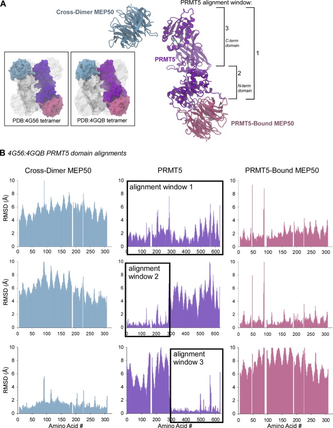
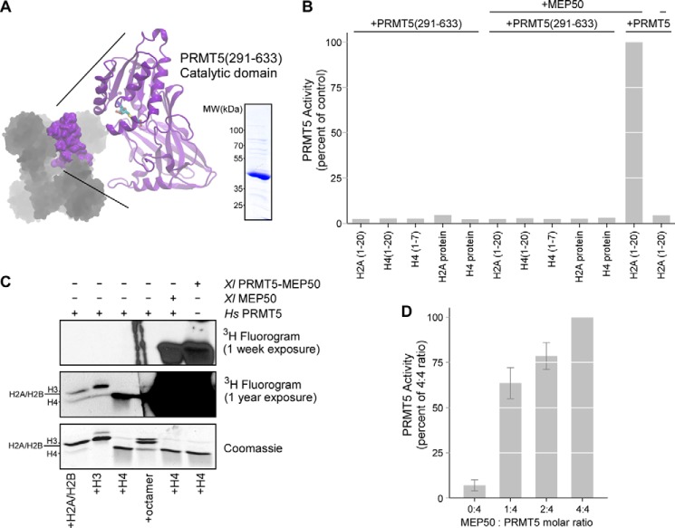
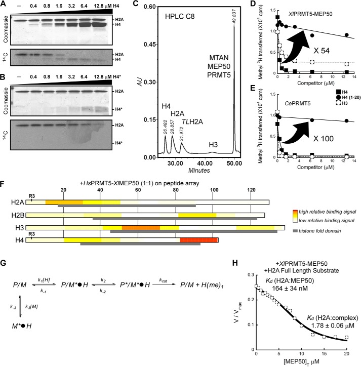
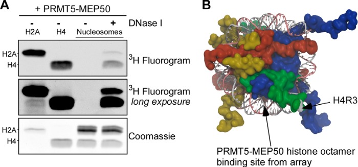
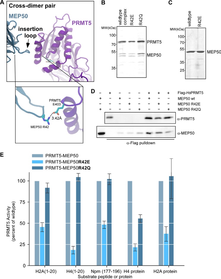
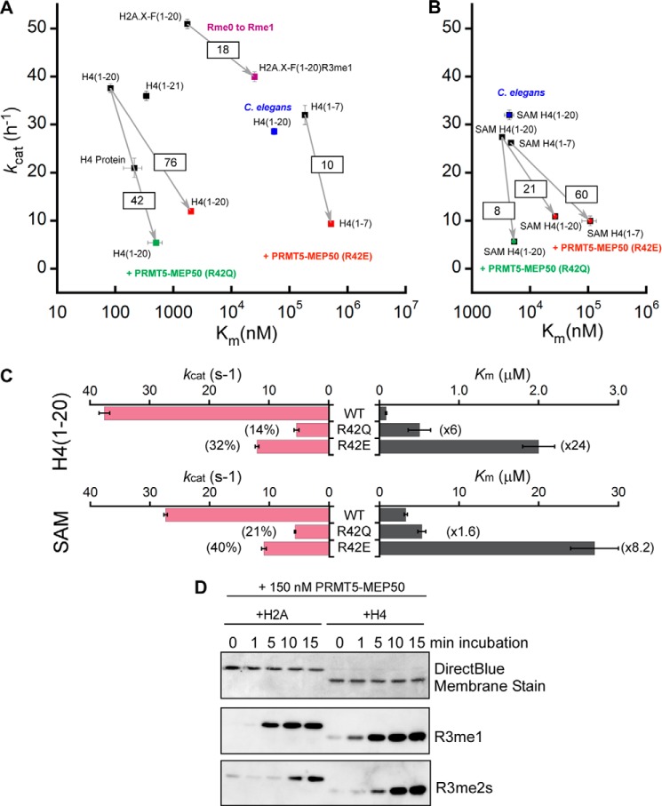
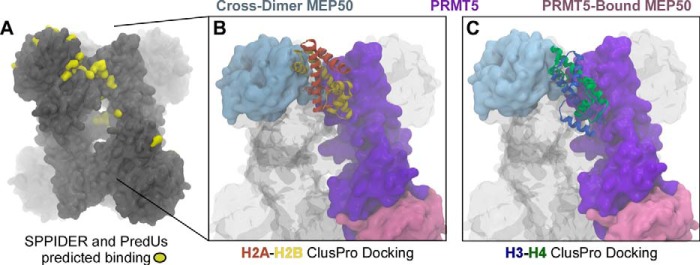
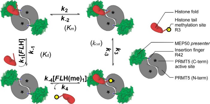
The protein arginine methyltransferase PRMT5 is complexed with the WD repeat protein MEP50 (also known as Wdr77 or androgen coactivator p44) in vertebrates in a tetramer of heterodimers. MEP50 is hypothesized to be required for protein substrate recruitment to the catalytic domain of PRMT5. Here we demonstrate that the cross-dimer MEP50 is paired with its cognate PRMT5 molecule to promote histone methylation. We employed qualitative methylation assays and a novel ultrasensitive continuous assay to measure enzyme kinetics. We demonstrate that neither full-length human PRMT5 nor the Xenopus laevis PRMT5 catalytic domain has appreciable protein methyltransferase activity. We show that histones H4 and H3 bind PRMT5-MEP50 more efficiently compared with histone H2A(1-20) and H4(1-20) peptides. Histone binding is mediated through histone fold interactions as determined by competition experiments and by high density histone peptide array interaction studies. Nucleosomes are not a substrate for PRMT5-MEP50, consistent with the primary mode of interaction via the histone fold of H3-H4, obscured by DNA in the nucleosome. Mutation of a conserved arginine (Arg-42) on the MEP50 insertion loop impaired the PRMT5-MEP50 enzymatic efficiency by increasing its histone substrate Km, comparable with that of Caenorhabditis elegans PRMT5. We show that PRMT5-MEP50 prefers unmethylated substrates, consistent with a distributive model for dimethylation and suggesting discrete biological roles for mono- and dimethylarginine-modified proteins. We propose a model in which MEP50 and PRMT5 simultaneously engage the protein substrate, orienting its targeted arginine to the catalytic site.

Keywords: Enzyme Kinetics; Enzyme Mechanism; Histone Methylation; Peptide Array; Protein Arginine N-methyltransferase 5 (PRMT5); WD Repeat.


Figures

FIGURE 1.
FIGURE 1.
Evolutionarily conserved spatial arrangement between the PRMT5 catalytic domain and its cross-dimer paired MEP50. A, ribbon diagram of the Xenopus PRMT5 monomer (purple), its directly bound MEP50 molecule (pink), and the cross-dimer MEP50 (blue). Structural alignment windows 1, 2, and 3 are indicated on the right. Inset boxes, surface diagrams of the Xenopus (Protein Data Bank code 4G56) and human (Protein Data Bank code 4GQB) PRMT5-MEP50 structures, with the analyzed PRMT5 and MEP50 molecules colored as in A; the remainder of the structures are shown in gray. B, per-residue Cα alignments between the Xenopus and human PRMT5 windows 1, 2, and 3 as calculated with VMD MultiSeq, plotted as RMSD (Å) against amino acid position in the sequence. The PRMT5 alignment windows are boxed.
FIGURE 2.
FIGURE 2.
Intact full-length PRMT5 complexed with MEP50 is necessary for histone methyltransferase activity. A, the catalytic C-terminal domain of XlPRMT5 (residues 291–633) is shown in purple and was expressed and purified. B, XlPRMT5(291–633) did not exhibit any activity toward histone tail peptide substrates of the intact complex (H2A(1–20), H4(1–20), H4(1–7)) or full-length histones H2A or H4. The addition of XlMEP50 to the catalytic domain did not stimulate activity, whereas the addition of XlMEP50 to full-length HsPRMT5 stimulated activity toward H2A(1–20) peptide. C, intact XlPRMT5-MEP50 complex or HsPRMT5 + XlMEP50 exhibited methyltransferase activity toward histone H4 (right two lanes). HsPRMT5 alone exhibited ultralow levels of activity toward H2A, H3, and H4, visible only after a 1-year exposure of the fluorogram (left four lanes, second panel). D, HsPRMT5 (200 nm) was preincubated with substoichiometric (1:4 and 2:4) and stoichiometric concentrations (4:4) of XlMEP50 and then assayed for methyltransferase activity against H4 peptide.
FIGURE 3.
FIGURE 3.
XlMEP50 is a presenter that primarily binds histone H4 through the histone fold and exposes its N-terminal tail for methylation by XlPRMT5. A, substrate competition experiment where XlPRMT5-MEP50 activity toward histone H2A was displaced as histone H4 was titrated into the reaction (experiment performed using 50 mm MOPS, pH 7.0, 100 nm XlPRMT5-MEP50, 20 microunits of SeMTAN, 25 μm [14C]-methyl-SAM, and a H2A concentration kept constant at 2 μm while H4 was added at 0, 0.4, 0.8, 1.6, 3.2, 6.4, and 12.8 μm final concentrations). B, similar competition experiment as in A where peptide H4(1–20) was used as competing substrate (experimental conditions identical to those in A). C, quantification of methyl transfer reactions using reversed-phase HPLC. Histones H4, H2A, tailless H2A (TLH2A), and H3 were separated from each other and from radiolabeled substrate SAM using a C8 reverse HPLC column (see “Experimental Procedures”). D, substrate competition experiment where XlPRMT5-MEP50 activity toward histone H2A was displaced as histone H4, peptide H4(1–20), and histone H3 were titrated into the reaction (experiment performed using 50 mm MOPS, pH 7.0, 100 nm XlPRMT5-MEP50, 20 microunits of SeMTAN, 25 μm [3H]methyl-SAM, and a H2A concentration kept constant at 2 μm while competitors were added at 0–12.6 μm final concentrations). Transfer of [3H]methyl was quantified by liquid scintillation counting after isolation of histones. E, competition experiment similar to that in D where XlPRMT5-MEP50 was replaced by its homologous enzyme from C. elegans (experimental conditions identical to those in D). F, FLAG-HsPRMT5-XlMEP50 complex was incubated on ultrahigh density histone peptide arrays. PRMT5-MEP50 binding on the histone peptide scan data was extracted, and relative binding levels were plotted as a heat map, with no to low signal as white to light yellow, and high relative binding was plotted in red. Histone amino acid sequence numbers are represented at the top of the plots. The histone fold domain is indicated as a gray box, and the substrate residue Arg-3 (R3) on both H2A and H4 is indicated. G, relief of methyltransferase activity through the addition of MEP50. In this model used to determine the affinities of histone H2A for MEP50, P/MH represents the PRMT5-MEP50·histone complex, where only the histone fold is bound to the MEP50 presenter. P*/MH represents the PRMT5-MEP50·histone complex, where the histone fold is bound to the MEP50 presenter and the histone tail is bound to the enzyme active site. MH represents the complex between histone and exogenous MEP50. Each step is characterized by a constant (i.e. k1, k−1, k2, k−2, kcat, k3, and k−3). G, transferase reaction catalyzed by XlPRMT5-MEP50 was followed continuously at pH 7.7 using the luciferase-based assay with histone H2A as substrate (H2A fixed at 2 μm; see “Experimental Procedures”); XlMEP50 was added to the reactions (0–20 μm), and resulting transferase activities were recorded. Exogenous XlMEP50 competes with XlPRMT5-MEP50 for H2A binding, and methyl transfer is inhibited with increasing concentrations of XlMEP50. A dissociation constant (Kd) of H2A for both exogenous XlMEP50 (Kd H2A:MEP50) and PRMT5-associated MEP50 (Kd H2A:complex) was determined.
FIGURE 4.
FIGURE 4.
XlPRMT5-MEP50 does not methylate histones embedded in DNA in nucleosomes. A, XlPRMT5-MEP50 was incubated with H2A, H4, or recombinant mononucleosomes, in the presence or absence of DNase I. PRMT5 methylated both H2A and H4 independently (lanes 1 and 2) but did not methylate histones in mononucleosomes (lane 3). Upon DNase I treatment, histone H4 and histone H2A activity was recovered (lane 4). B, schematic representation of the nucleosome core particle (Protein Data Bank code 1KX5), with H3 shown in blue, H4 in green, H2A in red, and H2B in yellow. The targeted H4 Arg-3 is shown (gray coloring and arrow), and the region on H3-H4 corresponding to the strongest sites of interaction on the peptide array is indicated.
FIGURE 5.
FIGURE 5.
Mutations of MEP50 insertion loop residue Arg-42 affect histone methylation. A, schematic representation of the insertion loop from the cross-dimer XlMEP50 positioned adjacent to the catalytic domain of the paired PRMT5 molecular. Inset enlarged view, the only contact the cross-dimer MEP50 makes with the catalytic domain is a putative salt bridge between XlMEP50 Arg-42 and XlPRMT5 Glu-403; no other contacting residues are found. B, Coomassie-stained gel of wild-type XlPRMT5-MEP50, XlPRMT5-MEP50R42E, and XlPRMT5-MEP50R42Q complexes. C, Coomassie-stained gel of wild type and XlMEP50R42E. D, FLAG-tagged HsPRMT5 captured on anti-FLAG resin after incubation alone or with XlMEP50, XlMEP50R42E, or XlMEP50R42Q and immunoblotted for HsPRMT5 and XlMEP50, demonstrating similar interactions for the wild-type and mutated MEP50 proteins. E, filter-binding activity assays of PRMT5-MEP50, PRMT5-MEP50R42E, or PRMT5-MEP50R42Q (100 nm complex) incubated with H2A(1–20), H4(1–20), and Npm(177–196) substrate peptides or H2A and H4 full-length protein substrates. PRMT5-directed activity is represented as a percentage of wild-type activity toward the peptide/protein substrate and is the average of three independent replicates.
FIGURE 6.
FIGURE 6.
Substrate specificities for XlPRMT5-MEP50 and the impact on enzymatic efficiency upon mutation of MEP50 insertion loop residue Arg-42. Kinetic parameters for the various tested substrates (histone H4, histone peptides, and SAM) are plotted, with the kcat (h−1) on the y axis and the Km (nm; logarithmic scale) on the x axis. Highest enzymatic efficiencies are obtained with substrates found in the top left quadrant, whereas low enzymatic efficiencies are obtained with substrates found in the opposite bottom right quadrant. Arrows indicate the loss (squared values) of enzymatic efficiency upon arginine monomethylation (purple) or upon mutation of MEP50 residue Arg-42 to glutamic acid (red) and to glutamine (green). For reference, enzymatic behavior of CePRMT5 is represented in blue. A, representation of kinetic parameters for histone substrates using saturating concentration of SAM. B, representation of kinetic parameters for SAM substrate using saturating concentration of histone substrates. C, impact of XlMEP50R42Q and XlMEP50R42E on catalytic turnover (kcat; pink bars) and substrates' affinities (Km; gray bars) for both peptide and SAM substrates. The decrease of methyl transfer is represented as a percentage of wild-type kcat, whereas the loss of affinity is given as -fold increase of wild-type Km. D, histones H2A or H4 were incubated with XlPRMT5-MEP50 and SAM. Reactions were stopped at 0, 1, 5, 10, and 15 min with the addition of SDS-polyacrylamide gel loading buffer and heating to 100 °C. Reaction products were immunoblotted with monomethylarginine (R3me1)- or symmetric dimethylarginine (R3me2s)-specific antibodies.
FIGURE 7.
FIGURE 7.
Prediction of histone binding sites onto the PRMT5-MEP50 complex. A, the predicted interacting residues on the cross-dimer pair of XlPRMT5-MEP50 was determined using the SPPIDER and PredUs algorithms and mapped onto the structure, as shown in yellow. Shown are docking of H2A-H2B dimer (orange/yellow) (B) and H3-H4 dimer (blue/green) (C) onto XlPRMT5-MEP50 (PRMT5 monomer (purple), its directly bound MEP50 molecule (pink), and the cross-dimer MEP50 (blue)), using ClusPro with attractive forces as determined by the peptide array and predictions in A.
FIGURE 8.
FIGURE 8.
Schematic model for the sequential binding of core histone onto the MEP50 presenter and favorable orientation of the N-terminal histone tail to the PRMT5 active site for methyl transfer. XlPRMT5-MEP50 is a tetramer of heterodimers, and the cross-dimer MEP50 (green) is paired with its cognate PRMT5 molecule (gray; C-terminal active site represented by an asterisk) to promote histone methylation according to a sequential mechanism with 1) binding of the full-length histone through its histone fold (FLH in red; dissociation constant Kd), 2) favorable orientation of the histone tail toward the PRMT5 cross-dimer active site (asterisk; step characterized by a Km value), 3) methylation of Arg-3 (yellow circle with methyl group transferred from S-adenosylmethionine; step characterized by a kcat value), and 4) release of the methylated histone tail and the histone fold from the active site and the MEP50, respectively.

Similar articles

Cited by

References

    1. Di Lorenzo A., Bedford M. T. (2011) Histone arginine methylation. FEBS Lett. 585, 2024–2031 - PMC - PubMed
    1. Wolf S. S. (2009) The protein arginine methyltransferase family: an update about function, new perspectives and the physiological role in humans. Cell Mol. Life Sci. 66, 2109–2121 - PMC - PubMed
    1. Wysocka J., Allis C. D., Coonrod S. (2006) Histone arginine methylation and its dynamic regulation. Front. Biosci. 11, 344–355 - PubMed
    1. Lee Y. H., Stallcup M. R. (2009) Minireview: protein arginine methylation of nonhistone proteins in transcriptional regulation. Mol. Endocrinol. 23, 425–433 - PMC - PubMed
    1. Wilczek C., Chitta R., Woo E., Shabanowitz J., Chait B. T., Hunt D. F., Shechter D. (2011) Protein arginine methyltransferase Prmt5-Mep50 methylates histones H2A and H4 and the histone chaperone nucleoplasmin in Xenopus laevis eggs. J. Biol. Chem. 286, 42221–42231 - PMC - PubMed

Publication types

MeSH terms

LinkOut - more resources