. 2015 Apr 10;290(15):9812-22.
doi: 10.1074/jbc.M114.616219. Epub 2015 Feb 24.

Transcriptional activity of the islet β cell factor Pdx1 is augmented by lysine methylation catalyzed by the methyltransferase Set7/9

Affiliations

Transcriptional activity of the islet β cell factor Pdx1 is augmented by lysine methylation catalyzed by the methyltransferase Set7/9

Aarthi V Maganti et al. J Biol Chem. .

Abstract

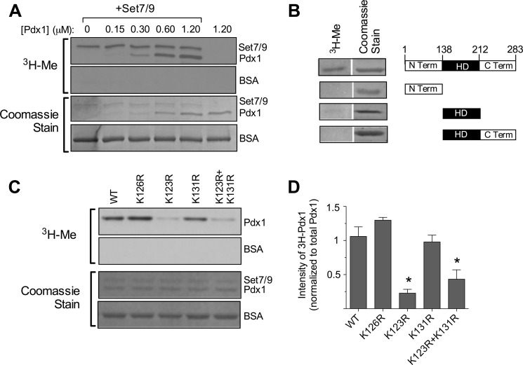
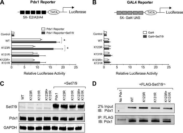
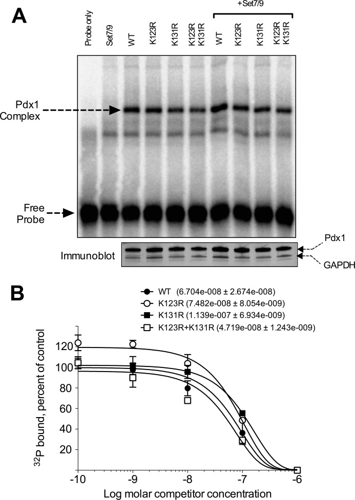
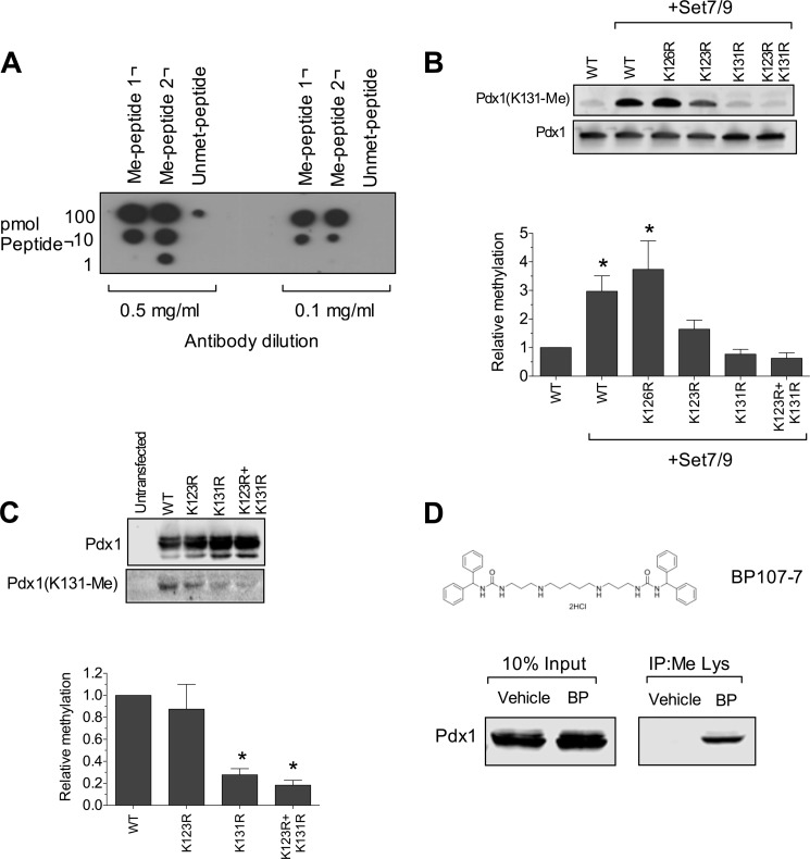
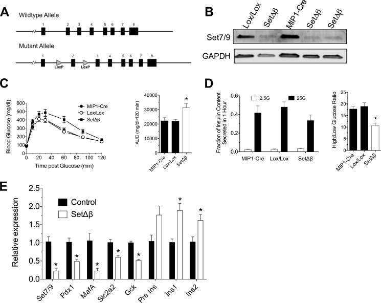
The transcription factor Pdx1 is crucial to islet β cell function and regulates target genes in part through interaction with coregulatory factors. Set7/9 is a Lys methyltransferase that interacts with Pdx1. Here we tested the hypothesis that Lys methylation of Pdx1 by Set7/9 augments Pdx1 transcriptional activity. Using mass spectrometry and mutational analysis of purified proteins, we found that Set7/9 methylates the N-terminal residues Lys-123 and Lys-131 of Pdx1. Methylation of these residues occurred only in the context of intact, full-length Pdx1, suggesting a specific requirement of secondary and/or tertiary structural elements for catalysis by Set7/9. Immunoprecipitation assays and mass spectrometric analysis using β cells verified Lys methylation of endogenous Pdx1. Cell-based luciferase reporter assays using wild-type and mutant transgenes revealed a requirement of Pdx1 residue Lys-131, but not Lys-123, for transcriptional augmentation by Set7/9. Lys-131 was not required for high-affinity interactions with DNA in vitro, suggesting that its methylation likely enhances post-DNA binding events. To define the role of Set7/9 in β cell function, we generated mutant mice in which the gene encoding Set7/9 was conditionally deleted in β cells (Set(Δ)β). Set(Δ)β mice exhibited glucose intolerance similar to Pdx1-deficient mice, and their isolated islets showed impaired glucose-stimulated insulin secretion with reductions in expression of Pdx1 target genes. Our results suggest a previously unappreciated role for Set7/9-mediated methylation in the maintenance of Pdx1 activity and β cell function.

Keywords: Diabetes; Gene Knockout; Pdx1; Protein Methylation; Set7/9; Transcription; pancreatic Islet.


Figures

FIGURE 1.
FIGURE 1.
Pdx1 methylation by Set7/9 in vitro. A, a methylation assay in vitro using recombinant Set7/9, [3H]AdoMet and increasing concentrations of Pdx1 protein was performed, and then reactions were subjected to polyacrylamide gel electrophoresis. The first and second panel show fluorography for 3H, and the third and fourth panels show corresponding Coomassie staining of the same gel. B, methylation assay in vitro using recombinant Set7/9, [3H]AdoMet, and full-length or truncated Pdx1 proteins was performed, followed by polyacrylamide gel electrophoresis. A schematic of truncated mutants of Pdx1 is shown in the right panel, and corresponding 3H fluorography and Coomassie stains are shown in the left panel. Term, terminus; HD, homeodomain. C, methylation assays in vitro using WT and mutated Pdx1 proteins were performed with corresponding quantitation of methylated Pdx1 protein intensities (normalized to total Pdx1 protein by Coomassie staining). All images shown are representative of at least three experiments. *, p < 0.05 compared with wild-type Pdx1.
FIGURE 2.
FIGURE 2.
MS/MS spectra of Pdx1 in vitro and in MIN6 cells. A, tandem mass spectrum of the peptide GQLPFPWMK showing modification of Lys-123 with monomethylation (+14 Da) following treatment with recombinant Set7/9 in vitro. The SEQUEST XCorr for this +2 peptide is 1.83, with a ppm (parts per million) of −1.65. (B) Tandem mass spectrum of the +3 peptide AHAWKGQWAGGAYAAEPEENKR showing monomethylation (+14 Da) of Lys-131 in Pdx1 following treatment with recombinant Set7/9 in vitro. The SEQUEST XCorr for this peptide is 6.19, with a ppm of 0.34. C, representative MS/MS spectrum from an LTQ ion trap for the +2 peptide AHAWKGQWAGGAYAAEPEENKR showing trimethylation at Lys-131 in Pdx1 in transduced MIN6 β cells. The SEQUEST XCorr for this peptide is 2.24, with a ppm of 19.25.
FIGURE 3.
FIGURE 3.
Transcription augmentation by Set7/9 is dependent upon the Pdx1 residue Lys-131. NIH3T3 cells were transiently cotransfected with Set7/9, WT and mutant Pdx1 proteins, and either the Pdx1 reporter plasmid or the Gal4 reporter plasmid. Cells were harvested 48 h later, and whole cell extracts were used to assay luciferase activity. A, luciferase activities (relative to control transfections without Pdx1) with the Pdx1 reporter plasmid, which contains tandem elements of the Ins E2/A3/A4 element. A schematic of reporter is shown in the top panel. n = 3 transfections in triplicate; *, p < 0.05 for the comparisons shown. B, luciferase activities (relative to control transfections without Pdx1) with the Gal4 reporter plasmid, which contains tandem elements of the yeast Gal4 DNA binding domain. A schematic of the reporter is shown in the top panel). n = 3 transfections in triplicate. UAS, upstream activating sequence. C, immunoblots (for the indicated proteins) from whole cell extract from transfected cells. Results are representative of three transfections. D, NIH3T3 cells were transfected with FLAG-Set7/9 and the indicated Pdx1 proteins, and then nuclear extracts were subjected to immunoprecipitation (IP) using FLAG antibody and immunoblotted (IB) using Pdx1 antibody. Results are representative of two experiments.
FIGURE 4.
FIGURE 4.
Point mutations do not affect DNA binding by Pdx1. A, NIH3T3 cells were cotransfected with the WT or mutant Pdx1 proteins with or without cotransfection of Set7/9. Nuclear extracts from transfected cells were harvested and subjected to EMSA using the 32P-labeled E-A DNA fragment containing a Pdx1 binding site derived from Ins E2/A3/A4. The positions of the Pdx1-containing complex and the unbound, free probe are indicated by arrows. Bottom panel, immunoblots for Pdx1 and GAPDH from the same nuclear extracts as used for the EMSA. B, quantitation and modeling of EMSA reactions (as in A), with increasing concentrations of unlabeled E-A probe. For each Pdx1 mutant, the control condition is the fraction of total probe bound in the absence of competitor. The ordinate represents probe binding (at the given competitor concentration) as a percentage of the control condition. The apparent dissociation constants of the WT and mutant proteins are shown in the inset, with no statistical differences seen. The data shown are from three EMSA experiments.
FIGURE 5.
FIGURE 5.
Residue Lys-131 of Pdx1 is methylated in cells. A, dot blot analysis using two dilutions of anti-Pdx1(Lys-131-Me) antibodies and methylated and unmethylated peptides corresponding to the N terminus of Pdx1 containing residue Lys-131. B, representative immunoblots (top panel) using anti-Pdx1(Lys-131-Me) antibody or anti-Pdx1 antibody on recombinant Pdx1 proteins methylated by Set7/9 in vitro. The quantitation of immunoblots (n = 3) is shown in the bottom panel. *, p < 0.05 compared with Pdx1 alone. C, HEK293 cells were transfected with Set7/9 and WT or mutant HA-tagged Pdx1 proteins, and then whole cell extracts were immunoprecipitated using anti-HA antibody, followed by immunoblot analysis using anti-Pdx1 and anti-Pdx1(Lys-131-Me) antibodies (top panel). The quantitation of methylated Pdx1 (relative to total immunoprecipitated Pdx1, n = 3) is shown in the bottom panel. *, p < 0.05 compared with WT Pdx1. D, top panel, chemical structure of the Lys demethylase inhibitor BP-107-7 (BP). Bottom panel, INS-1 832/13 β cells were treated with vehicle or BP-107-7 (10 μm) overnight, and then nuclear extracts were immunoprecipitated (IP) using anti-methyl Lys antibody followed by immunoblot using anti-Pdx1 antibody.
FIGURE 6.
FIGURE 6.
β Cell-specific deletion of Set7/9 in mice. A, schematic of WT and mutant alleles present in control and mutant mice, respectively, showing positions of the exons (numbered) and the Loxp sites. B, Immunoblot analysis of whole cell lysates of islets isolated from Setd7+/+;MIP1-CreERT (MIP1-Cre), Setd7Loxp/Loxp (Lox/Lox), and MIP1-CreERT;Setd7Loxp/Loxp (SetΔβ) mice for Set7/9 (top panel) and GAPDH (bottom panel). C, results of intraperitoneal glucose tolerance test on MIP1-Cre (n = 12), Lox/Lox (n = 9), and SetΔβ (n = 12) mice (left panel) with a corresponding area under the curve (AUC) analysis (right panel). *, p < 0.05 compared with either MIP1-Cre or Lox/Lox. D, results of glucose-stimulated insulin release assays from islets isolated from MIP1-Cre, Lox/Lox, and SetΔβ mice (left panel), and the corresponding ratio of insulin secreted at high glucose relative to low glucose is shown in the right panel. n = 3, *, p < 0.05 compared with either MIP1-Cre or Lox/Lox. E, results of real-time RT-PCR for the indicated genes from islets isolated from control (MIP1-Cre and Lox/Lox) versus SetΔβ mice. Data are normalized to Actb mRNA levels; n = 4 independent islet isolations; *, p < 0.05 compared with control.

References

    1. Ferrannini E., Mari A., Nofrate V., Sosenko J. M., Skyler J. S., and DPT-1 Study Group (2010) Progression to diabetes in relatives of type 1 diabetic patients: mechanisms and mode of onset. Diabetes 59, 679–685 - PMC - PubMed
    1. Ferrannini E., Gastaldelli A., Miyazaki Y., Matsuda M., Mari A., DeFronzo R. A. (2005) β-Cell function in subjects spanning the range from normal glucose tolerance to overt diabetes: a new analysis. J. Clin. Endocrinol. Metab. 90, 493–500 - PubMed
    1. Jonsson J., Carlsson L., Edlund T., Edlund H. (1994) Insulin-promoter-factor 1 is required for pancreas development in mice. Nature 371, 606–609 - PubMed
    1. Offield M. F., Jetton T. L., Labosky P. A., Ray M., Stein R. W., Magnuson M. A., Hogan B. L., Wright C. V. (1996) PDX-1 is required for pancreatic outgrowth and differentiation of the rostral duodenum. Development 122, 983–995 - PubMed
    1. Stoffers D. A., Zinkin N. T., Stanojevic V., Clarke W. L., Habener J. F. (1997) Pancreatic agenesis attributable to a single nucleotide deletion in the human IPF1 gene coding sequence. Nat. Genet. 15, 106–110 - PubMed

Publication types

MeSH terms

LinkOut - more resources