. 2015 Apr 10;290(15):9387-98.
doi: 10.1074/jbc.M114.631804. Epub 2015 Feb 20.

Structure of a BAG6 (Bcl-2-associated athanogene 6)-Ubl4a (ubiquitin-like protein 4a) complex reveals a novel binding interface that functions in tail-anchored protein biogenesis

Affiliations

Structure of a BAG6 (Bcl-2-associated athanogene 6)-Ubl4a (ubiquitin-like protein 4a) complex reveals a novel binding interface that functions in tail-anchored protein biogenesis

Naoyuki Kuwabara et al. J Biol Chem. .

Abstract

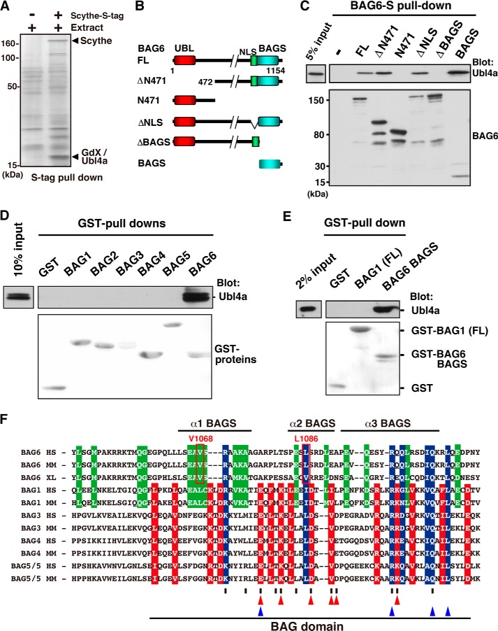
BAG6 is an essential protein that functions in two distinct biological pathways, ubiquitin-mediated protein degradation of defective polypeptides and tail-anchored (TA) transmembrane protein biogenesis in mammals, although its structural and functional properties remain unknown. We solved a crystal structure of the C-terminal heterodimerization domains of BAG6 and Ubl4a and characterized their interaction biochemically. Unexpectedly, the specificity and structure of the C terminus of BAG6, which was previously classified as a BAG domain, were completely distinct from those of the canonical BAG domain. Furthermore, the tight association of BAG6 and Ubl4a resulted in modulation of Ubl4a protein stability in cells. Therefore, we propose to designate the Ubl4a-binding region of BAG6 as the novel BAG-similar (BAGS) domain. The structure of Ubl4a, which interacts with BAG6, is similar to the yeast homologue Get5, which forms a homodimer. These observations indicate that the BAGS domain of BAG6 promotes the TA protein biogenesis pathway in mammals by the interaction with Ubl4a.

Keywords: Membrane Protein; Protein Assembly; Small-angle X-ray Scattering (SAXS); Tail Anchor; Ubiquitin; X-ray Crystallography.


Figures

FIGURE 1.
FIGURE 1.
The BAGS domain of BAG6 is a specific binding site for Ubl4a. A, Scythe/BAG6-associated proteins in Xenopus embryos identified by PMF analysis. Precipitates obtained with S-tagged Scythe were subjected to SDS-PAGE, and the gel was silver-stained. B, schematic representation of the BAG6-deletion mutant proteins used in this study. Numbers denote corresponding amino acids. C, Ubl4a binding assay with a series of BAG6 deletion mutants. Full-length (FL) 2S-tagged BAG6 and its truncated derivatives were expressed in HeLa cells. Each form of BAG6 was affinity-purified using S-protein-agarose beads. After washing the beads, recombinant FLAG-tagged Ubl4a was mixed and precipitated, and bound materials were immunoblotted with anti-Ubl4a and anti-S-peptide antibodies. The BAGS domain of BAG6 is necessary and sufficient for Ubl4a binding. D and E, bacterially produced GST-tagged BAG domains derived from BAG1, BAG2, BAG3, BAG4, or BAG5 or the BAGS domains of BAG6 were mixed with purified FLAG-tagged Ubl4a in vitro. After GST pull-down, the precipitates were probed with an anti-FLAG antibody to detect Ubl4a co-precipitation (top). Precipitated GST proteins were stained with Ponceau S (bottom). Full-length BAG1 (FL) was also examined. F, multiple amino acid sequence alignments of the BAGS domain of BAG6 and BAG domains of BAG1, BAG3, BAG4, and BAG5. We exclude BAG2 from this alignment because of its extremely low amino acid conservation relative to other BAG family proteins. Red box, conserved residues in all BAG family proteins except for BAG6. Green box, conserved residues between BAG1 and BAG6 (showing validity of BAG1-BAG6 alignment). Blue box, conserved residues in all of these proteins. More than half of the residues reported to be involved in the Hsp70 interaction in BAG1 are not conserved in the BAG6 BAGS domain (shown as red triangles). Vertical slash, residues in human BAG1 that interact with Hsp70 (defined by Briknarova et al. (53)). Blue triangles, four strictly conserved residues in BAG family proteins (defined by Thress et al. (6)). Three α-helical regions of the BAGS domain are indicated by bars. HS, H. sapiens; MM, M. musculus; XL, Xenopus laevis.
FIGURE 2.
FIGURE 2.
The C-terminal TUGS-sequence of Ubl4a is essential for BAG6 binding. A and C, schematic representation of full-length (FL) and a series of Ubl4a deletion mutants used in this study. B, binding assay of Ubl4a deletion mutants. The E. coli-expressed GST-BAG domain of BAG6 was incubated with a series of deletion mutants of purified 5HA-tagged Ubl4a. Material obtained by GST pull-down was probed with anti-HA (for Ubl4a) and anti-GST (for BAG6) antibodies. D, C-terminal deletion analysis of the minimal region of Ubl4a necessary for BAG6 interaction. A series of FLAG-tagged truncated Ubl4a were expressed in HeLa cells, and anti-FLAG immunoprecipitation was performed. Immunoprecipitates were then probed with anti-BAG6 antibody to examine their interaction. A 40-amino acid-long region (residues 99–138) of Ubl4a was identified as the sequence necessary for BAG6 interaction. E, sequence alignment of the C-terminal half (from the end of the UBL to the C terminus) of vertebrate Ubl4a family proteins revealed a highly conserved island (indicated by red characters). This region covers the entire stretch necessary for BAG6 binding (boxed), and hence we designated it the TUGS (tethering Ubl4a to BAGS) sequence. Three α-helical regions are indicated by bars. Hs, H. sapiens; Mm, Mus musculus; Md, Monodelphis domestica (opossums); Xt, Xenopus tropicalis; Dr, Danio rerio.
FIGURE 3.
FIGURE 3.
Complex formation of the BAG6 BAGS domain and Ubl4a. A, SPR analysis of binding of Ubl4a to immobilized BAG6 BAGS domain. SPR sensorgrams for various concentrations of full-length Ubl4a wild type (0.1, 0.2, 0.5, 1, 2, 3, 5, 7.5, 10, and 20 nm) and H106E mutant (0.01, 0.02, 0.05, 0.1, 0.2, 0.5, 1, 5, 10, 20, and 50 μm) are shown as black curves. A two-state reaction model was fitted to the data, and the resulting fit is shown in red. B, CD spectra of BAG6 BAGS domain (dotted line), full-length Ubl4a (gray line), and the complex (black continuous line) are shown. Concentrations of recombinant proteins were 100 μm each.
FIGURE 4.
FIGURE 4.
Structure of the complex containing the TUGS region of Ubl4a and the BAG6 BAGS domain. A, the overall structures of human Ubl4a and BAG6 are represented in a ribbon model. TUGS and BAGS domains are shown in magenta and cyan, respectively. Ubl4a TUGS consists of three helices named Ubl4a-α1 (α1TUGS), -α2 (α2TUGS), and -α3 (α3TUGS). BAG6 BAGS also consists of three helices named BAG6-α1 (α1BAGS), -α2 (α2BAGS), and -α3 (α3BAGS). One CHAPS molecule in the crystal structure is shown as a green stick model. B, structural comparison of the TUGS region of the BAGS-TUGS complex with the yeast Get5 C terminus homodimer (Protein Data Bank code 3VEJ, chain A). The root mean square deviation between the two models is 0.732 Å. TUGS is shown in magenta, and Get5 is shown in gray. C, comparison of the BAG6 BAGS domain and mouse BAG1 BAG domain (Protein Data Bank code 1I6Z). The homologous region of the mouse BAG1 domain (residues 135–196), according to the alignment in Fig. 1F, is presented as an orange schematic model. The BAG6 BAGS domain is shown in cyan. D, open book presentation of the hydrophobic interface of Ubl4a TUGS (left) and BAG6 BAGS (right) domains. The hydrophobic side chains, located on the interface, are shown as stick models. E, close-up view of the loop region between α1TUGS and α2BAGS. His-106 of Ubl4a TUGS makes hydrogen bonds with Thr-1089 (2.7 Å). Asp-1089 of BAG6 BAGS (2.7 Å), respectively. Asp-1089 of BAG6 BAGS also weakly interacts with Arg-105 of Ubl4a TUGS (3.5 Å) via a hydrogen bond. 2FoFc omit map at contoured 1.5σ is shown in blue.
FIGURE 5.
FIGURE 5.
Importance of conserved residues at the interface of the BAGS-TUGS interaction. A, a series of bacterially expressed GST-tagged Ubl4a (full-length) variants containing point mutations were immobilized to glutathione-Sepharose 4B and then incubated with His-tagged BAGS domain fragment (residues 1048–1124). After the beads were washed several times, the samples were analyzed by SDS-PAGE, and precipitated proteins were detected by Coomassie Brilliant Blue (CBB) staining. GST-Ubl4a (40 kDa) and His-tagged BAG6 (10 kDa) are indicated. B and C, a series of GST-tagged BAGS domain mutants were incubated with full-length Ubl4a. GST precipitants were subjected to Western blotting with the indicated antibodies.
FIGURE 6.
FIGURE 6.
BAGS-Ubl4a complex forms extended structure in solution. A, pair-distance distribution (P(r)) functions of BAGS-TUGS (residues 95–147) and BAGS-Ubl4a (residues 1–157) complexes are shown by black and gray lines, respectively. These functions were calculated from the x-ray scattering intensity profile. Dmax values of the complexes are 55 Å (BAGS-TUGS) and 93 Å (BAGS-Ubl4a), respectively. B, SAXS envelopes calculated using GASBOR, of the BAGS-TUGS complex (top) and BAGS-Ubl4a complex (bottom). Each GASBOR model is shown as a gray surface model. Rigid body docking of the BAGS-TUGS complex structural model into the BAGS-TUGS complex model using SUPCOMB is shown (top). In the case of the BAGS-Ubl4a complex, the NMR structure of Ubl4a UBL domain (Protein Data Bank code 2DZI) and the BAGS-TUGS complex structure were manually docked into the model (bottom).
FIGURE 7.
FIGURE 7.
BAG6 association stabilizes the Ubl4a protein. A, Ubl4a protein is destabilized by loss of its C-terminal BAG6 binding region. Expression vectors encoding FLAG-tagged wild-type Ubl4a and its ΔC29 derivative were transfected into HeLa cells, and CHX chase analysis was performed, following Western blotting with anti-FLAG and anti-β-actin antibodies. Actin loading control for wild type (a) and for ΔC29 (b) are indicated. B, CHX chase analysis of wild-type and H106E mutant Ubl4a in HeLa cells.

References

    1. Lüders J., Demand J., Höhfeld J. (2000) The ubiquitin-related BAG-1 provides a link between the molecular chaperone Hsc70/Hsp70 and the proteasome. J. Biol. Chem. 275, 4613–4617 - PubMed
    1. Demand J., Alberti S., Patterson C., Höhfeld J. (2001) Cooperation of a ubiquitin domain protein and E3 ubiquitin ligase during chaperone/proteasome coupling. Curr. Biol. 11, 1569–1577 - PubMed
    1. Takayama S., Reed J. C. (2001) Molecular chaperone targeting and regulation by BAG family proteins. Nat. Cell Biol. 3, E237–E241 - PubMed
    1. Kabbage M., Dickman M. B. (2008) The BAG proteins: a ubiquitous family of chaperone regulators. Cell. Mol. Life Sci. 65, 1390–1402 - PMC - PubMed
    1. Banerji J., Sands J., Strominger J. L., Spies T. (1990) A gene pair from the human major histocompatibility complex encodes large proline-rich proteins with multiple repeated motifs and a single ubiquitin-like domain. Proc. Natl. Acad. Sci. U.S.A. 87, 2374–2378 - PMC - PubMed

Publication types

MeSH terms

Associated data