. 2015 Feb 23;107(5):djv034.
doi: 10.1093/jnci/djv034.

The role of BPTF in melanoma progression and in response to BRAF-targeted therapy

Affiliations

The role of BPTF in melanoma progression and in response to BRAF-targeted therapy

Altaf A Dar et al. J Natl Cancer Inst. .

Abstract

Background: Bromodomain PHD finger transcription factor (BPTF) plays an important role in chromatin remodeling, but its functional role in tumor progression is incompletely understood. Here we explore the oncogenic effects of BPTF in melanoma.

Methods: The consequences of differential expression of BPTF were explored using shRNA-mediated knockdown in several melanoma cell lines. Immunoblotting was used to assess the expression of various proteins regulated by BPTF. The functional role of BPTF in melanoma progression was investigated using assays of colony formation, invasion, cell cycle, sensitivity to selective BRAF inhibitors, and in xenograft models of melanoma progression (n = 12 mice per group). The biomarker role of BPTF in melanoma progression was assessed using fluorescence in situ hybridization and immunohistochemical analyses. All statistical tests were two-sided.

Results: shRNA-mediated BPTF silencing suppressed the proliferative capacity (by 65.5%) and metastatic potential (by 66.4%) of melanoma cells. Elevated BPTF copy number (mean ≥ 3) was observed in 28 of 77 (36.4%) melanomas. BPTF overexpression predicted poor survival in a cohort of 311 melanoma patients (distant metastasis-free survival P = .03, and disease-specific survival P = .008), and promoted resistance to BRAF inhibitors in melanoma cell lines. Metastatic melanoma tumors progressing on BRAF inhibitors contained low BPTF-expressing, apoptotic tumor cell subclones, indicating the continued presence of drug-responsive subclones within tumors demonstrating overall resistance to anti-BRAF agents.

Conclusions: These studies demonstrate multiple protumorigenic functions for BPTF and identify it as a novel target for anticancer therapy. They also suggest the combination of BPTF targeting with BRAF inhibitors as a novel therapeutic strategy for melanomas with mutant BRAF.


Figures

Figure 1.
Figure 1.
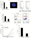
Effects of suppression of bromodomain PHD finger transcription factor (BPTF) expression on 1205-Lu melanoma cells. A) Expression of BPTF mRNA in 1205-Lu cells following BPTF suppression. B) Fluorescence in situ hybridization analysis showing probe for BPTF (red) and probe for chromosome 17 (green). C) Cell cycle analysis of 1205-Lu cells following BPTF suppression. D) Colony formation assay following BPTF suppression in 1205-Lu cells. E) Apoptotic activity in 1205-Lu cells following BPTF knockdown. F) In vivo tumor cell growth (n = 10 mice per group) following BPTF knockdown (mean tumor volume of control shRNA on day 32 = 1770.3mm3, 95% confidence interval [CI] =257.4 to 3035.5; vs mean of BPTF shRNA 3 on day 32 = 841.6mm3, 95% CI = 332.1 to 1527.1; P = .02). G) Metastatic lung tumor count following BPTF knockdown (n = 12 mice per group). In panels (A), (C), (D), (E), and (G), data presented reflect mean ± SD. Statistical significance was calculated by the two-tailed Student’s t test. *P < .05. In panel (B), scale bar reflects 20 μm. BPTF = bromodomain PHD finger transcription factor.
Figure 2.
Figure 2.
Effects of bromodomain PHD finger transcription factor (BPTF) suppression on C8161.9 human melanoma cells. A) BPTF mRNA expression following BPTF suppression in C8161.9 melanoma cells. B) Fluorescence in situ hybridization analysis showing probe for BPTF (red) and probe for chromosome 17 (green). C) Proliferative activity following BPTF knockdown as determined by a colony formation assay. D) Apoptotic activity in C8161.9 melanoma cells following BPTF suppression. E) In vivo tumor growth following BPTF shRNA expression. In panels (A), (C), and (D), data presented reflect mean ± SD. In panel (E), n = 10 for each group (mean tumor volume of control shRNA at day 28 = 1538.5, 95% confidence interval [CI] = 770.1 to 2567.9; vs mean of BPTF shRNA 3 at day 28 = 457.5, 95% CI = 200.9 to 744.5). Statistical significance was calculated by the two-tailed Student’s t test. *P < .05. In panel (B), scale bar reflects 20 μm. BPTF = bromodomain PHD finger transcription factor.
Figure 3.
Figure 3.
Effects of regulation of bromodomain PHD finger transcription factor (BPTF) expression on expression of genes involved in tumor cell proliferation and apoptosis. A-B) Expression of CCND2, BCL-XL, and BCL-2 at the mRNA level following BPTF suppression in 1205-Lu and C8161.9 cells, respectively. C-D) Western analysis showing expression of CCND2, BCL-XL, and BCL2 after BPTF silencing in 1205-Lu and C8161.9 cells, respectively. E) Proliferative activity of 1205-Lu cells following BPTF overexpression. F) Expression levels of various genes following BPTF overexpression in 1205-Lu cells. G) Western blots showing expression of various proteins following BPTF overexpression in 1205-Lu cells. In panels (A), (B), (E), and (F), data presented reflect mean ± SD. Statistical significance was calculated by the two-tailed Student’s t test. *P < .05. BPTF = bromodomain PHD finger transcription factor.
Figure 4.
Figure 4.
Effects of modulation of expression of various genes in 1205-Lu cells. A-B) Western blot analysis of total ERK and pERK levels in 1205-Lu and C8161.9 cells, respectively, following bromodomain PHD finger transcription factor (BPTF) silencing. C-D) Luciferase activity in 1205-Lu and C8161.9 cells, respectively, after cotransfection of BPTF cDNA along with a vector encoding the luciferase gene driven by the ERK promoter. E) Effects of overexpression of cDNAs encoding ERK or BCL-XL in 1205-Lu expressing BPTF shRNA 3. In panels (C), (D), and (E), data presented reflect mean ± SD. Statistical significance was calculated by the two-tailed Student’s t test. *P < .05. BPTF = bromodomain PHD finger transcription factor.
Figure 5.
Figure 5.
Effects of modulation of bromodomain PHD finger transcription factor (BPTF) expression on sensitivity to selective BRAF inhibitors. Sensitivity of 1205-Lu melanoma cells expressing BPTF shRNA3 to vemurafenib (A) or dabrafenib (B) treatment. C-D) Sensitivity of 1205-Lu melanoma cells following BPTF overexpression to vemurafenib (C) or dabrafenib (D) treatment. BPTF = bromodomain PHD finger transcription factor.
Figure 6.
Figure 6.
Bromodomain PHD finger transcription factor (BPTF) expression in human tissue specimens prior to and following progression on selective BRAF inhibitor treatment. A) Immunohistochemical staining of BPTF expression in a melanoma metastasis resected prior to initiation of vemurafenib treatment. Three regions (Ι-ΙΙΙ) were randomly selected for quantification of BPTF copy number (Supplementary Table 2, available online). B-C) Dual-color fluorescence in situ hybridization (FISH) for BPTF locus (red) and centromere of chromosome 17 (green) from selected regions (I and II), representing the signal distribution from each probe. D) Immunohistochemical staining of BPTF in a metastasis from the same patient resected following progression on vemurafenib. E) Higher magnification (40x) of black insert from (D) (black arrows show the higher BPTF-expressing regions, whereas the red arrow shows the lower BPTF-expressing region). F) Immunostaining using a cocktail of antibodies against HMB45, MART-1, and tyrosinase, detecting cells of melanocytic lineage in black insert from (D). G) TUNEL staining (green), including counterstaining with DAPI, (corresponding to region captured by red insert from panel D). Quantification of BPTF copy number in different regions (i-vii) (Supplementary Table 2, available online) of the tumor resected following progression on vemurafenib. H-J) Dual-color FISH for BPTF locus (red) and centromere of chromosome 17 from a region with low staining for BPTF (region i, panel H), a region with high BPTF staining (region v, panel I), and a region representing the transition between these two regions (region i→v, panel J). The green background is typical of autofluorescence from lipofuscin, the breakdown product of red blood cells. In panels (H-J), the mean BPTF copy number for each region is indicated within the figure. In panels (A), (D), (E), (F), and (G), the scale bars reflect 2000 μm. In panels (B), (C), (H), (I), and (J), they reflect 20 μm. BPTF = bromodomain PHD finger transcription factor.

References

    1. Clapier CR, Cairns BR. The biology of chromatin remodeling complexes. Annu Rev Biochem. 2009;78;273–304. - PubMed
    1. Tsukiyama T, Wu C. Purification and properties of an ATP-dependent nucleosome remodeling factor. Cell. 1995;83(6):1011–1020. - PubMed
    1. Xiao H, Sandaltzopoulos R, Wang HM, et al. Dual functions of largest NURF subunit NURF301 in nucleosome sliding and transcription factor interactions. Mol Cell. 2001;8(3):531–543. - PubMed
    1. Landry J, Sharov AA, Piao Y, et al. Essential role of chromatin remodeling protein Bptf in early mouse embryos and embryonic stem cells. PLoS Genet. 2008;4(10):e1000241. - PMC - PubMed
    1. Kwon SY, Xiao H, Wu C, Badenhorst P. Alternative splicing of NURF301 generates distinct NURF chromatin remodeling complexes with altered modified histone binding specificities. PLoS Genet. 2009;5(7):e1000574. - PMC - PubMed

Publication types

MeSH terms