Review
. 2015;67(2):310-37.
doi: 10.1124/pr.114.009464.

International Union of Basic and Clinical Pharmacology. XCIII. The parathyroid hormone receptors--family B G protein-coupled receptors

Affiliations
Review

International Union of Basic and Clinical Pharmacology. XCIII. The parathyroid hormone receptors--family B G protein-coupled receptors

Thomas J Gardella et al. Pharmacol Rev. 2015.

Abstract

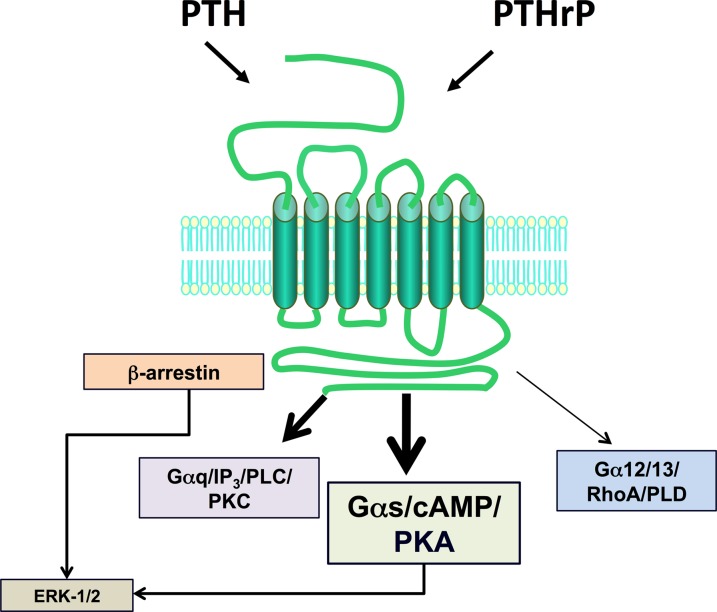
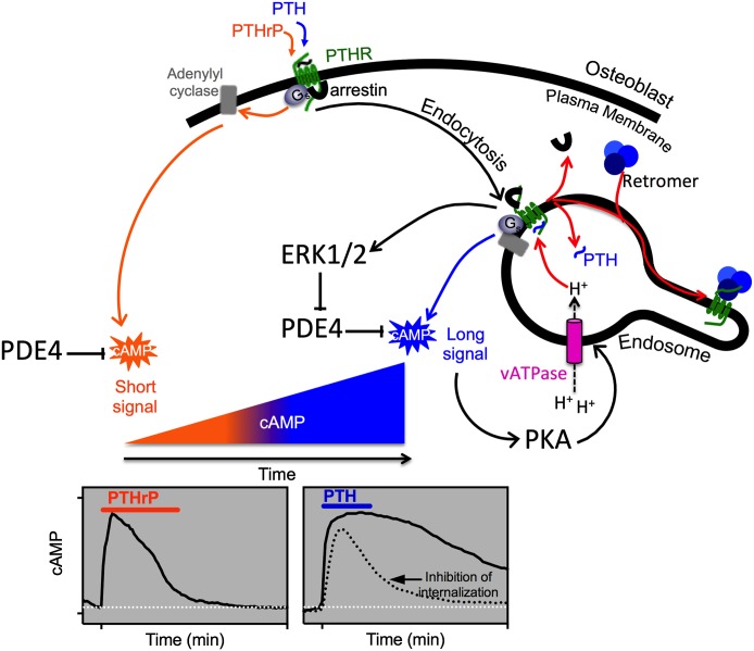
The type-1 parathyroid hormone receptor (PTHR1) is a family B G protein-coupled receptor (GPCR) that mediates the actions of two polypeptide ligands; parathyroid hormone (PTH), an endocrine hormone that regulates the levels of calcium and inorganic phosphate in the blood by acting on bone and kidney, and PTH-related protein (PTHrP), a paracrine-factor that regulates cell differentiation and proliferation programs in developing bone and other tissues. The type-2 parathyroid hormone receptor (PTHR2) binds a peptide ligand, called tuberoinfundibular peptide-39 (TIP39), and while the biologic role of the PTHR2/TIP39 system is not as defined as that of the PTHR1, it likely plays a role in the central nervous system as well as in spermatogenesis. Mechanisms of action at these receptors have been explored through a variety of pharmacological and biochemical approaches, and the data obtained support a basic "two-site" mode of ligand binding now thought to be used by each of the family B peptide hormone GPCRs. Recent crystallographic studies on the family B GPCRs are providing new insights that help to further refine the specifics of the overall receptor architecture and modes of ligand docking. One intriguing pharmacological finding for the PTHR1 is that it can form surprisingly stable complexes with certain PTH/PTHrP ligand analogs and thereby mediate markedly prolonged cell signaling responses that persist even when the bulk of the complexes are found in internalized vesicles. The PTHR1 thus appears to be able to activate the Gα(s)/cAMP pathway not only from the plasma membrane but also from the endosomal domain. The cumulative findings could have an impact on efforts to develop new drug therapies for the PTH receptors.


Figures

Fig. 1.
Fig. 1.
Ligand recognition and second-messenger signaling at PTHR1. The PTHR1 is a family B GPCR that mediates the actions of two peptide ligands—parathyroid hormone and PTH-related protein. In response to either ligand, the PTHR1 can couple to a variety of signal transduction pathways, including most prominently, the Gαs/cAMP/PKA pathway but also the Gαq/PLC/PKC pathway, the Gα12/13/RhoA/PLD pathway, and the ERK-1/2–MAP-kinase pathway, the latter via G protein–dependent and G protein–independent/β-arrestin–dependent mechanisms.
Fig. 2.
Fig. 2.
Sequences of PTH, PTHrP TIP39. Sequences of the bioactive 1–34 portions of PTH and PTHrP and the intact TIP39 sequence are shown with residues that are identical in PTH and PTHrP and retained in TIP39 shown red fill. The bars represent peptide fragment lengths that correspond to approximate minimum-length functional domains of the bioactive peptides.
Fig. 3.
Fig. 3.
Bioactive domains and key functional determinants of PTH(1–34) receptor ligands. (A) Shown is the sequence of the bioactive (1–34) segment of parathyroid hormone in linear format with residues in the amino-terminal portion that are critical for signaling shown in blue and those in the C-terminal domain that are critical for binding shown in red. (B) The PTH(1–34) ligand is presented in three-dimensional ribbon format to display the two-helical-domain structure typically observed in solution-phase NMR studies, with the side chains shown of the key binding and signaling residues. The structure was generated using the solution-phase NMR-derived coordinate file PDB ID 1HPH deposited in the Protein Data Bank (Marx et al., 1995).
Fig. 4.
Fig. 4.
Optimized PTH(1–14) domain. Shown is the sequence of the human PTH(1–14) peptide and the six amino acid substitutions that comprise the “M” set of modifications that together increase cAMP signaling potency of the PTH(1–14) fragment by five orders of magnitude and stabilize α-helical structure in the otherwise disordered PTH(1–14) peptide (Tsomaia et al., 2004). The graphs depict dose-response curves for cAMP generation obtained for PTH(1–34), M-PTH(1–14), and native PTH(1–14) in COS-7 cells transiently transfected to express either the intact wild-type PTHR1 (left) or the PTHR1-delNT construct (right), which is deleted for most of the ECD. These data reveal that whereas potency of PTH(1–34) is about 100-fold weaker on PTHR1-delNT than on the wild-type PTHR1, the PTH(1–14) fragments exhibit the same potency on the two receptors. The data thus demonstrate that whereas PTH(1–34) requires both the ECD and the TMD regions to obtain full potency, the PTH(1–14) portion of the ligand only interacts with the TMD region of the receptor (Shimizu et al., 2001b).
Fig. 5.
Fig. 5.
The PTH receptor type 1. This “snake” diagram of the human the PTHR1 illustrates the receptor's 593 amino acids in a topological arrangement typical of the family B GPCRs. The receptor thus has a relatively large amino ECD of about 160 amino acids (minus the 23 amino acids of the N-terminal signal sequence) that are involved in initial ligand binding, the seven helical transmembrane domains and connecting loops that mediate agonist-induced receptor activation and signal transduction events, and a C-terminal tail of about 130 amino acids that contains sites involved in mediating ligand-induced receptor internalization, trafficking, and signal termination events. Key specific amino acids identified include the four pairs of extracellular cysteine (C) residues that form a disulfide bond network that is conserved in the family B GPCRs and maintains receptor structure and function (Lee et al., 1994; Pioszak and Xu, 2008; Pioszak et al., 2009); four glycosylated asparagine (N) residues in the ECD (Zhou et al., 2000); Thr33 and Gln37, which modulate interaction with tryptophan-23 in the ligand (Mannstadt et al., 1998; Mann et al., 2008); Phe184 and Arg186, which mediate interactions involving ligand residues at or near lysine-13 (Adams et al., 1998; Carter et al., 1999a); Ser370, Ile371, Met425, Trp437, and Gln440, which contribute interactions involving ligand residues at or near valine-2 and likely play a role in receptor activation (Gardella et al., 1994; Lee et al., 1995; Bisello et al., 1998; Behar et al., 1999; Gensure et al., 2001a); Arg233 and Gln451, which participate in an interhelical interaction network (dashed connectors) that likely helps modulate PTHR activation (Gardella et al., 1996a) and is conserved in the family B GPCRs (Hollenstein et al., 2013); conserved Pro132 in the ECD, which is the site of an inactivating mutation (Leu) in Blomstrand’s chondrodysplasia (Zhang et al., 1998); His223, Thr410, and Ile458, at which mutations result in constitutive signaling activity and in patients result in Jansen’s chondrodysplasia (Schipani et al., 1999); Lys319, at which mutations impair Gαq signaling (Iida-Klein et al., 1997); Lys388, at which mutations impair Gαq and Gαs signaling (Huang et al., 1996). Key residues in the C-tail include the seven serine (S) residues that are phosphorylated upon agonist activation and mediate recruitment of β-arrestins (Malecz et al., 1998; Qian et al., 1998; Tawfeek et al., 2002; Vilardaga et al., 2002; Rey et al., 2006) and the C-terminal ETVM sequence that mediates interaction with the NHERF family of proteins (Mahon et al., 2002, 2003; Ardura et al., 2011; Mamonova et al., 2012). In each transmembrane domain, the residue identified as the most conserved residue among the family B GPCRs is enclosed in a hexagon (Wootten et al., 2013).
Fig. 6.
Fig. 6.
Phylogenetic relationships among PTH receptors and ligands from different species. (A) The amino acid sequences of PTH receptors from humans, zebrafish (Danio rerio) and chicken were aligned after removal of the predicted signal peptides and the segment of the human PTH1R encoded by exon E2 using the ClustalW(2.012) program (gap penalties: opening, 10; extending, 0.2 multiple, 0.1 pairwise) and an unrooted tree in which branch distances indicate amino acid sequence divergence was generated using the Phylip(3.67) DrawTree program. The diagram illustrates the three subtypes of PTH receptors, for which the PTH1R is present in all vertebrates, the PTH2R is present in humans and fish (Danio) but absent in birds, and the PTH3R is present in birds and fish but not in higher vertebrates. The tree includes a PTHR-like sequence that was identified in the genome of the tunicate Ciona intestinalis and which shows ∼35% overall identity to the human PTHR1. The Protein database accession numbers for the sequences used are as follows: human PTH1R, Q03431; chicken PTH1R, 418507; Danio PTH1R, Q9PVD3; chicken PTH3R, Danio PTH3R, Q9PVD2; Ciona PTHR-L, ci0100139945; human PTH2R, P49190; Danio PTH2R, Q9PWB7. (B) Also shown is an alignment of the (1–37) portions of PTH and PTHrP ligands and full-length TIP39 from human, zebrafish (D. rerio), and chicken species. Key residues involved in signaling, aligning with Val2, Ile5, and Met8 in the human PTH sequence, and binding, aligning with Arg20, Trp23, Leu24, and Leu28, are colored green and blue, respectively. Database accession numbers are shown for each ligand along with the specifies-ligand identifying label.
Fig. 7.
Fig. 7.
Two-site model of the PTH/PTHR1 interaction mechanism. Illustrated is the two-site mechanism of PTH-PTHR1 interaction, according to which the C-terminal portion of PTH(1–34), in α-helical conformation, first interacts with the amino-terminal extracellular domain (ECD) of the PTHR1, and then the N-terminal portion of the ligand binds to the transmembrane domain (TMD) region of the receptor, leading to conformational changes involved in receptor activation and coupling to heterotrimeric G proteins. Whereas the C-terminal portion of PTH(1–34) binds as a preformed α-helix, the N-terminal portion of the ligand is shown to undergo a coil-helix transition during the binding process, as suggested by structure-activity studies on PTH(1–14) peptide analogs (Shimizu et al., 2001b). The resulting folding cooperativity could contribute to the overall affinity of binding, as also suggested for other family B GPCRs (Parthier et al., 2009).
Fig. 8.
Fig. 8.
Molecular model of the PTH(1–34)•PTHR1 complex. Shown is a plausible model of PTH(1–34) bound to the PTHR1. The protein backbone chains are shown in ribbon format with the ligand colored magenta and the receptor colored by segment with the amino-terminal extracellular domain (ECD) colored blue-green and the transmembrane domain (TMD) colored by transmembrane helices as follows: purple (TM1), blue (TM2), cyan (TM3), green (TM4), yellow (TM5), orange (TM6), and red (TM7). The structure of the PTH(15–34) in complex with the ECD is according to the structural coordinates reported by Pioszak and Xu (2008) (PDB ID 3C4M). The TMD region of the PTHR1, spanning residues Thre175 to Ser491, was modeled by homology using the crystal structure coordinates of the CRFR1 (Hollenstein et al., 2013) (PDB ID 4K5Y) as a template. The PTH(1–14) segment, modeled as a partial α-helix, was docked to the TMD region manually, placing Val2 and Lys13 of the ligand near the extracellular ends of TMs 6 and 1, respectively. The ECD•PTH(15–34) component was positioned onto the TMD•PTH(1–14) component, manually, allowing for a bend in the ligand between the amino-terminal and carboxyl-terminal domain, as suggested by NMR structural studies on PTH peptide ligands and photoaffinity cross-linking and mutational data on PTH•PTHR1 complexes (Adams et al., 1998; Behar et al., 2000; Marx et al., 2000; Gensure et al., 2001a, 2003; Peggion et al., 2002; Wittelsberger et al., 2006). The side chains of ligand residues Val2, Lys13, Arg20, Trp23, Leu24, and Leu28 are shown in dotted surface format, and the three disulfide bonds in the ECD are in red. The insets show the unliganded ECD and two views of the TMD, rotated 180° relative to each other, which highlight the V-shaped putative ligand-binding groove that forms between the extracellular ends of the TM helices.
Fig. 9.
Fig. 9.
Properties of a long-acting PTH analog. Shown is the capacity of the long-acting analog, LA-PTH, a hybrid peptide consisting of a modified M-PTH(1–14) segment joined to a modified PTHrP(15–36) segment, to bind with high affinity to the R0 PTHR1 conformation and thus mediate prolonged signaling responses in cells and in animals. The analog is compared with PTH(1–34) for (A) binding to the PTHR1 in the G protein–independent conformation, R0; (B) inducing dose-dependent cAMP signaling responses in HEK-293 cells expressing the rat PTHR1 and the luciferase-based glosensor cAMP reporter; (C) capacity to maintain cAMP responses in the same cells after an initial application of ligand (0.3 nM/15 minutes) and then a washout (at t = 0') of unbound ligand; and (D) stimulating blood ionized calcium responses in mice [after injections with either vehicle, PTH(1–34) at 50 nmol/kg or LA-PTH at 10 nmol/kg]. Not shown is that LA-PTH bound to the G protein–coupled PTHR1 conformation, RG, with an affinity only threefold higher than that of PTH(1–34); this similar affinity for RG likely explains the similar potencies observed in the cAMP dose-response assays, whereas the greater difference in binding to the R0 PTHR1 conformation likely explains the more prolonged cAMP responses in cells and calcemic responses in animals. Adapted from Maeda et al. (2013).
Fig. 10.
Fig. 10.
Canonical and noncanonical G protein signaling at the PTHR1. The cAMP signaling responses induced by PTH and PTHrP peptides are typically of similar potency and efficacy when measured acutely; however, recent kinetic signaling and fluorescent imaging studies revealed differences in the response mechanisms used. Thus studies performed in HEK-293 cells transfected with green fluorescent protein–tagged PTHR1 thus shows that whereas PTHrP forms complexes with Gαs that are active and thus signal through cAMP only at the plasma membrane and then dissociate, PTH forms complexes that not only signal at the cell surface but remain associated as the complexes internalize. Moreover, as shown in the graphs depicting FRET-based cAMP biosensor responses, the response induced by PTH(1–34) persists for at least 30 minutes after initial binding (the ligand is applied during the time indicated by the bar at the top of the graph and then washed out), whereas the response induced by PTHrP(1–36) is transient and decays after ligand wash out. Prolonged cAMP signaling responses that correlate temporally with bulk internalization of ligand-receptor complexes also correlate positively with the capacity of a given ligand to bind with high affinity to the R0 PTHR1 conformation. The overall results lead to the hypothesis the PTHR1 can mediate cAMP signaling responses both by canonical mechanisms operating at the plasma membrane as well as by noncanonical mechanisms that operate from within the internalized endosomal domain (Dean et al., 2006b, 2008; Okazaki et al., 2008; Ferrandon et al., 2009; Feinstein et al., 2011).

References

    1. Abou-Samra AB, Jüppner H, Force T, Freeman MW, Kong XF, Schipani E, Urena P, Richards J, Bonventre JV, Potts JT, Jr, et al. (1992) Expression cloning of a common receptor for parathyroid hormone and parathyroid hormone-related peptide from rat osteoblast-like cells: a single receptor stimulates intracellular accumulation of both cAMP and inositol trisphosphates and increases intracellular free calcium. Proc Natl Acad Sci USA 89:2732–2736. - PMC - PubMed
    1. Adams AE, Bisello A, Chorev M, Rosenblatt M, Suva LJ. (1998) Arginine 186 in the extracellular N-terminal region of the human parathyroid hormone 1 receptor is essential for contact with position 13 of the hormone. Mol Endocrinol 12:1673–1683. - PubMed
    1. Alexander SP, Benson HE, Faccenda E, Pawson AJ, Sharman JL, Spedding M, Peters JA, Harmar AJ, CGTP Collaborators (2013) The Concise Guide to PHARMACOLOGY 2013/14: G protein-coupled receptors. Br J Pharmacol 170:1459–1481. - PMC - PubMed
    1. Ardura JA, Wang B, Watkins SC, Vilardaga JP, Friedman PA. (2011) Dynamic Na+-H+ exchanger regulatory factor-1 association and dissociation regulate parathyroid hormone receptor trafficking at membrane microdomains. J Biol Chem 286:35020–35029. - PMC - PubMed
    1. Aubry L, Guetta D, Klein G. (2009) The arrestin fold: variations on a theme. Curr Genomics 10:133–142. - PMC - PubMed

MeSH terms