. 2015 Apr;8(4):403-15.
doi: 10.1242/dmm.018960. Epub 2015 Feb 20.

Modeling autosomal recessive cutis laxa type 1C in mice reveals distinct functions for Ltbp-4 isoforms

Affiliations

Modeling autosomal recessive cutis laxa type 1C in mice reveals distinct functions for Ltbp-4 isoforms

Insa Bultmann-Mellin et al. Dis Model Mech. 2015 Apr.

Abstract

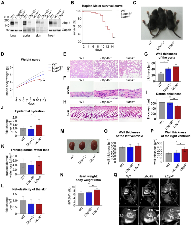
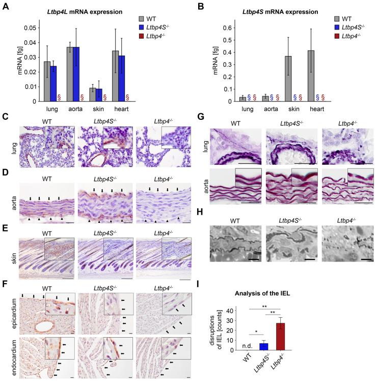
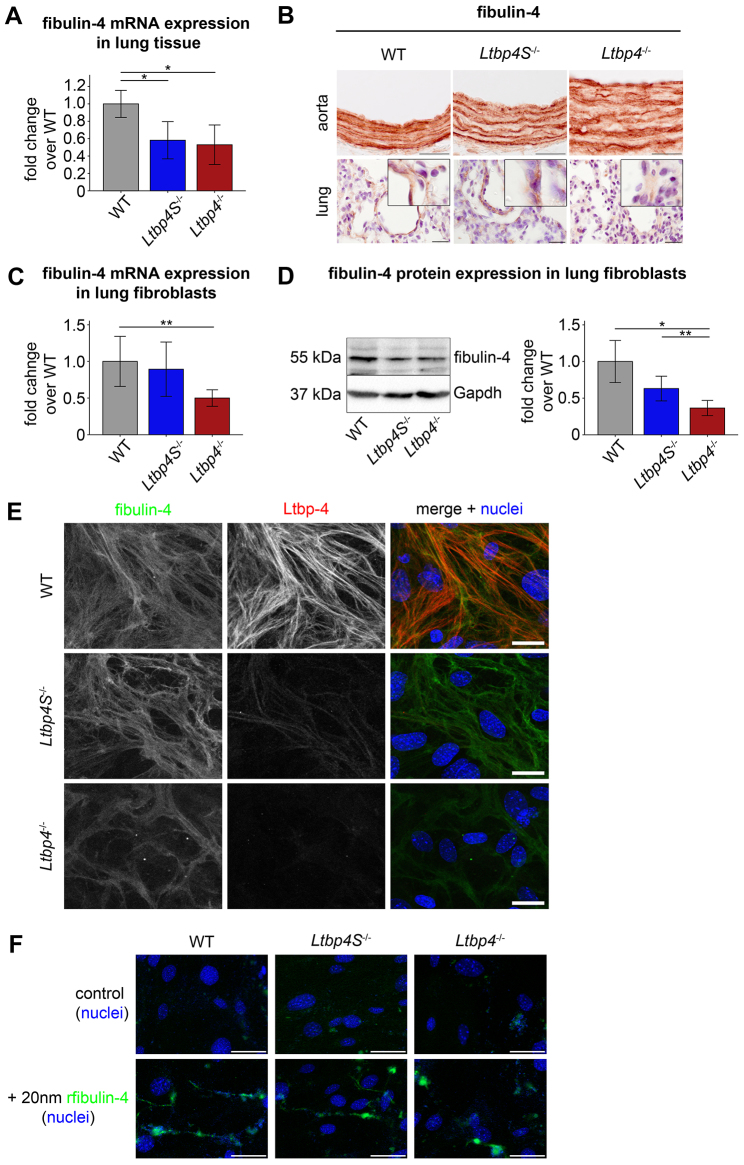
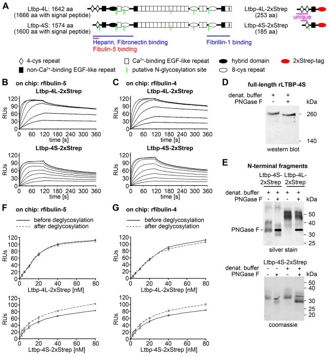
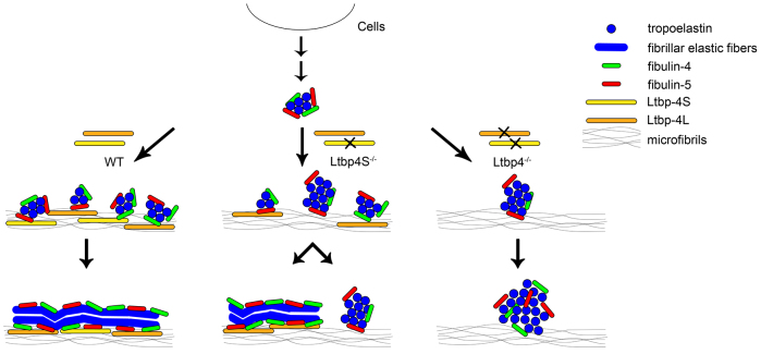
Recent studies have revealed an important role for LTBP-4 in elastogenesis. Its mutational inactivation in humans causes autosomal recessive cutis laxa type 1C (ARCL1C), which is a severe disorder caused by defects of the elastic fiber network. Although the human gene involved in ARCL1C has been discovered based on similar elastic fiber abnormalities exhibited by mice lacking the short Ltbp-4 isoform (Ltbp4S(-/-)), the murine phenotype does not replicate ARCL1C. We therefore inactivated both Ltbp-4 isoforms in the mouse germline to model ARCL1C. Comparative analysis of Ltbp4S(-/-) and Ltbp4-null (Ltbp4(-/-)) mice identified Ltbp-4L as an important factor for elastogenesis and postnatal survival, and showed that it has distinct tissue expression patterns and specific molecular functions. We identified fibulin-4 as a previously unknown interaction partner of both Ltbp-4 isoforms and demonstrated that at least Ltbp-4L expression is essential for incorporation of fibulin-4 into the extracellular matrix (ECM). Overall, our results contribute to the current understanding of elastogenesis and provide an animal model of ARCL1C.

Keywords: ARCL1C; Autosomal recessive cutis laxa type 1C; ECM; Elastogenesis; Extracellular matrix; Fibulin-4; Fibulin-5; Latent transforming growth factor β-binding protein 4; Ltbp-4; Ltbp-4L; Ltbp-4S.


Figures

Fig. 1.
Fig. 1.
Clinical and morphological analyses of Ltbp-4 deficient mice. (A) Representative immunoblots of lung, aorta, skin and hearts showing the reduced or absent expression of Ltbp-4 in Ltbp4S−/− and Ltbp4−/− mice compared to WT mice. (B) Kaplan–Meier survival curve revealing the significantly higher neonatal mortality in Ltbp4−/− mice compared to Ltbp4S−/− and WT mice (n≥23; **P<0.01 versus WT). (C) Ltbp4−/− mice showed reduced body size compared to Ltbp4S−/− and WT mice. Scale bar: 0.5 cm. (D) The P4-P12 weight curve showed significantly reduced body weight of Ltbp4−/− mice compared to Ltbp4S−/− and WT mice (n≥8; **P<0.01 versus WT). (E) In Ltbp4S−/− mice, the pulmonary parenchyma showed enlarged alveolar spaces with reduced numbers of alveoli and multifocal areas of atelectasis compared to WT mice. Ltbp4−/− lungs revealed lack of lobular architecture, severely enlarged alveolar spaces and emphysematous areas compared to WT mice. Scale bars: 40 μm. (F,G) Aortas showed marked thickening of the aortic wall in Ltbp4S−/− and Ltbp4−/− mice compared to WT mice (n≥3; *P<0.05, **P<0.01). Scale bars: 40 μm. (H,I) Ltbp4−/− mice showed reduced dermal thickness compared to Ltbp4S−/− and WT mice (n≥5;**P<0.01). Scale bars: 200 μm. (J) The epidermal hydration was increased in Ltbp4−/− mice compared to Ltbp4S−/− and WT mice (n≥9; *P<0.05). (K) The transepidermal water loss tended to be higher in Ltbp4−/− mice compared to Ltbp4S−/− and WT mice (n≥9; not significant). (L) The net-elasticity of the skin of both Ltbp-4-deficient mice tended to be lower compared to WT mice (n≥9; not significant). (M) Representative images showing the increased size of hearts of Ltbp4−/− mice compared to hearts of WT and Ltbp4S−/− mice. Scale bar: 0.1 cm. (N) Heart weight:body weight ratios showing that hearts of Ltbp4−/− mice were significantly heavier than hearts of WT and Ltbp4S−/− mice (n≥7; **P<0.01). (O) Wall thickness of the left ventricle was not changed in Ltbp4S−/− and Ltbp4−/− mice compared to WT mice (n≥6; not significant). (P) Wall thickness of the right ventricle was significantly increased in Ltbp4−/− mice compared to Ltbp4S−/− and WT mice (n≥6; *P<0.05). (Q) Representative short-axis images from MRI analysis revealing a flattened interventricular septum resulting in a more oval shape of the left ventricle in Ltbp4S−/− and Ltbp4−/− mice compared to WT mice.
Fig. 2.
Fig. 2.
Localization patterns of Ltbp-4L, Ltbp-4S and elastin. (A) Quantitative PCRs of lung, aorta, skin and heart of WT and Ltbp4S−/− mice showing that each tissue had different amounts of Ltbp4L mRNA. There was no mRNA expression of Ltbp4L in lung, aorta, skin and heart of Ltbp4−/− mice (n≥3; §, no expression detectable). (B) Quantitative PCRs of lung, aorta, skin and heart of WT mice showing that each tissue had different amounts of Ltbp4S mRNA. There was no mRNA expression of Ltbp4S in lung, aorta, skin and heart of Ltbp4S−/− and Ltbp4−/− mice (n≥3; §, no expression detectable). (C) Representative images of Ltbp-4 immunoreactivity of lungs from WT, Ltbp4S−/− and Ltbp4−/− mice. Ltbp-4 was localized particularly in bronchial and bronchiolar walls and in vascular walls of WT and Ltbp4S−/− mice. Lungs of Ltbp4−/− mice were negative for Ltbp-4 immunoreactivity. Scale bars: 20 μm. (D) Representative images of Ltbp-4 immunoreactivity of aortas from WT, Ltbp4S−/− and Ltbp4−/− mice. Black arrows point to the aortic luminal side and black arrowheads to the adventitia. Ltbp-4 immunoreactivity was present in the vicinity of aortic elastic lamella throughout the entire aorta (from the endothelial lining to the adventitia) of WT mice and in the vicinity of the internal elastic lamella (IEL) and in the adventitia of Ltbp4S−/− mice. The aortic intramural elastic lamella of Ltbp4S−/− mice and the entire aorta of Ltbp4−/− mice showed no immunoreactivity for Ltbp-4. Scale bars: 20 μm. (E) Representative images of Ltbp-4 immunoreactivity of skin from WT, Ltbp4S−/− and Ltbp4−/− mice. In WT skin, Ltbp-4 immunoreactivity was present in the entire dermis, whereas it was completely absent in the epidermis. There was no difference in the tissue distribution of Ltbp-4 between WT and Ltbp4S−/− skin. The skin of Ltbp4−/− mice expressed no Ltbp-4. Scale bars: 20 μm. (F) Representative images of Ltbp-4 immunoreactivity of hearts from WT, Ltbp4S−/− and Ltbp4−/− mice. Upper panel: black arrows point to the epicardium of the heart. Ltbp-4 immunoreactivity was present within the myocardium and in the epicardium of WT mice. The myocardium and the epicardium of Ltbp4S−/− and Ltbp4−/− mice were negative for Ltbp-4 immunoreactivity. Lower panel: black arrows point to the endocardium. The endocardium of WT and Ltbp4S−/− mice clearly has Ltbp-4 immunoreactivity, whereas Ltbp4−/− mice were negative for Ltbp-4 immunoreactivity. Scale bars: 20 μm. (G) Representative histochemical elastica stainings of lungs (upper panels) and aortas (lower panels) displaying moderate elastic fiber fragmentation with intact and disrupted elastic fibers in Lbp4S−/− mice compared to WT mice and an increased degree of fragmentation of the elastic fibers in Ltbp4−/− mice compared to Lbp4S−/− mice. Scale bars: 20 μm. (H) Representative semi-thin sections of lungs showing elastic fibers with fragmented and intact parts in Lbp4S−/− mice and total disruption of elastic fibers in Ltbp4−/− mice compared to WT mice. Scale bars: 6 μm. (I) Quantitative analysis of disruptions of the IEL showing significantly higher numbers of disruptions in Ltbp4−/− mice compared to WT and Ltbp4S−/− mice and significantly higher numbers of disruptions in Ltbp4S−/− mice compared to WT mice (n=6; n.d., not detectable; *P<0.05, **P<0.01).
Fig. 3.
Fig. 3.
Ltbp-4L is necessary for fibrillar matrix deposition of fibulin-4. (A) Fibulin-4 mRNA expression showed significant downregulation in lungs from Ltbp4S−/− and Ltbp4−/− mice compared to WT mice (n=4; *P<0.05). (B) Representative images showing fibulin-4 immunoreactivity and disruption of the fibrillar structure of fibulin-4 fibers in aortas (upper panel) and lungs (lower panel) from Ltbp4−/− mice compared to WT and Ltbp4S−/− mice. Scale bars: 20 μm (upper panels), 50 μm (lower panels). (C) Fibulin-4 mRNA expression showed significant downregulation in lung fibroblasts isolated from Ltbp4−/− mice compared to Ltbp4S−/− and WT mice (n≥5; **P<0.01). (D) Representative immunoblot of lung fibroblasts (left) and its densitometric analysis (right) revealing significant downregulation of fibulin-4 in Ltbp4S−/− and Ltbp4−/− mice compared to WT mice (n≥4; *P<0.05; **P<0.01). Protein as well as mRNA expression of fibulin-4 of the WT was set to 1. (E) Representative immunofluorescence staining of Ltbp-4 and fibulin-4 revealing reduced expression and disrupted fibrillar structure of fibulin-4 in primary lung fibroblasts from Ltbp4−/− mice compared to Ltbp4S−/− and WT mice. Scale bars: 100 μm. (F) Representative immunofluorescence staining revealing fibrillar deposition of recombinant full-length fibulin-4 (rfibulin-4; 20 nm) in primary lung fibroblasts from Ltbp4S−/− and WT mice, whereas rfibulin-4 appeared scattered and not fibrillar in primary lung fibroblasts from Ltbp4−/− mice. For detection of rfibulin-4, an anti-strep antibody was used. Scale bars: 50 μm.
Fig. 4.
Fig. 4.
Interaction studies of the Ltbp-4L and Ltbp-4S N-terminal regions with full-length fibulin-4 and fibulin-5. (A) Domain structure of full-length Ltbp-4L and Ltbp-4S and the recombinantly expressed Ltbp-4L (Ltbp-4L-2xStrep) and Ltbp-4S (Ltbp-4S-2xStrep) N-terminal fragments. The full-length proteins consist of 4-cystein (4-cys) repeats (white rhombi), non-Ca2+-binding EGF-like repeats (black rectangles), Ca2+-binding EGF-like repeats (white rectangles), hybrid domains (black ellipses) and 8-cystein (8-cys) repeats (white ellipses). The N-terminal fragments consist of two (Ltbp-4L-2xStrep) or one (Ltbp-4S-2xStrep) unique 4-cys repeats, the common non-Ca2+-binding EGF-like repeat and a C-terminal 2xStrep tag (red ellipses). Binding sites for ECM proteins, putative N-glycosylation sites (green lines) as well as the amino acid (aa) lengths are indicated. (B,C) Sensorgrams from surface-plasmon resonance interaction experiments showed a stronger binding affinity of Ltbp-4L-2xStrep (0–320 nM) ‘flown’ over immobilized recombinant full-length fibulin-5 (B; rfibulin-5) or immobilized recombinant full-length fibulin-4 (C; rfibulin-4) compared to Ltbp-4S-2xStrep (0–80 nM) flown over immobilized rfibulin-5 (B) or rfibulin-4 (C). The results are expressed as resonance units (RUs; n=2). (D) Deglycosylation digest with PNGase F of denatured recombinant full-length human LTBP-4S (rLTBP-4S) showing that there is a shift towards lower molecular mass positions. (E) Upper panel: deglycosylation of Ltbp-4L-2xStrep and Ltbp-4S-2xStrep. Ltbp-4L-2xStrep was unaffected, whereas Ltbp-4S-2xStrep showed a shift towards lower molecular weight positions. Lower panel: Ltbp-4S-2xStrep was digested with PNGase F under native (left lanes) and denaturing (right lanes) conditions. Both conditions resulted in a shift towards lower molecular weight positions. (F,G) After digest under non-denaturing conditions Ltbp-4L-2xStrep and Ltbp-4S-2xStrep were both able to bind to rfibulin-4 and -5 immobilized on a Biacore chip. Ltbp-4L-2xStrep binding was not affected, whereas Ltbp-4S-2xStrep showed an increase in binding of 15% to 20% after deglycosylation. The continuous lines represented the response before and the dashed lines after deglycosylation (n=2).
Fig. 5.
Fig. 5.
Proposed model for the role of Ltbp-4L and Ltbp-4S in elastogenesis. In the presence of Ltbp-4L and Ltbp-4S, microaggregation of tropoelastin, which is tethered to fibulin-4 and fibulin-5, deposits linearly onto microfibrils. Subsequent coalescence of tropoelastin takes place on microfibrils, resulting in fibrillar deposition of the tropoelastin–fibulin-4–fibulin-5 complex. In the absence of Ltbp-4S, the tropoelastin–fibulin-4–fibulin-5 complex only partly deposits on microfibrils, resulting in scattered fibrillar elastic fibers as well as amorphous aggregates of tropoelastin–fibulin-4–fibulin-5, which grow to form globular structures. In the absence of Ltbp-4L and Ltbp-4S, all tropoelastin–fibulin-4–fibulin-5 complexes form dysmorphous, non-fibrillar globular structures. Modified from Noda et al. (Noda et al., 2013).

References

    1. Abe M., Harpel J. G., Metz C. N., Nunes I., Loskutoff D. J., Rifkin D. B. (1994). An assay for transforming growth factor-beta using cells transfected with a plasminogen activator inhibitor-1 promoter-luciferase construct. Anal. Biochem. 216, 276–284. - PubMed
    1. Annes J., Vassallo M., Munger J. S., Rifkin D. B. (2004). A genetic screen to identify latent transforming growth factor beta activators. Anal. Biochem. 327, 45–54. - PubMed
    1. Bleeker G. B., Steendijk P., Holman E. R., Yu C. M., Breithardt O. A., Kaandorp T. A., Schalij M. J., van der Wall E. E., Bax J. J., Nihoyannopoulos P. (2006). Acquired right ventricular dysfunction. Heart 92 Suppl. 1, i14–i18. - PMC - PubMed
    1. Bultmann I., Conradi A., Kretschmer C., Sterner-Kock A. (2013). Latent transforming growth factor β-binding protein 4 is downregulated in esophageal cancer via promoter methylation. PLoS ONE 8, e65614. - PMC - PubMed
    1. Bunck A. C., Engelen M. A., Schnackenburg B., Furkert J., Bremer C., Heindel W., Stypmann J., Maintz D. (2009). Feasibility of functional cardiac MR imaging in mice using a clinical 3 Tesla whole body scanner. Invest. Radiol. 44, 749–756. - PubMed

Publication types

MeSH terms