. 2015 Apr;8(4):373-84.
doi: 10.1242/dmm.018648. Epub 2015 Feb 20.

Nuclear p120-catenin regulates the anoikis resistance of mouse lobular breast cancer cells through Kaiso-dependent Wnt11 expression

Affiliations

Nuclear p120-catenin regulates the anoikis resistance of mouse lobular breast cancer cells through Kaiso-dependent Wnt11 expression

Robert A H van de Ven et al. Dis Model Mech. 2015 Apr.

Abstract

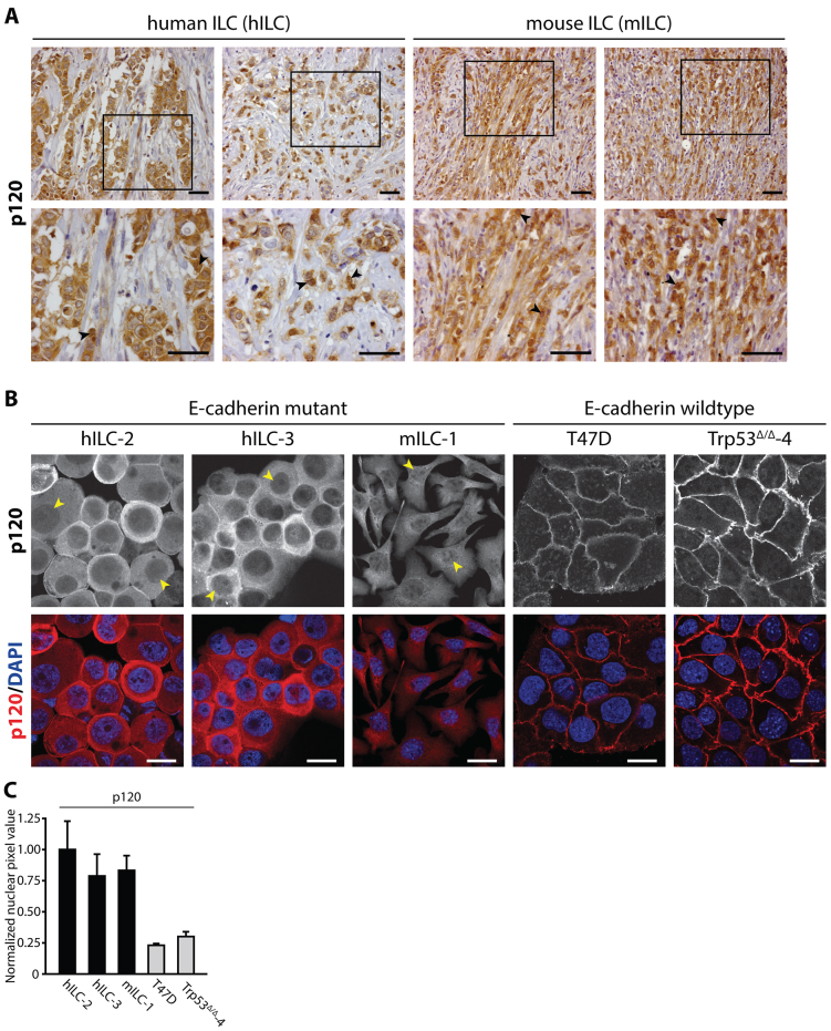
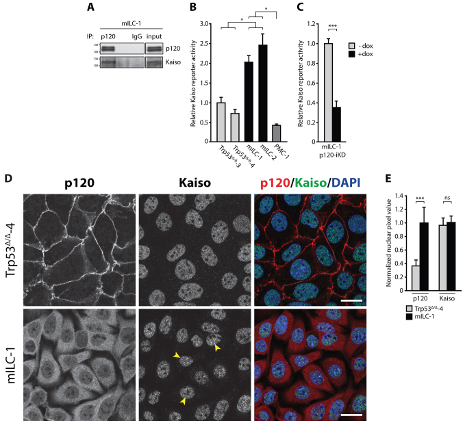
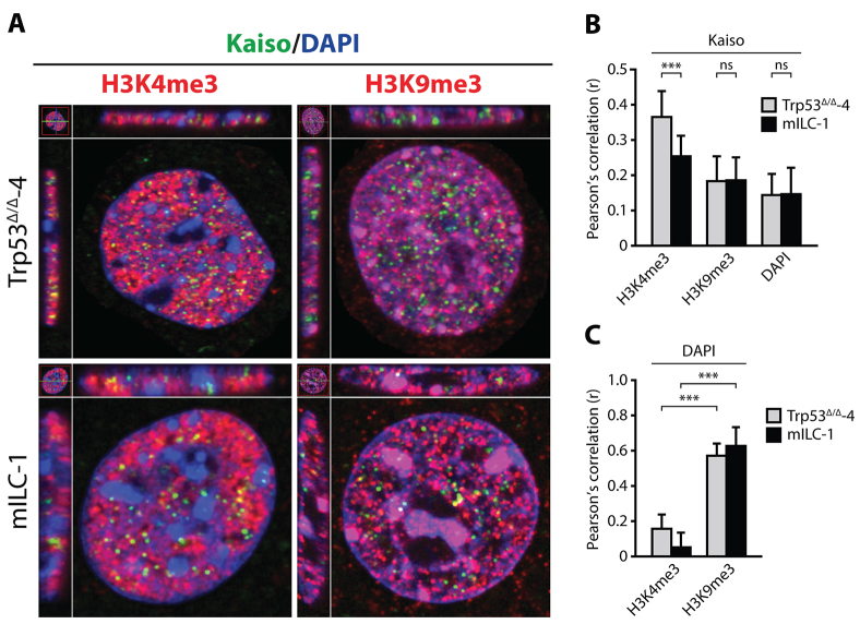
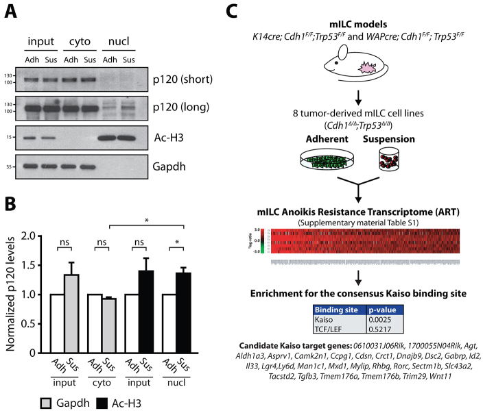
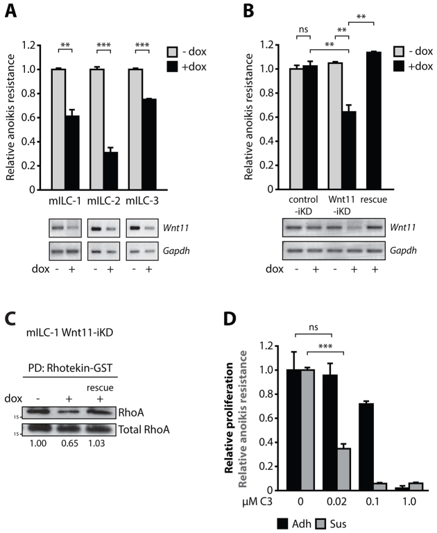
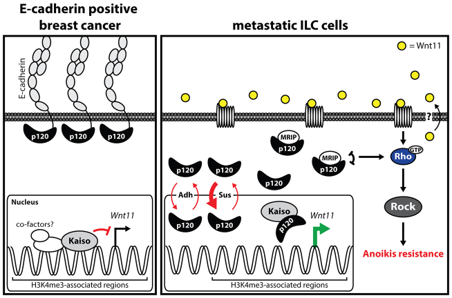
E-cadherin inactivation underpins the progression of invasive lobular breast carcinoma (ILC). In ILC, p120-catenin (p120) translocates to the cytosol where it controls anchorage independence through the Rho-Rock signaling pathway, a key mechanism driving tumor growth and metastasis. We now demonstrate that anchorage-independent ILC cells show an increase in nuclear p120, which results in relief of transcriptional repression by Kaiso. To identify the Kaiso target genes that control anchorage independence we performed genome-wide mRNA profiling on anoikis-resistant mouse ILC cells, and identified 29 candidate target genes, including the established Kaiso target Wnt11. Our data indicate that anchorage-independent upregulation of Wnt11 in ILC cells is controlled by nuclear p120 through inhibition of Kaiso-mediated transcriptional repression. Finally, we show that Wnt11 promotes activation of RhoA, which causes ILC anoikis resistance. Our findings thereby establish a mechanistic link between E-cadherin loss and subsequent control of Rho-driven anoikis resistance through p120- and Kaiso-dependent expression of Wnt11.

Keywords: Anoikis resistance; Breast cancer metastasis; Kaiso; p120-catenin.


Figures

Fig. 1.
Fig. 1.
p120 translocates to the cytosol and nucleus in mouse and human ILC cells. (A) p120 localization in ILC. Immunohistochemistry showing expression of p120 in primary human ILC (left panels) and primary mouse ILC cells (right panels). Lower panels are magnifications that correspond to the area indicated in the upper panel. Arrowheads denote representative cells with pronounced nuclear p120 expression. Scale bars: 50 μm. (B) Nuclear p120 expression in primary metastatic human ILC and mouse ILC cell lines. Immunofluorescence staining for p120 (upper panels) shows nuclear and cytosolic p120 in the majority of two independent primary human metastatic ILC samples (hILC-2 and hILC-3) and the tumor-derived mouse ILC cell line (mILC-1). Arrowheads denote representative cells with nuclear p120 expression in E-cadherin mutant ILC. Lower panels show the merge with DAPI (blue). The human breast cancer cell line T47D and mouse mammary carcinoma cells (Trp53Δ/Δ-4) were used to exemplify the near absence of nuclear p120 in E-cadherin-expressing breast carcinoma cells. Scale bars: 10 μm. (C) Quantifications of nuclear p120 in cells shown in B. At least 10 cells per cell line were quantified. Results are expressed as mean±s.d.
Fig. 2.
Fig. 2.
Kaiso-mediated transcriptional repression is relieved by nuclear p120 in mILC. (A) Kaiso interacts with p120 in mILC. p120 was immunoprecipitated to show the interaction between Kaiso and p120 (lane 1). Mouse IgG was used as a nonspecific immunoprecipitation (IP) control (lane 3). The total cell lysate (input) is shown in a separate panel. (B) Kaiso-mediated transcriptional repression is relieved in mILC. Renilla-based and Kaiso-specific reporter assays were performed on lysates from Trp53Δ/Δ, mILC and PMC-1 cells. (C) Nuclear p120 controls relief of Kaiso-mediated transcriptional repression in mILC. Findings in B were substantiated by inducible knockdown of p120 (p120-iKD) in mILC-1 cells. (D) Localization of p120 and Kaiso in the presence and absence of the epithelial adherens junction. E-cadherin-proficient Trp53Δ/Δ-4 cells and E-cadherin mutant mILC-1 cells were analyzed by immunofluorescence. Although p120 is predominantly localized to the plasma membrane in Trp53Δ/Δ-4 cells (upper panels), mILC-1 cells are characterized by cytosolic and nuclear p120 expression (lower panels). In contrast, the subcellular localization of Kaiso is not altered in mILC compared to Trp53Δ/Δ-4 cells. Note the punctate accumulation of Kaiso in mILC-1 nuclei (arrowheads). Scale bars: 10 μm. (E) Quantification of nuclear p120 and Kaiso in cells shown in D. Note the increase in nuclear p120 in mILC-1 compared to Trp53Δ/Δ-4. In contrast, no difference in nuclear Kaiso levels was observed between the two models. Shown are data from three independent experiments. Results are expressed as mean±s.d. *P<0.05; ***P<0.001; ns, not significant (Student’s t-test).
Fig. 3.
Fig. 3.
Kaiso is displaced from genomic regions of active transcription in mILC cells. (A) Colocalization of Kaiso with the active transcriptional mark H3K4me3, the inactive transcriptional mark H3K9me3 and heterochromatin (DAPI) was assessed by means of immunofluorescence. (B) Colocalization of Kaiso with the active transcription mark H3K4me3-positive genomic regions is decreased in mILC, whereas colocalization with the inactive transcription mark H3K9me3 is unaltered between cell models. Shown is a quantification of colocalization using the Pearson’s correlation coefficient (r). (C) Colocalization of DAPI with H3K4me3 and H3K9me3 was used as a validation of the method used in B. Results are expressed as mean±s.d. (n=40 or more analyzed nuclei). ***P<0.001; ns, not significant (Student’s t-test).
Fig. 4.
Fig. 4.
Identification of candidate Kaiso target genes in anchorage-independent mILC. (A) Nuclear p120 is enriched in anchorage-independent mILC cells. Fractionation was performed on adherent (Adh) and anchorage-independent (Sus) mILC-1 cells and the cytosolic and nuclear fractions were analyzed for p120 expression using western blotting. The purity of the lysate samples was confirmed by using the nuclear marker acetylated histone H3 (Ac-H3) and the cytosolic marker Gapdh (bottom panels). Subsequent analysis of the nuclear fractions showed enrichment for p120 in anchorage-independent mILC-1 cells (compare lane 5 and 6). Shown are two exposure times for the p120 blot (short and long). (B) Quantification of p120 in cytosolic and nuclear fractions. The signal for p120 was quantified and normalized to the appropriate marker (Gapdh for the cytosolic pool; Ac-H3 for the nuclear pool). Note the significant and specific increase in nuclear p120 in anchorage-independence compared to adherent conditions. Shown are the pooled data from three experiments. Results are expressed as mean±s.d. *P<0.05; ns, not significant (Student’s t-test). (C) The mILC Anoikis Resistance Transcriptome. To identify candidate Kaiso target genes specifically upregulated in anchorage-independent conditions, eight independent mILC cell lines were cultured in adherent and suspension and subjected to genome-wide microarray analysis. Using an arbitrary 2log1.5 cut-off we identified 249 genes that were upregulated in mILC cells cultured under anchorage-independent conditions. We termed this gene list the mILC Anoikis Resistance Transcriptome (ART). Subsequent TFBS promoter analysis of genes within the mILC ART revealed enrichment for the consensus binding sequence of Kaiso (KBS). No enrichment for TCF/LEF-binding sites was observed. Based on the presence of KBS sequences in the promoter regions of genes within the mILC ART we identified 29 candidate Kaiso target genes including Wnt11. The P-values reported in this figure are the result of initial testing for the KBS and TCF/LEF consensus sites by a Student’s t-test.
Fig. 5.
Fig. 5.
Anchorage-independent upregulation of the Kaiso target gene Wnt11 in mILC is p120-dependent. (A) Anchorage-independent upregulation of Wnt11 in mILC cell lines. Three independent mILC cell lines were cultured in the presence of cell-matrix anchorage (Adh) or in suspension (Sus). Wnt11 expression was quantified using qPCR. No Wnt11 upregulation was observed in mILC-2 cells, most likely due to high basal levels in anchorage-dependent conditions. The fold increase Wnt11 expression derived from the microarray analyses (Fig. 3B) is shown in italic. (B) Upregulation of Wnt11 is dependent on p120. Wnt11 expression was assayed in mILC p120-iKD cells in the absence or presence of doxycycline (dox) and under adherent (Adh) and suspension (Sus) conditions using qPCR. (C,D) Kaiso binds to the Wnt11 promotor. Kaiso-specific ChIP was performed on Trp53Δ/Δ-4 cells and mouse IgG was used as negative control (C). No input (H2O) and genomic DNA (gDNA) served as PCR controls. Specificity of the ChIP data shown in C was validated by amplification of a β-globin intronic promoter region (D). Fold increase of the KBS-specific signal compared to the β-globin signal was determined for the Kaiso-specific and the control ChIP using qPCR. (E) Expression of p120 in PMC-1 induces Wnt11 expression. PMC-1 (Ctnd1Δ/Δ;Trp53Δ/Δ) cells were transduced with either a control vector or a vector expressing p120-1A. (F,G) Kaiso knockdown in Trp53Δ/Δ-4 Kaiso-iKD cells using western blotting (F) and qPCR (G). (H) Kaiso knockdown in Trp53Δ/Δ-4 Kaiso-iKD cells induces Wnt11 expression. Shown are qPCR data from three independent experiments. Results are expressed as mean±s.d. *P<0.05; **P<0.01; ***P<0.001; ns, not significant (Student’s t-test).
Fig. 6.
Fig. 6.
Wnt11 regulates RhoA-dependent anoikis resistance in mILC. (A) Knockdown of Wnt11 in mILC-1 Wnt11-iKD cells reduces anoikis resistance of anchorage-independent mILC cell lines. Knockdown of Wnt11 was validated by RT-PCR. Gapdh was used as a loading control. (B) Introduction of a non-targetable Wnt11 cDNA in mILC-1 Wnt11-iKD restored anoikis resistance in mILC-1 cells (rescue). A scrambled shRNA (control-iKD; left lanes) was used as control for dox and shRNA expression. (C) Wnt11 regulates RhoA activation in mILC. Shown is a western blot depicting the effect of Wnt11-iKD on endogenous RhoA-GTP levels. GTP-loaded Rho was pulled down from the lysates using agarose-coupled Rhotekin-GST. Total RhoA was used as loading control. Note the rescue of Wnt11-iKD-induced RhoA inhibition (rescue). Shown below the blot are quantifications of RhoA-GTP levels. (D) Rho controls anoikis resistance of mILC cells. Inhibition of Rho by cell-permeable C3 transferase leads to inhibition of anoikis resistance of mILC-1 (Sus), but does not affect adherent cellular viability (Adh). Note the differential inhibitory effect at low molar concentrations. Shown are representative data from three independent experiments. Results are expressed as mean±s.d. **P<0.01; ***P<0.001; ns, not significant (Student’s t-test).
Fig. 7.
Fig. 7.
A model for p120-dependent relief of Kaiso transcriptional Wnt11 repression in ILC. In E-cadherin-expressing cancer cells, p120 resides at the plasma membrane in the adherens junction (AJ) complex with E-cadherin (left panel). In this scenario, Kaiso is repressing or dampening expression of its target genes in transcriptionally active regions by KBS-dependent binding and recruitment of repressive machinery. Upon loss of E-cadherin, p120 translocates to the cytosol and the nucleus (Adh), a shuttling process that is exacerbated by unknown mechanisms when cells lose cell-matrix interactions (Sus). In the nucleus p120 binds Kaiso and relieves Kaiso-mediated transcriptional repression. As a consequence, Kaiso target genes including Wnt11 are expressed. Autocrine Wnt11 will subsequently activate the small GTPase RhoA, which is a crucial event in the regulation of anchorage-independent tumor growth and metastasis of ILC.

References

    1. Arpino G., Bardou V. J., Clark G. M., Elledge R. M. (2004). Infiltrating lobular carcinoma of the breast: tumor characteristics and clinical outcome. Breast Cancer Res. 6, R149–R156. - PMC - PubMed
    1. Behrens J., von Kries J. P., Kühl M., Bruhn L., Wedlich D., Grosschedl R., Birchmeier W. (1996). Functional interaction of beta-catenin with the transcription factor LEF-1. Nature 382, 638–642. - PubMed
    1. Berx G., Cleton-Jansen A. M., Nollet F., de Leeuw W. J., van de Vijver M., Cornelisse C., van Roy F. (1995). E-cadherin is a tumour/invasion suppressor gene mutated in human lobular breast cancers. EMBO J. 14, 6107–6115. - PMC - PubMed
    1. Bhadriraju K., Yang M., Alom Ruiz S., Pirone D., Tan J., Chen C. S. (2007). Activation of ROCK by RhoA is regulated by cell adhesion, shape, and cytoskeletal tension. Exp. Cell Res. 313, 3616–3623. - PMC - PubMed
    1. Blattler A., Yao L., Wang Y., Ye Z., Jin V. X., Farnham P. J. (2013). ZBTB33 binds unmethylated regions of the genome associated with actively expressed genes. Epigenetics Chromatin 6, 13. - PMC - PubMed

Publication types

MeSH terms

LinkOut - more resources