. 2015 Mar 10;112(10):3086-91.
doi: 10.1073/pnas.1414272112. Epub 2015 Feb 23.

Thiopeptide antibiotics stimulate biofilm formation in Bacillus subtilis

Affiliations

Thiopeptide antibiotics stimulate biofilm formation in Bacillus subtilis

Rachel Bleich et al. Proc Natl Acad Sci U S A. .

Abstract

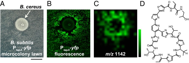
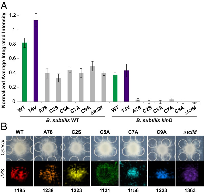
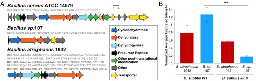
Bacteria have evolved the ability to produce a wide range of structurally complex natural products historically called "secondary" metabolites. Although some of these compounds have been identified as bacterial communication cues, more frequently natural products are scrutinized for antibiotic activities that are relevant to human health. However, there has been little regard for how these compounds might otherwise impact the physiology of neighboring microbes present in complex communities. Bacillus cereus secretes molecules that activate expression of biofilm genes in Bacillus subtilis. Here, we use imaging mass spectrometry to identify the thiocillins, a group of thiazolyl peptide antibiotics, as biofilm matrix-inducing compounds produced by B. cereus. We found that thiocillin increased the population of matrix-producing B. subtilis cells and that this activity could be abolished by multiple structural alterations. Importantly, a mutation that eliminated thiocillin's antibiotic activity did not affect its ability to induce biofilm gene expression in B. subtilis. We go on to show that biofilm induction appears to be a general phenomenon of multiple structurally diverse thiazolyl peptides and use this activity to confirm the presence of thiazolyl peptide gene clusters in other bacterial species. Our results indicate that the roles of secondary metabolites initially identified as antibiotics may have more complex effects--acting not only as killing agents, but also as specific modulators of microbial cellular phenotypes.

Keywords: Bacillus cereus; Bacillus subtilis; biofilm formation; thiazolyl antibiotics; thiopeptides.


Conflict of interest statement

The authors declare no conflict of interest.

Figures

Fig. 1.
Fig. 1.
IMS used to identify a matrix-inducing compound produced by B. cereus. (A) B. cereus grown as a colony on a microcolony lawn of B. subtilis. (Scale bar: 0.5 cm.) (B) This B. subtilis strain contains a fluorescent transcriptional reporter for biofilm matrix gene expression (PtapAyfp); the B. subtilis colonies closest to B. cereus are fluorescing, indicating that matrix gene expression is activated. (C) IMS data showing the distribution of an ion with m/z = 1,142 (linear negative mode) that corresponds to the area of fluorescence observed in B; relative scale is from 0.7 to 1.8. (D) Structure of micrococcin P1, a thiazolyl peptide antibiotic produced by B. cereus with a molecular weight consistent with it being the ion observed in C.
Fig. 2.
Fig. 2.
Thiocillin contributes to the ability of B. cereus to induce PtapAyfp gene expression in B. subtilis in a kinD- and antibiotic-independent manner. (A) Colonies of WT and ΔtclE-H B. cereus spotted onto a lawn of WT or kinD B. subtilis PtapAyfp microcolonies and quantification of the fluorescence (n = 3). (B) Colonies of the thiocillin T4V mutant of B. cereus spotted onto the same B. subtilis lawns, as well as quantification of the fluorescence (n = 3). (C) A total of 450 ng of purified thiocillin or T4V thiocillin spotted onto a lawn of WT B. subtilis PtapAyfp microcolonies and quantification of the fluorescence (n = 3). The halos visible in C are not due to B. subtilis cells dying, but by being physically moved during spotting of the compound. *P < 0.1; **P < 0.05.
Fig. 3.
Fig. 3.
Purified thiocillin increases the proportion of matrix-producing B. subtilis cells in liquid culture, even when not inhibiting growth. (A) Growth curves of B. subtilis from growth in shaking liquid culture with 12.5 nM YM-266183 or an equivalent volume of DMSO (gray diamonds, WT with DMSO; black squares, WT with YM-266183; light green triangles, PtapAyfp with DMSO; dark green circles, PtapAyfp with YM-266183). (B) Flow cytometry of the fluorescence intensity of B. subtilis cells harvested from the 9-h time point from A (front, WT cells with DMSO; middle, PtapAyfp cells with DMSO; back, PtapAyfp cells with YM-266183). A total of 30,000 cells were quantified for each sample.
Fig. 4.
Fig. 4.
Purified thiazolyl peptides induce PtapAyfp gene expression in B. subtilis. Quantification of fluorescence intensity resulting from spotting 450 ng of the purified thiazolyl peptides onto a lawn of B. subtilis PtapAyfp microcolonies (n = 3–13) is shown. There is no significant difference in PtapAyfp fluorescence induction between the B. subtilis WT and kinD lawns.
Fig. 5.
Fig. 5.
Structural modifications to thiocillin abolish its matrix-induction activity. (A) Quantification of fluorescence intensity of thiocillin mutant producers spotted onto a lawn of B. subtilis WT (Left) or kinD PtapAyfp (Right) microcolonies (n = 3). None of the mutants (gray bars) were significantly different from one another on either the WT or kinD B. subtilis lawns, but each were significantly different from both WT and T4V B. cereus (by at least P < 0.05 on WT and at least P < 0.0001 on kinD). (B) Images of B. cereus colonies producing the mutant thiocillins indicated grown on agar on a MALDI plate with corresponding IMS data; colors represent ions of m/z indicated: WT [M+H+Na]+; A78 [M+H+Na]+; C2S [M+Na+K]+; C5A [M+H]+; C7A [M+H+Na]+; C9A [M+H+Na+K]+; ∆tclM [M+H]+.
Fig. 6.
Fig. 6.
Bacterial strains containing cryptic thiazolyl peptide biosynthesis genes induce PtapAyfp gene expression in B. subtilis. (A) Gene clusters comparing enzyme and peptide sequences for B. cereus, B. atrophaeus, and Bacillus sp. 107. The known precursor peptide sequence is shown in red, with the cleavage site marked with an asterisk. (B) Quantification of the fluorescence intensity induced by WT B. cereus, B. atrophaeus 1942, and Bacillus sp. 107 in WT and kinD B. subtilis microcolony lawns (n = 3). Bacillus sp. 107 induction of PtapAyfp WT and kinD B. subtilis lawns are significantly different (P = 0.0094), but B. atrophaeus 1942 induction in these two strains is not significantly different, indicating a kinD-independent mechanism of induction. **P < 0.01.

Similar articles

Cited by

References

    1. Romero D, Traxler MF, López D, Kolter R. Antibiotics as signal molecules. Chem Rev. 2011;111(9):5492–5505. - PMC - PubMed
    1. Straight PD, Kolter R. Interspecies chemical communication in bacterial development. Annu Rev Microbiol. 2009;63:99–118. - PubMed
    1. Ryan RP, Dow JM. Diffusible signals and interspecies communication in bacteria. Microbiology. 2008;154(Pt 7):1845–1858. - PubMed
    1. Fajardo A, Martínez JL. Antibiotics as signals that trigger specific bacterial responses. Curr Opin Microbiol. 2008;11(2):161–167. - PubMed
    1. Williams P. Quorum sensing, communication and cross-kingdom signalling in the bacterial world. Microbiology. 2007;153(Pt 12):3923–3938. - PubMed

Publication types