. 2015 Mar 10;112(10):3164-9.
doi: 10.1073/pnas.1422373112. Epub 2015 Feb 23.

Cardiac ferroportin regulates cellular iron homeostasis and is important for cardiac function

Affiliations

Cardiac ferroportin regulates cellular iron homeostasis and is important for cardiac function

Samira Lakhal-Littleton et al. Proc Natl Acad Sci U S A. .

Erratum in

  • Correction.
    [No authors listed] [No authors listed] Proc Natl Acad Sci U S A. 2015 Apr 7;112(14):E1812. doi: 10.1073/pnas.1504383112. Epub 2015 Mar 11. Proc Natl Acad Sci U S A. 2015. PMID: 25762073 Free PMC article. No abstract available.

Abstract

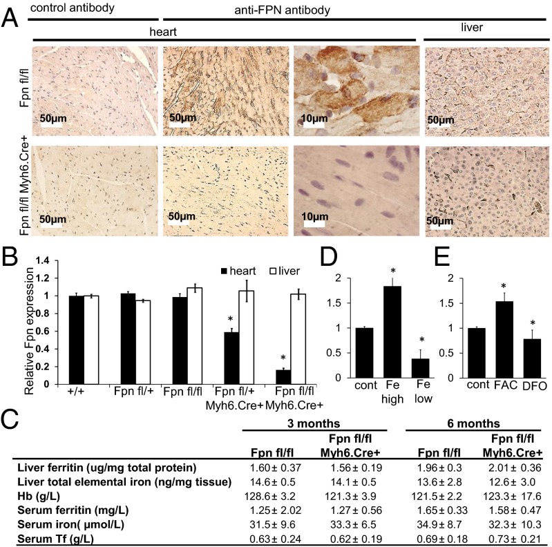
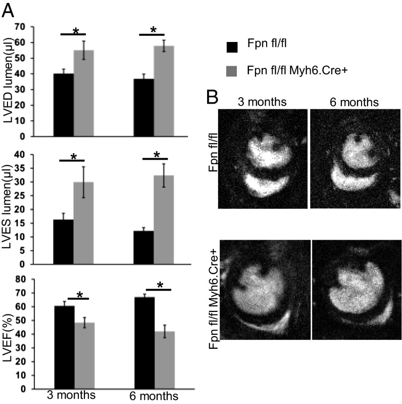
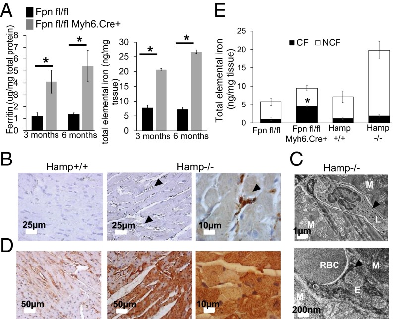
Iron is essential to the cell. Both iron deficiency and overload impinge negatively on cardiac health. Thus, effective iron homeostasis is important for cardiac function. Ferroportin (FPN), the only known mammalian iron-exporting protein, plays an essential role in iron homeostasis at the systemic level. It increases systemic iron availability by releasing iron from the cells of the duodenum, spleen, and liver, the sites of iron absorption, recycling, and storage respectively. However, FPN is also found in tissues with no known role in systemic iron handling, such as the heart, where its function remains unknown. To explore this function, we generated mice with a cardiomyocyte-specific deletion of Fpn. We show that these animals have severely impaired cardiac function, with a median survival of 22 wk, despite otherwise unaltered systemic iron status. We then compared their phenotype with that of ubiquitous hepcidin knockouts, a recognized model of the iron-loading disease hemochromatosis. The phenotype of the hepcidin knockouts was far milder, with normal survival up to 12 mo, despite far greater iron loading in the hearts. Histological examination demonstrated that, although cardiac iron accumulates within the cardiomyocytes of Fpn knockouts, it accumulates predominantly in other cell types in the hepcidin knockouts. We conclude, first, that cardiomyocyte FPN is essential for intracellular iron homeostasis and, second, that the site of deposition of iron within the heart determines the severity with which it affects cardiac function. Both findings have significant implications for the assessment and treatment of cardiac complications of iron dysregulation.

Keywords: cardiomyocyte; ferroportin; heart; hepcidin; iron.


Conflict of interest statement

The authors declare no conflict of interest.

Figures

Fig. 1.
Fig. 1.
FPN expression in the heart and its regulation by iron. (A) Immunohistochemical staining for FPN in heart (ventricular myocardium) and liver sections from Fpn fl/fl Myh6.Cre+ and Fpn fl/fl littermate control mice. (B) Relative expression of Fpn in hearts and livers of Fpn+/+ (wild type), Fpn fl/+ (heterozygous floxed), Fpn fl/fl (homozygous floxed), Fpn fl/+ Myh6.Cre+ (heterozygous cardiac knockout), and Fpn fl/fl Myh6.Cre+ (homozygous cardiac knockout) mice. n = 6 per group determined by quantitative PCR. (C) Indices of iron homeostasis in the liver and circulation of Fpn fl/fl Myh6.Cre+ mice and Fpn fl/fl littermate controls at age 3 and 6 mo. n = 6 per group. (D) Quantitative PCR expression of Fpn in hearts of wild-type C57B6 mice after 8 wk of a low-iron diet or 6 wk of a high-iron diet, relative to mice on the respective control diets. n = 6 per group. (E) Quantitative PCR expression of Fpn in isolated adult cardiomyocytes following 8-h treatment with ferric citrate (FAC) or deferoxamine (DFO). n = 3 biological replicates. All values are plotted as mean ± SEM. *P < 0.05 relative to control.
Fig. 2.
Fig. 2.
Reduced survival of Fpn fl/fl Myh6.Cre+ mice is associated with abnormal heart morphology. (A) Cumulative survival of Fpn fl/fl Myh6.Cre+ mice and Fpn fl/fl littermates over 40 wk. *P < 0.05. n = 20 per group. (B) Representative images of gross morphology in longitudinal cardiac sections and cardiomyocyte histology in a 6-mo-old Fpn fl/fl Myh6.Cre+ mouse (Lower) and an Fpn fl/fl littermate control (Upper). The arrowhead indicates an elongated nucleus. (C) Representative electron microscopy images of cardiac myofibril organization in Fpn fl/fl mice and Fpn fl/fl Myh6.Cre+ mice at age 3 mo. Note the loss of myofibrillar regularity and increased mitochondrial numbers in cardiomyocytes in Fpn fl/fl Myh6.Cre+ hearts.
Fig. 3.
Fig. 3.
Cardiac performance and morphology in vivo by cine MRI. (A) LV lumen size at end-systole (LVES) and end-diastole (LVED) and LVEF in Fpn fl/fl Myh6.Cre+ mice and Fpn fl/fl controls at age 3 mo (n = 12 per group) and 6 mo (n = 6 per group). Values are plotted as mean ± SEM. *P < 0.05. (B) Representative midventricular MR images of hearts from Fpn fl/fl Myh6.Cre+ mice and Fpn fl/fl controls at age 3 and 6 mo.
Fig. 4.
Fig. 4.
Retention of iron in cardiomyocytes of Fpn fl/fl Myh6.Cre+ mice. (A) Ferritin concentration in cardiac lysates from Fpn fl/fl Myh6.Cre+ mice and Fpn fl/fl controls at age 3 wk (n = 3 per group), 6 wk (n = 3 per group), 3 mo (n = 12 per group), and 6 mo (n = 6 per group). (B) Total elemental iron concentration in cardiac lysates from Fpn fl/fl Myh6.Cre+ mice and Fpn fl/fl controls at age 3 wk (n = 3 per group), 6 wk (n = 3 per group), 3 mo (n = 12 per group), and 6 mo (n = 6 per group). (C) T*2 in Fpn fl/fl Myh6.Cre+ mice and Fpn fl/fl controls at age 6 mo (n = 6 per group). (D) Representative images of DAB-enhanced Perls iron staining in ventricular myocardium from Fpn fl/fl Myh6.Cre+ mice and Fpn fl/fl controls at age 6 mo. (E) Representative electron microscopy images of ventricular myocardium from 3-mo-old Fpn fl/fl Myh6.Cre+ mice and Fpn fl/fl controls. Arrowheads indicate sites of iron deposition. (F) Levels of elemental zinc, cobalt, manganese, and copper in hearts of Fpn fl/fl Myh6.Cre+ mice and Fpn fl/fl controls at age 6 mo (n = 6 per group); below DL, below detection limit. (G) Parameters of cardiac function as measured by cine MRI in 3-mo-old mice after 8 wk on the control diet or the low-iron diet (n = 6 per group). All values are plotted as mean ± SEM. *P < 0.05.
Fig. 5.
Fig. 5.
Iron distribution in Hamp−/− mice. (A) Ferritin and total elemental iron concentrations in cardiac lysates from Hamp−/− mice and Hamp+/+ controls at age 3 and 6 mo (n = 12 per group). *P < 0.05 relative to Fpn fl/fl. (B) Representative images of DAB-enhanced Perls staining in ventricular myocardium of Hamp−/− mice and Hamp+/+ littermate controls at age 6 mo. Arrowheads indicate sites of iron accumulation. (C) Representative electron microscopy images of ventricular myocardium from 6-mo-old Hamp−/− mice. Arrowheads indicate sites of iron deposition. E, endothelial cell; L, leukocyte; M, myocyte; RBC, red blood cell. (D) Representative images of FPN immunohistochemical staining in ventricular myocardium of 6-mo-old Hamp−/− mice and Hamp+/+ littermate controls. (E) Levels of elemental iron as measured by ICP-MS in cardiomyocyte fractions (CF) and noncardiomyocyte fractions (NCF) from hearts of 6-mo-old Fpn fl/fl Myh6.Cre+ mice, Hamp−/− mice, and respective littermate controls (n = 3 per group). *P < 0.05 relative to cardiomyocyte fractions in Hamp−/− mice. Values are shown as mean ± SEM.
Fig. 6.
Fig. 6.
The role of FPN in cardiac iron homeostasis. (A) Integrated model of cellular iron homeostasis. FPN in cardiomyocytes is central to their cellular iron homeostasis. Together with TfR1-mediated iron uptake and ferritin storage, it controls iron levels within cardiomyocytes. FPN itself is regulated by intracellular iron levels in a manner dependent on IRP1 and IRP2 and by hepcidin derived from the liver and/or the heart itself. (B) FPN determines the site of cardiac iron deposition and the severity of the associated cardiac phenotype. Cardiomyocyte iron homeostasis occurs at the point of balance between iron entry and iron efflux. Because FPN is the only known mammalian iron-export protein, its expression in cardiomyocytes promotes iron efflux and a reduction in intracellular iron content. Loss of cardiomyocyte FPN leads to a severe cardiac phenotype, despite a mild degree of total cardiac iron loading. Up-regulation of FPN in Hamp−/− mice appears to be protective against the effects of systemic iron overload by enabling the release of iron from cardiomyocytes.

References

    1. Anker SD, et al. FAIR-HF Trial Investigators Ferric carboxymaltose in patients with heart failure and iron deficiency. N Engl J Med. 2009;361(25):2436–2448. - PubMed
    1. Okonko DO, et al. Effect of intravenous iron sucrose on exercise tolerance in anemic and nonanemic patients with symptomatic chronic heart failure and iron deficiency FERRIC-HF: A randomized, controlled, observer-blinded trial. J Am Coll Cardiol. 2008;51(2):103–112. - PubMed
    1. Klip IT, et al. Iron deficiency in chronic heart failure: An international pooled analysis. Am Heart J. 2013;165(4):575–582, e3. - PubMed
    1. Lekawanvijit S, Chattipakorn N. Iron overload thalassemic cardiomyopathy: Iron status assessment and mechanisms of mechanical and electrical disturbance due to iron toxicity. Can J Cardiol. 2009;25(4):213–218. - PMC - PubMed
    1. Murphy CJ, Oudit GY. Iron-overload cardiomyopathy: Pathophysiology, diagnosis, and treatment. J Card Fail. 2010;16(11):888–900. - PubMed

Publication types

LinkOut - more resources