. 2015 Mar 24;112(12):E1433-42.
doi: 10.1073/pnas.1418401112. Epub 2015 Feb 23.

Differential fates of biomolecules delivered to target cells via extracellular vesicles

Affiliations

Differential fates of biomolecules delivered to target cells via extracellular vesicles

Masamitsu Kanada et al. Proc Natl Acad Sci U S A. .

Abstract

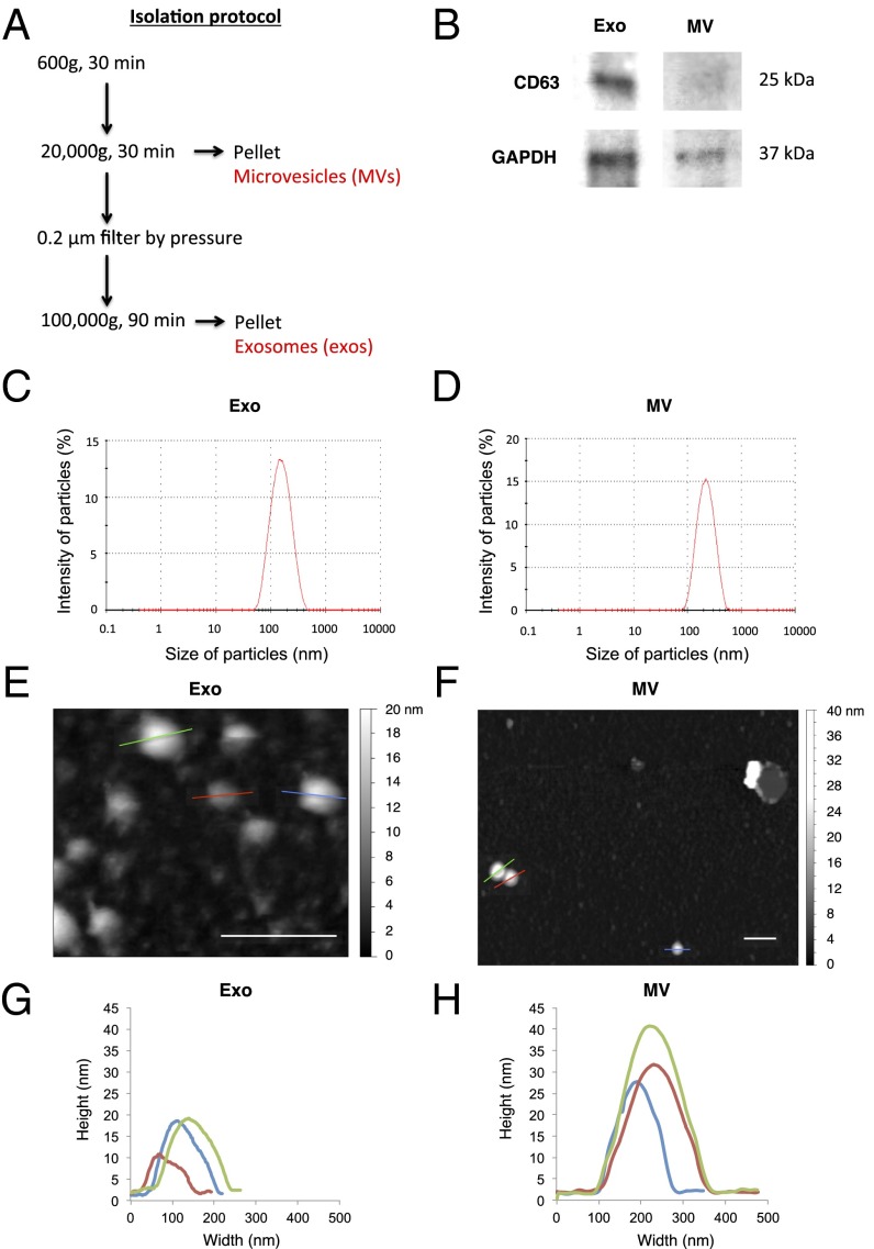
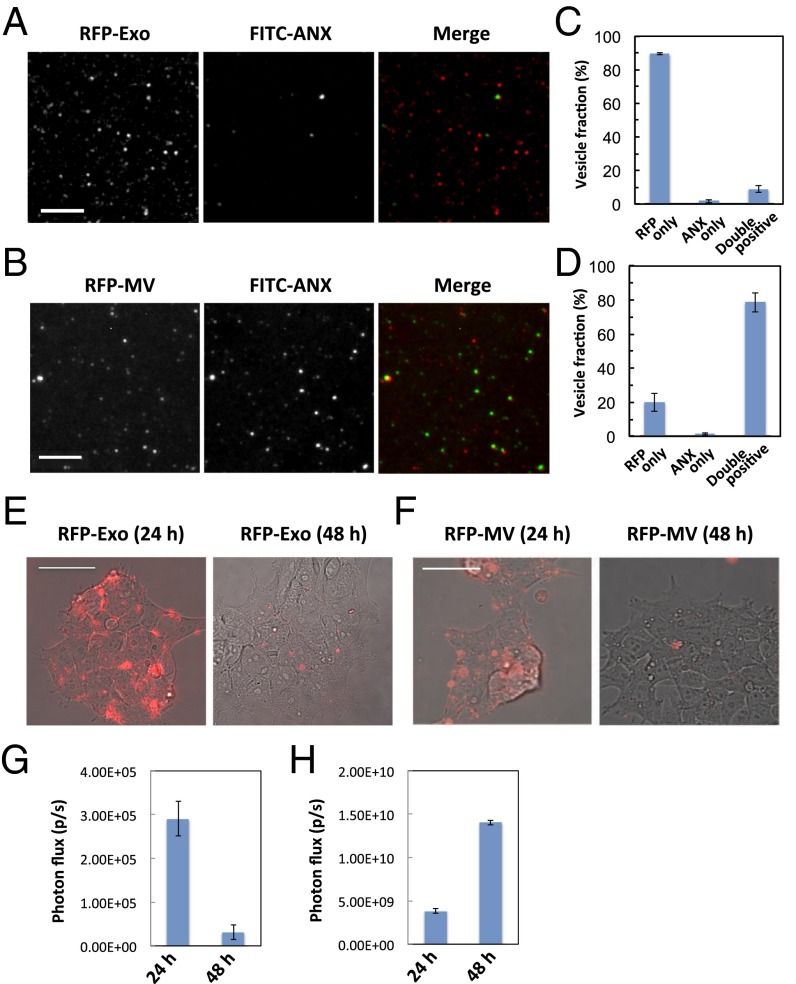
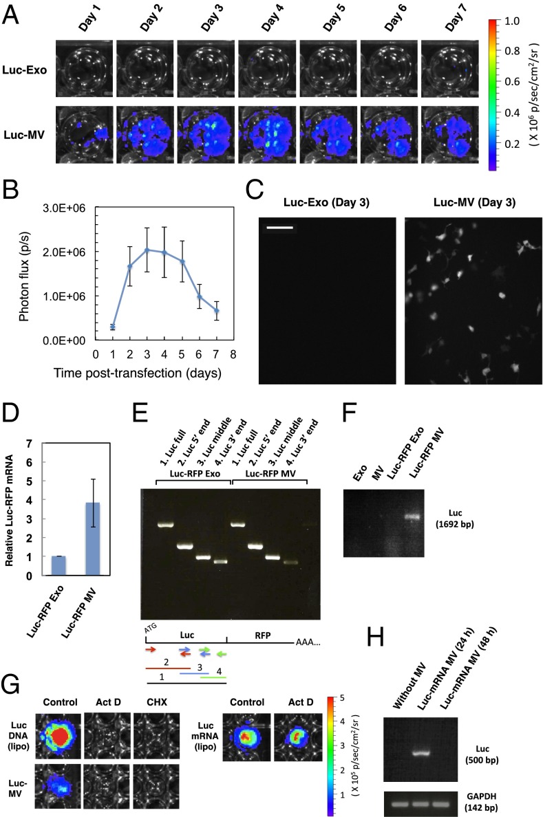
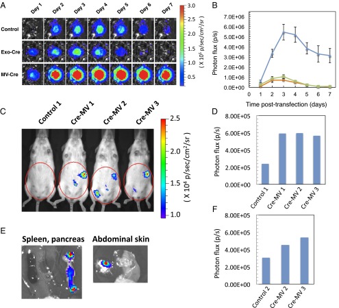
Extracellular vesicles (EVs), specifically exosomes and microvesicles (MVs), are presumed to play key roles in cell-cell communication via transfer of biomolecules between cells. The biogenesis of these two types of EVs differs as they originate from either the endosomal (exosomes) or plasma (MVs) membranes. To elucidate the primary means through which EVs mediate intercellular communication, we characterized their ability to encapsulate and deliver different types of macromolecules from transiently transfected cells. Both EV types encapsulated reporter proteins and mRNA but only MVs transferred the reporter function to recipient cells. De novo reporter protein expression in recipient cells resulted only from plasmid DNA (pDNA) after delivery via MVs. Reporter mRNA was delivered to recipient cells by both EV types, but was rapidly degraded without being translated. MVs also mediated delivery of functional pDNA encoding Cre recombinase in vivo to tissues in transgenic Cre-lox reporter mice. Within the parameters of this study, MVs delivered functional pDNA, but not RNA, whereas exosomes from the same source did not deliver functional nucleic acids. These results have significant implications for understanding the role of EVs in cellular communication and for development of EVs as delivery tools. Moreover, studies using EVs from transiently transfected cells may be confounded by a predominance of pDNA transfer.

Keywords: apoptotic body; cell communication; exosome; extracellular vesicle; microvesicle.


Conflict of interest statement

The authors declare no conflict of interest.

Figures

Fig. 1.
Fig. 1.
Size, surface markers, and physical characteristics of EVs. (A) Schematic depiction of the isolation protocol for exosomes and MVs. (B) Western blot analysis of the exosome marker protein, CD63. Detection of GAPDH was used as a loading control. (C and D) Size distribution of exosomes and MVs measured by DLS. (E and F) Topographic AFM images of exosomes and MVs adsorbed to the mica surface. Arrow indicates collapsed MV and lipid-bilayer spreading on the mica surface. (Scale bars, 400 nm.) (G and H) Size distribution of three randomly chosen exosomes and MVs imaged by AFM.
Fig. 2.
Fig. 2.
Membrane lipid composition of EVs and uptake by recipient cells. (A and B) Detection of fluorescent protein and phosphatidylserine (PS) in EVs. Loaded RFP was visualized by fluorescence (Left) and pseudocolor rendered red in the merged images (Right). PS in the outer membranes of EVs derived from HEK293FT cells was stained with FITC-annexin V (Middle, green in the merged images). (Scale bars, 10 µm.) (C and D) Fraction of the EVs that contain RFP only, stain with annexin V (ANX) only, or exhibiting both signals. Two hundred vesicles were counted. Error bars represent SD (n = 3). (E and F) Merged images of fluorescence of RFP-containing EVs and phase contrast images of the recipient HEK293FT cells. The EVs were cultured with the recipient cells for 24 h (labeled as 24 h). The recipient cells were cultured for another 24 h after removing the nonadherent EVs (labeled as 48 h). (Scale bars, 50 µm.) (G and H) Evaluation of delivered Luc protein in the recipient cells. A total of 0.5 µg of the Luc–RFP-containing EVs were cultured with the cells as described in E and F. The recipient cells were lysed at 24 or 48 h, respectively, and Luc activity was measured. Error bars represent SD (n = 3).
Fig. 3.
Fig. 3.
MV-mediated delivery of pDNA. (A) Bioluminescence in EV-treated HEK293FT cells. (Upper) HEK293FT cells were treated with 0.4 µg exosomes derived from HEK293FT cells transiently transfected with Luc–RFP expression vector. (Lower) Recipient cells were treated with 0.4 µg MVs from the same donor cells. The color scale indicates radiance (x106 photons/cm2/s/sr). (B) Time course of bioluminescence in the recipient cells that took up Luc–RFP-containing MVs. Photon flux (photons/s) is plotted over time (days). Error bars represent SEM. (n = 8). (C) Bioluminescence microscopic images of the recipient HEK293FT cells treated with the Luc–RFP-containing EVs. (Scale bar, 100 µm.) (D) The amount of Luc–RFP mRNA in EVs was determined by qRT-PCR. GAPDH was used as an internal control. Error bars represent SD (n = 3). (E) Analysis of fragmentation of Luc–RFP-encoding mRNAs in EVs by RT-PCR. Four primer sets and their amplified products are indicated below. (F) PCR amplification of the entire Luc ORF in EVs with and without Luc–RFP-encoding pDNAs. Equal amounts of OD260 were PCR amplified. (G) Analysis of MV-mediated biomolecule transfer. Recipient cells were treated with actinomycin D (1.0 µg/mL) or cycloheximide (100 µM) to inhibit transcription or translation, respectively. Transfection with purified Luc mRNA was used as a control of Act D treatment. Color scale: radiance (x105 photons/cm2/s/sr). (H) Analysis of degradation of delivered mRNA in the recipient cells. HEK293FT cells were incubated for 24 h with MVs derived from 4T1 cells stably expressing Luc, and, after removing nonadherent MVs, cultured for another 24 h. GAPDH was amplified as an internal control of recipient cell mRNA.
Fig. 4.
Fig. 4.
Imaging of functional biomolecule transfer by tumor cell-derived MVs. (A) In vitro activation of Cre-lox reporter cells with MVs from Cre recombinase-expressing cells. Reporter HEK293 cells were treated with 0.5 µg MVs derived from 4T1 cells transiently expressing Cre recombinase. (B) Time course of A. HEK293 Cre-lox Luc reporter cells were treated with Cre-containing exosomes (green), MVs (blue), or left untreated (orange), and photon emission from individual wells was imaged and quantitated as described above. Error bars represent SEM (n = 8). (C) Approximately 6 µg Met-1 cell-derived MVs that contain Cre recombinase were injected i.v. into Cre-lox Luc reporter mice. Color scale is radiance (x104 photons/cm2/s/sr). (D) Analysis of the bioluminescence from the abdominal regions (red circles indicated in C) 48 h after the MV injection. (E) Luc expression was measured in excised spleen, pancreas, and abdominal skin. (F) Analysis of the bioluminescence from the abdominal regions (red circles indicated in SI Appendix, Fig. S13) on day 21.

Comment in

References

    1. Raposo G, Stoorvogel W. Extracellular vesicles: Exosomes, microvesicles, and friends. J Cell Biol. 2013;200(4):373–383. - PMC - PubMed
    1. EL Andaloussi S, Mäger I, Breakefield XO, Wood MJ. Extracellular vesicles: Biology and emerging therapeutic opportunities. Nat Rev Drug Discov. 2013;12(5):347–357. - PubMed
    1. Lee Y, El Andaloussi S, Wood MJA. Exosomes and microvesicles: Extracellular vesicles for genetic information transfer and gene therapy. Hum Mol Genet. 2012;21(R1):R125–R134. - PubMed
    1. Valadi H, et al. Exosome-mediated transfer of mRNAs and microRNAs is a novel mechanism of genetic exchange between cells. Nat Cell Biol. 2007;9(6):654–659. - PubMed
    1. Al-Nedawi K, et al. Intercellular transfer of the oncogenic receptor EGFRvIII by microvesicles derived from tumour cells. Nat Cell Biol. 2008;10(5):619–624. - PubMed

Publication types

MeSH terms