. 2015 Jun;28(6):163-70.
doi: 10.1093/protein/gzv009. Epub 2015 Feb 23.

Specific disulfide cross-linking to constrict the mobile carrier domain of nonribosomal peptide synthetases

Affiliations

Specific disulfide cross-linking to constrict the mobile carrier domain of nonribosomal peptide synthetases

Michael J Tarry et al. Protein Eng Des Sel. 2015 Jun.

Abstract

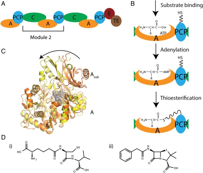
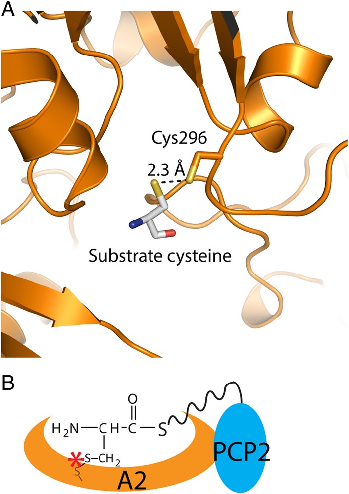
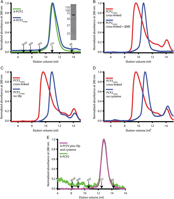
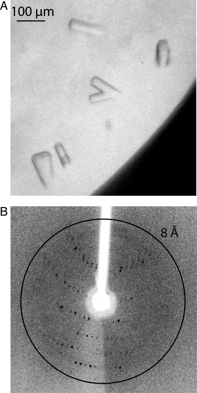
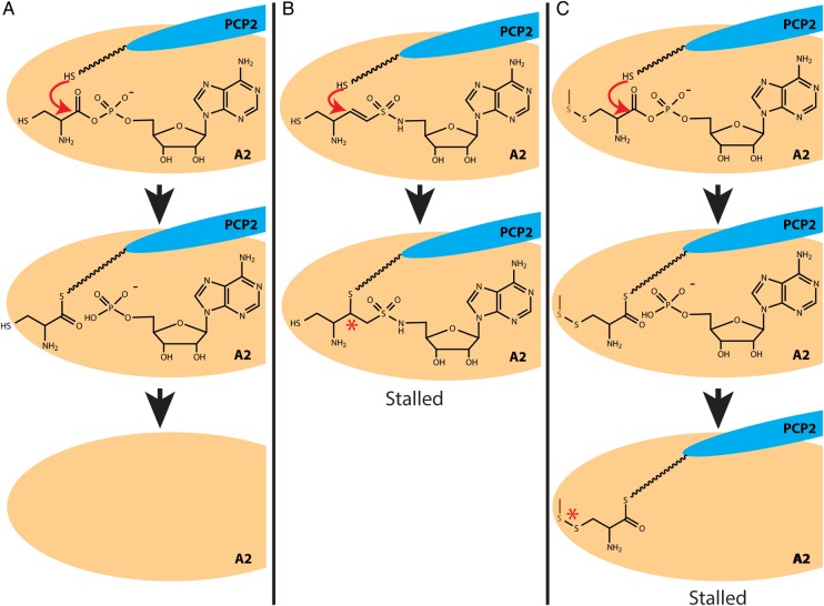
Nonribosomal peptide synthetases are large, multi-domain enzymes that produce peptide molecules with important biological activity such as antibiotic, antiviral, anti-tumor, siderophore and immunosuppressant action. The adenylation (A) domain catalyzes two reactions in the biosynthetic pathway. In the first reaction, it activates the substrate amino acid by adenylation and in the second reaction it transfers the amino acid onto the phosphopantetheine arm of the adjacent peptide carrier protein (PCP) domain. The conformation of the A domain differs significantly depending on which of these two reactions it is catalyzing. Recently, several structures of A-PCP di-domains have been solved using mechanism-based inhibitors to trap the PCP domain in the A domain active site. Here, we present an alternative strategy to stall the A-PCP di-domain, by engineering a disulfide bond between the native amino acid substrate and the A domain. Size exclusion studies showed a significant shift in apparent size when the mutant A-PCP was provided with cross-linking reagents, and this shift was reversible in the presence of high concentrations of reducing agent. The cross-linked protein crystallized readily in several of the conditions screened and the best crystals diffracted to ≈8 Å.

Keywords: crystallization; disulfide cross-link; nonribosomal peptide synthetase; protein engineering; thioesterification.


Figures

Fig. 1
Fig. 1
Schematic diagrams for ACV synthetase NRPS and products. (A) Schematic diagram showing the domain organization of P. chrysogenum ACV synthetase (E: epimerization domain, TE: thioesterase domain). (B) Schematic diagrams showing the two reactions performed by the A domain. (C) Ribbon diagram showing the 140° rotation of the A subdomain (labeled Asub) between the adenylation (orange) and thioesterification (yellow) conformations. A domains of PheA (PDBID:1AMU, orange) and PA1221 (PDBID:4DG9, yellow) were superimposed using the main body of the A domain (labeled A). PheA substrates are shown in gray spheres to mark the active site. (D) The chemical structures of (i) the tri-peptide product of ACV synthetase and (ii) penicillin G.
Fig. 2
Fig. 2
Engineered disulfide cross-linking strategy to stall the A–PCP di-domain. (A) Homology model of A–PCP2V296C showing cysteine 296 and the substrate cysteine. (B) Schematic diagram of the A–PCP2V296C trapped engineered disulfide cross-link. The dark orange sulfur atom represents the sulfur of the cysteine introduced into the amino acid binding site of the A domain by site-directed mutagenesis.
Fig. 3
Fig. 3
Specific cross-linking alters the elution profile of the A–PCP2 di-domain in size exclusion chromatography. (A) Elution profiles of the wild type and A–PCP2V296C proteins show no shift in apparent molecular weight. The elution volume and weight (kDa) of protein standards are shown. Inset: SDS-PAGE of purified A–PCP2V296C, with molecular weight makers (kDa) given to the left. (B) Elution profiles of cross-linked A–PCP2V296C before and after incubation with 10 mM βME show the shift is dependent on oxidizing conditions. (C) Elution profiles of A–PCP2V296C after undergoing the cross-linking protocol with and without the PPTase Sfp show the shift is dependent on the phosphopantetheinyl arm. (D) Elution profiles of A–PCP2V296C after undergoing the cross-linking protocol with and without the cysteine substrate show the shift is dependent on cysteine. (E) The wild-type A–PCP2 protein eluted at the same volume under cross-linking and non-cross-linking conditions. A newer size exclusion column was used in panel E.
Fig. 4
Fig. 4
Crystallography of cross-linked A–PCP2V296C. (A) Crystals of A–PCP2V296C grown in 3.5 M sodium formate. The biggest crystals had dimensions of ∼100 µm × 20 µm × 20 µm and appeared after several days. (B) X-ray diffraction image of a crystal of A–PCP2V296C showing a diffraction limit of ∼8 Å.
Fig. 5
Fig. 5
Comparison of two techniques to stall A–PCP di-domains. (A) The native thioesterification reaction catalyzed by the A domain. (B) How the thioesterification reaction is stalled by vinyl sulfonamide adenylate analogs. A cysteinyl sulfonamide adenylate analog is shown in this example. (C) How the thioesterization reaction is stalled by the engineered disulfide cross-link approach. The dark orange sulfur atom represents the sulfur of the cysteine introduced into the amino acid binding site of the A domain by site-directed mutagenesis.

Similar articles

Cited by

References

    1. Ashworth J., Baker D. (2009) Nucleic Acids Res., 37, e73. - PMC - PubMed
    1. Biasini M., Bienert S., Waterhouse A., et al. (2014) Nucleic Acids Res., 42, W252–W258. - PMC - PubMed
    1. Bloudoff K., Rodionov D., Schmeing T.M. (2013) J. Mol. Biol., 425, 3137–3150. - PMC - PubMed
    1. Caboche S., Pupin M., Leclere V., Fontaine A., Jacques P., Kucherov G. (2008) Nucleic Acids Res., 36, D326–D331. - PMC - PubMed
    1. Challis G.L., Ravel J., Townsend C.A. (2000) Chem. Biol., 7, 211–224. - PubMed

Publication types

MeSH terms

LinkOut - more resources