

A cone-beam computed tomographic study of root canal systems in mandibular premolars in a Turkish population: Theoretical model for determining orifice shape
- PMID: 25713478
- PMCID: PMC4319286
- DOI: 10.4103/1305-7456.149632
A cone-beam computed tomographic study of root canal systems in mandibular premolars in a Turkish population: Theoretical model for determining orifice shape
Abstract
Objective: The purposes of this retrospective study were to represent a newly designed theoretical model for determining orifice shape and morphologic properties of mandibular premolars and to correlate these findings with each other.
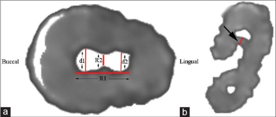
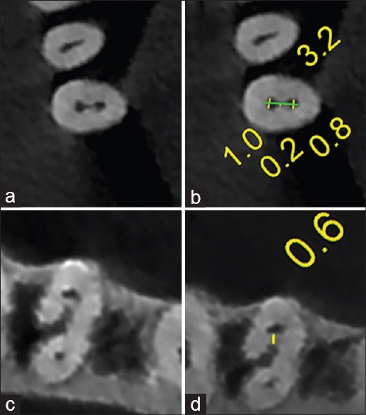
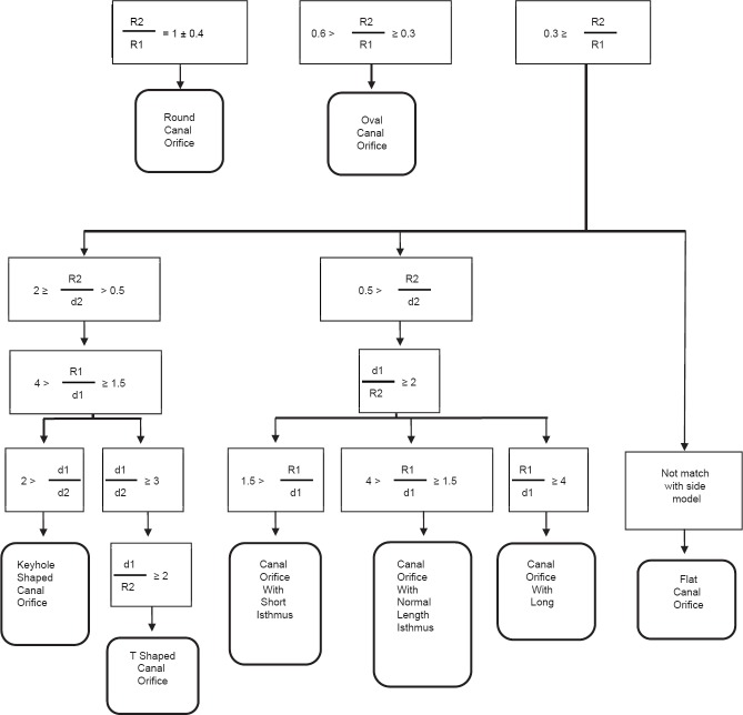
Materials and methods: A total of 287 mandibular premolar images obtained from 88 patients by cone-beam computed tomography were included in this study. The measurements were performed below the cementoenamel junction, and different orifice configurations were defined in accordance with various ratios. The age and gender of the patient, the tooth type and position, the number of roots, orifice configuration, root canal configuration, presence of C-shaped canal, and the presence of radicular groove were recorded. It was also recorded whether the root canal becomes round or not and if any, length of the root canal from the orifice to the section in which it becomes round. Furthermore, the theoretical model for determining orifice shape was defined after measurements. The orifice shape was determined as round, oval, flat, keyhole-shaped, and T-shaped, and orifices with short, normal length, and long isthmus. Statistical analyses were performed using Chi-square and Spearman's rank correlation tests (P = 0.05).
Results: Orifice configurations were, usually, flat (37%), or keyhole-shaped (23%). The prevalence of T-shaped was found to be 3.8%. The prevalence of C-shaped canals was found to be 2.1%. The percentage of root canals that became round in the middle or apical thirds was 95.1%. Radicular grooves were detected in 37 (24%) of first premolars and six (4.5%) of second premolars. Statistical analysis revealed that the mean length of distance until the canal reached a round shape varied according to age group (r = -0.270; P < 0.001). There was a statistically significant difference between radicular groove and tooth type (P < 0.001).
Conclusions: The mean length of distance until the canal reached a round shape correlated with the patient's age. The new theoretical model could be beneficial to determine orifice configurations.
Keywords: Cone-beam computed tomography; Turkish; isthmus; mandibular premolars; orifice; theoretical modeling.
Conflict of interest statement
Figures
References
-
- Sachdeva GS, Ballal S, Gopikrishna V, Kandaswamy D. Endodontic management of a mandibular second premolar with four roots and four root canals with the aid of spiral computed tomography: A case report. J Endod. 2008;34:104–7. - PubMed
-
- Barbizam JV, Ribeiro RG, Tanomaru Filho M. Unusual anatomy of permanent maxillary molars. J Endod. 2004;30:668–71. - PubMed
-
- Poorni S, Karumaran CS, Indira R. Mandibular first premolar with two roots and three canals. Aust Endod J. 2010;36:32–4. - PubMed
-
- Awawdeh LA, Al-Qudah AA. Root form and canal morphology of mandibular premolars in a Jordanian population. Int Endod J. 2008;41:240–8. - PubMed
LinkOut - more resources
Full Text Sources
Other Literature Sources