

Impaired translocation of GLUT4 results in insulin resistance of atrophic soleus muscle
- PMID: 25713812
- PMCID: PMC4332754
- DOI: 10.1155/2015/291987
Impaired translocation of GLUT4 results in insulin resistance of atrophic soleus muscle
Abstract
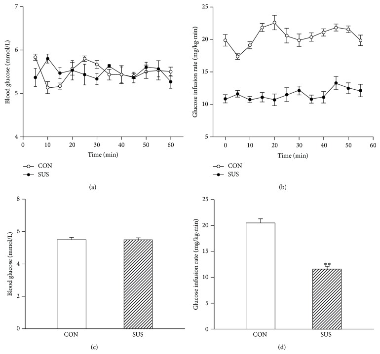
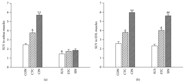
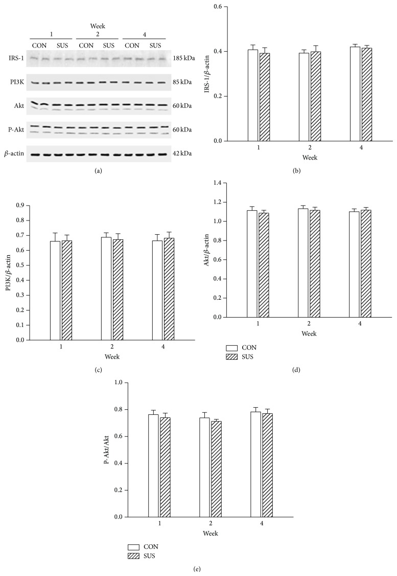
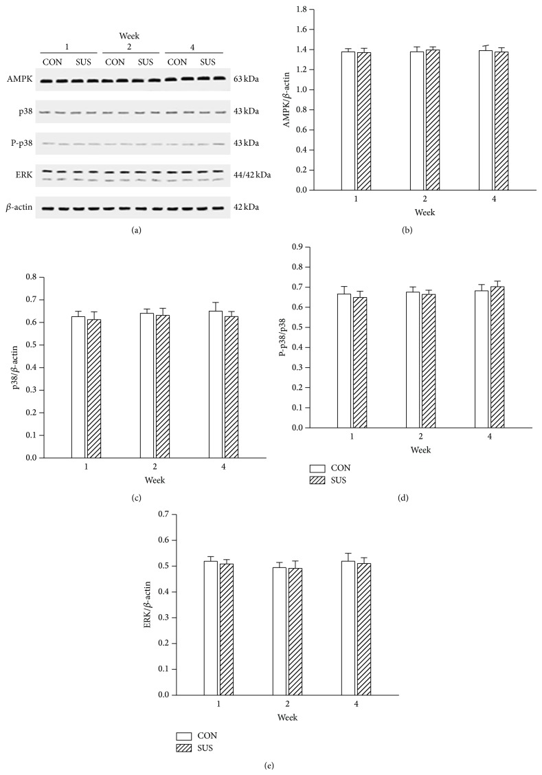
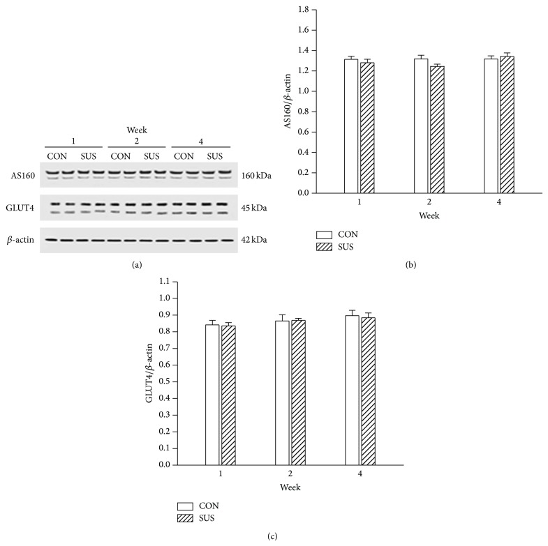
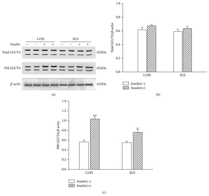
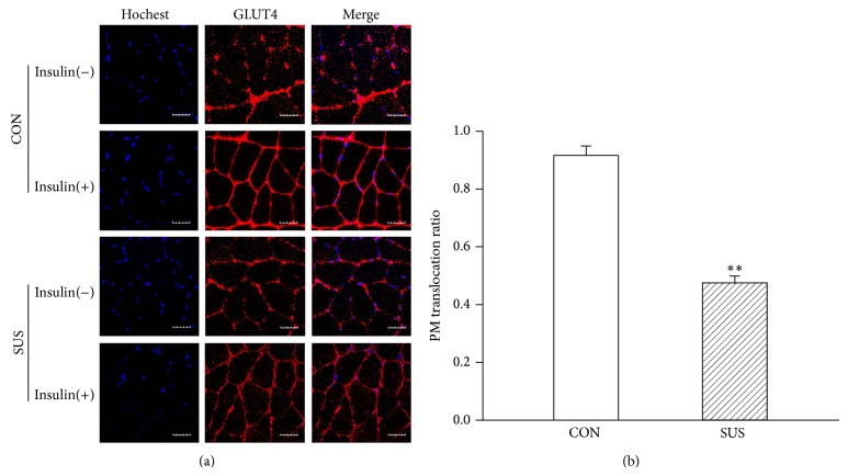
Whether or not the atrophic skeletal muscle induces insulin resistance and its mechanisms are not resolved now. The antigravity soleus muscle showed a progressive atrophy in 1-week, 2-week, and 4-week tail-suspended rats. Hyperinsulinemic-euglycemic clamp showed that the steady-state glucose infusion rate was lower in 4-week tail-suspended rats than that in the control rats. The glucose uptake rates under insulin- or contraction-stimulation were significantly decreased in 4-week unloaded soleus muscle. The key protein expressions of IRS-1, PI3K, and Akt on the insulin-dependent pathway and of AMPK, ERK, and p38 on the insulin-independent pathway were unchanged in unloaded soleus muscle. The unchanged phosphorylation of Akt and p38 suggested that the activity of two signal pathways was not altered in unloaded soleus muscle. The AS160 and GLUT4 expression on the common downstream pathway also was not changed in unloaded soleus muscle. But the GLUT4 translocation to sarcolemma was inhibited during insulin stimulation in unloaded soleus muscle. The above results suggest that hindlimb unloading in tail-suspended rat induces atrophy in antigravity soleus muscle. The impaired GLUT4 translocation to sarcolemma under insulin stimulation may mediate insulin resistance in unloaded soleus muscle and further affect the insulin sensitivity of whole body in tail-suspended rats.
Figures
Similar articles
-
Insulin-independent pathways mediating glucose uptake in hindlimb-suspended skeletal muscle.J Appl Physiol (1985). 2005 Dec;99(6):2181-8. doi: 10.1152/japplphysiol.00743.2005. Epub 2005 Aug 11. J Appl Physiol (1985). 2005. PMID: 16099889
-
Rat hindlimb unloading down-regulates insulin like growth factor-1 signaling and AMP-activated protein kinase, and leads to severe atrophy of the soleus muscle.Appl Physiol Nutr Metab. 2007 Dec;32(6):1115-23. doi: 10.1139/H07-102. Appl Physiol Nutr Metab. 2007. PMID: 18059585
-
Tetanic contraction induces enhancement of fatigability and sarcomeric damage in atrophic skeletal muscle and its underlying molecular mechanisms.Zhongguo Ying Yong Sheng Li Xue Za Zhi. 2013 Nov;29(6):525-33. Zhongguo Ying Yong Sheng Li Xue Za Zhi. 2013. PMID: 24654535
-
Exercise and GLUT4.Exerc Sport Sci Rev. 2020 Jul;48(3):110-118. doi: 10.1249/JES.0000000000000224. Exerc Sport Sci Rev. 2020. PMID: 32568924 Review.
-
Insulin- and contraction-induced glucose transporter 4 traffic in muscle: insights from a novel imaging approach.Exerc Sport Sci Rev. 2013 Apr;41(2):77-86. doi: 10.1097/JES.0b013e318275574c. Exerc Sport Sci Rev. 2013. PMID: 23072821 Free PMC article. Review.
Cited by
-
Understanding the Mechanism Underlie the Antidiabetic Activity of Oleuropein Using Ex-Vivo Approach.Rep Biochem Mol Biol. 2022 Apr;11(1):146-156. doi: 10.52547/rbmb.11.1.146. Rep Biochem Mol Biol. 2022. PMID: 35765534 Free PMC article.
-
Involvement of MrgprC in Electroacupuncture Analgesia for Attenuating CFA-Induced Thermal Hyperalgesia by Suppressing the TRPV1 Pathway.Evid Based Complement Alternat Med. 2018 Feb 12;2018:9102107. doi: 10.1155/2018/9102107. eCollection 2018. Evid Based Complement Alternat Med. 2018. PMID: 29619074 Free PMC article.
-
Metformin and Insulin Resistance: A Review of the Underlying Mechanisms behind Changes in GLUT4-Mediated Glucose Transport.Int J Mol Sci. 2022 Jan 23;23(3):1264. doi: 10.3390/ijms23031264. Int J Mol Sci. 2022. PMID: 35163187 Free PMC article. Review.
-
Insulin Secretory Actions of Ethanol Extract of Eucalyptus citriodora Leaf, including Plasma DPP-IV and GLP-1 Levels in High-Fat-Fed Rats, as Well as Characterization of Biologically Effective Phytoconstituents.Metabolites. 2022 Aug 17;12(8):757. doi: 10.3390/metabo12080757. Metabolites. 2022. PMID: 36005629 Free PMC article.
-
Preventive effect of oleate on palmitate-induced insulin resistance in skeletal muscle and its mechanism of action.J Physiol Biochem. 2017 Nov;73(4):605-612. doi: 10.1007/s13105-017-0594-9. Epub 2017 Oct 3. J Physiol Biochem. 2017. PMID: 28971334
References
-
- Bertelli D. F., Ueno M., Amaral M. E. C., et al. Reversal of denervation-induced, insulin resistance by SHIP2 protein synthesis blockade. American Journal of Physiology—Endocrinology and Metabolism. 2003;284(4):E679–E687. - PubMed
-
- Hirose M., Kaneki M., Sugita H., Yasuhara S., Martyn J. A. J. Immobilization depresses insulin signaling in skeletal muscle. The American Journal of Physiology—Endocrinology and Metabolism. 2000;279(6):E1235–E1241. - PubMed
-
- Stein T. P., Schluter M. D., Boden S. G. Development of insulin resistance by astronauts during spaceflight. Aviation Space and Environmental Medicine. 1994;65(12):1091–1096. - PubMed
Publication types
MeSH terms
Substances
LinkOut - more resources
Full Text Sources
Other Literature Sources
Medical
Miscellaneous
