. 2015 Feb 28;6(6):4066-79.
doi: 10.18632/oncotarget.2926.

miR-106b-5p targets tumor suppressor gene SETD2 to inactive its function in clear cell renal cell carcinoma

Affiliations

miR-106b-5p targets tumor suppressor gene SETD2 to inactive its function in clear cell renal cell carcinoma

Wei Xiang et al. Oncotarget. .

Abstract

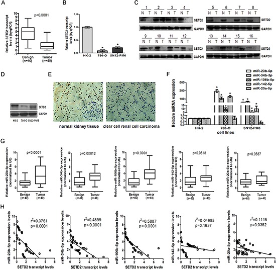
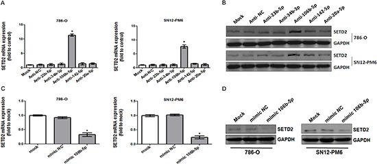
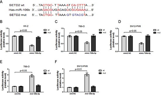
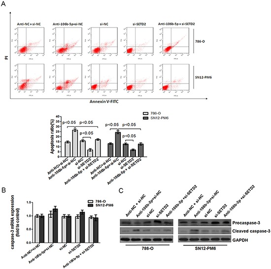
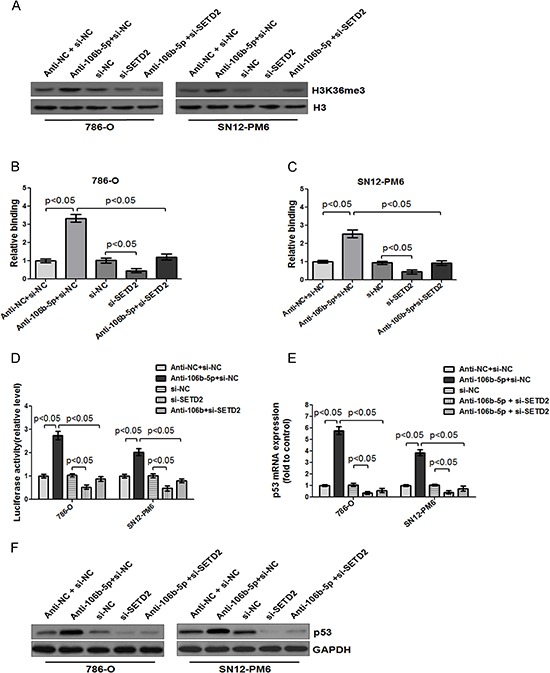
Inactivation of human SET domain containing protein 2 (SETD2) is a common event in clear cell renal cell carcinoma (ccRCC). However, the mechanism underlying loss of SETD2 function, particularly the post-transcriptional regulatory mechanism, still remains unclear. In the present study, we found that SETD2 was downregulated and inversely correlated with high expression of miR-106b-5p in ccRCC tissues and cell lines. Over-expression of miR-106b-5p resulted in the decreased mRNA and protein levels of SETD2 in ccRCC cells. In an SETD2 3'-UTR luciferase reporter system, miR-106b-5p downregulated the luciferase activity, and the effects were abolished by mutating the predicted miR-106b-5p binding site. Moreover, attenuation of miR-106b-5p induced cell cycle arrest at G0/G1 phase, suppressed cell proliferation, enhanced processing of caspase-3, and promoted cell apoptosis in ccRCC cells, whereas these effects were reversed upon knockdown of SETD2. In addition, transfection of miR-106b-5p antagomir resulted in the increased binding of H3K36me3 to the promoter of p53 and enhanced its activity, as well as upregulated the mRNA and protein levels of p53, and the effects were also abolished by cotransfection with si-SETD2. Collectively, our findings extend the knowledge about the regulation of SETD2 at the posttranscriptional level by miRNA and regulatory mechanism downstream of SETD2 in ccRCC.


Conflict of interest statement

Conflict of interest

The authors declare no conflict of interest.

Figures

Figure 1
Figure 1. SETD2 was lowly expressed and inversely correlated with endogenous miR-23b-5p, miR-34b-3p and miR-106b-5p in ccRCC tissues and cell lines
The expression of SETD2 mRNA in 40 pairs of human ccRCC samples (A) and human proximal tubule epithelial cell line HK-2 and ccRCC cell lines 786-O, SN12-PM6 (B) were examined by real-time RT-PCR, using GAPDH as the internal control. Western blot was used to detect the expression of SETD2 protein in human ccRCC samples (C) and HK-2, 786-O, and SN12-PM6 cells (D). Immunohistochemistry was executed to check the SETD2 protein expression in ccRCC samples and surrounding normal kidney tissues (40 ×) (E). The microRNAs including miR-23b-5p, miR-34b-3p, miR-106b-5p, miR-142–5p and miR-20a-5p were tested among HK-2,786-O and SN12-PM6 cell lines (F) as well as ccRCC tissues (G) by real-time RT-PCR, using U6 as an internal control. The linear regression between the expression of SETD2 mRNA and miRNAs in 40 ccRCC cases were analyzed respectively (H). *P < 0.05 compared with HK-2 cells. Results are the means ± SD.
Figure 2
Figure 2. miR-106b-5p downregulated the expression of SETD2 in ccRCC cells
786-O and SN12-PM6 cells were transfected for 72 h with 100 nM anti-miR negative control, anti-miR-23b-5p, anti-miR-34b-3p, anti-miR-106b-5p, anti-miR-142–5p or anti-miR-20a-5p. SETD2 mRNA and protein expression levels were tested by real-time RT–PCR (A) and western blot (B), respectively. 786-O and SN12-PM6 cells were transfected for 72 h with 50 nM negative control mimic or miR-106b-5 mimic, real-time RT–PCR and western blot were used to analyze the expression of SETD2 mRNA (C) and SETD2 protein (D). *P < 0.05 versus anti-miR negative control, **P < 0.05 versus negative control mimic. Results are the means ± SD in triplicate.
Figure 3
Figure 3. miR-106b-5p directly interacts with 3′UTR of SETD2 mRNA
(A) Chematic representation of miR-106b-5p target binding site in the SETD2 mRNA 3′UTR identified by the microinspector prediction program. Wild type (wt) and mutation (mut) of 3′UTR in seed sequences were indicated. Transfection of miR-106b-5p mimic into HK-2 (B), 786-O (C) and SN12-PM6 cells (D) resulted in decreased luciferase activities of SETD2 3′-UTR reporter, when compared with those transfected with mimic negative control (mimic NC). These effects were abolished by the mutation in the putative miR-106b-5p binding site within the 3′-UTR of SETD2. Transfection of anti-miR-106b-5p inhibitor (100 nM) into 786-O (E) and SN12-PM6 (F) cells increased the luciferase activity when compared with those transfected with negative control inhibitor (anti-NC), whereas mutation of miR-106b-5p recognition site abolished these effects. Results are the means ± SD in triplicate.
Figure 4
Figure 4. MiR-106b-5p antagomir induced up-regulation of SETD2 expression was reversed by knockdown of SETD2 in ccRCC cells
The effects of si-SETD2 on SETD2 expression were confirmed first in HK-2 cells through real-time PCR (A) and western blot (B) respectively. Transfection of antagomir against miR-106b-5p resulted in the increased level of SETD2 mRNA in both 786-O (C) and SN12-PM6 (D) cells, which was blocked by co-transfection of si-SETD2. The changes of SETD2 protein levels in both 786-O (E) and SN12-PM6 (F) cells were further confirmed by Western blot. *P < 0.05 versus negative control. Results are the means ± SD in triplicate.
Figure 5
Figure 5. MiR-106b-5p antagomir induced cell cycle arrest and proliferation suppression through up-regulation of SETD2 expression in ccRCC cells
(A) Flow cytometry indicated that transfection of miR-106b-5p antagomir resulted in cell cycle arrest at G0/G1 phase in both 786-O and SN12-PM6 cells compared with negative control ( p < 0.05), while transfection of si-SETD2 decreased the basal rate of cells in G0/G1 phase and effectively reversed the miR-106b-5p antagomir induced G0/G1 phase arrest (p < 0.05). EdU assay showed that transfection of miR-106b-5p antagomir inhibited proliferation of 786-O (B) and SN12-PM6 (C) cells compared with negative control ( p < 0.05), which was reversed by the transfection of si-SETD2. Results are the means ± SD in triplicate.
Figure 6
Figure 6. Attenuation of miR-106b-5p promoted caspase-3 mediated apoptosis through up-regulation of SETD2 expression
(A) Annexin V and PI staining flow cytometry assay revealed that transfection of miR-106b-5p antagomir increased the rate of apoptosis in 786-O and SN12-PM6 cells compared with negative control group, while transfection of si-SETD2 decreased the apoptotic cells, and significantly inhibited miR-106b-5p antagomir induced cell apoptosis. (B) The effects of miR-106b-5p antagomir, si-SETD2 and their cotransfection on the expression of caspase-3 mRNA in 786-O and SN12-PM6 cells were examined by real-time RT-PCR. (C) The effects of miR-106b-5p antagomir, si-SETD2 and their cotransfection on the processing of caspase-3 were detected by Western blot. Results are the means ± SD in triplicate.
Figure 7
Figure 7. Inhibition of miR-106b-5p enhanced the binding of H3K36me3 to the promoter of p53, and upregulated p53 transcription through a SETD2 dependent way
(A) Western blot showed that transfection of miR-106b-5p antagomir up-regulated the level of H3K36me3 in 786-O and SN12-PM6 cells compared with negative control group, which was reversed by cotransfection of si-SETD2. ChIP assay indicated that transfection of miR-106b-5p antagomir enhanced binding of H3K36me3 to the promoter of p53 in 786-O (B) and SN12-PM6 (C) cells, which was abolished by cotransfection of si-SETD2. (D) Luciferase activity assay demonstrated that a significant increase of p53 promoter activity was induced by the attenuation of miR-106b-5p, while it was reversed by cotransfection of si-SETD2 in 786-O and SN12-PM6 cells. Real-time RT-PCR and western blot analysis indicated that transfection of miR-106b-5p antagomir increased the p53 expression in both mRNA (E) and protein (F) levels, whereas knockdown of SETD2 reversed the effects. Results are the means ± SD in triplicate.

References

    1. Leibovich BC, Lohse CM, Crispen PL, Boorjian SA, Thompson RH, Blute ML, Cheville JC. Histological subtype is an independent predictor of outcome for patients with renal cell carcinoma. The Journal of urology. 2010;183:1309–1315. - PubMed
    1. Klatte T, Rao PN, de Martino M, LaRochelle J, Shuch B, Zomorodian N, Said J, Kabbinavar FF, Belldegrun AS, Pantuck AJ. Cytogenetic profile predicts prognosis of patients with clear cell renal cell carcinoma. J Clin Oncol. 2009;27:746–753. - PubMed
    1. Chino K, Esumi M, Ishida H, Okada K. Characteristic loss of heterozygosity in chromosome 3P and low frequency of replication errors in sporadic renal cell carcinoma. The Journal of urology. 1999;162:614–618. - PubMed
    1. Nickerson ML, Jaeger E, Shi Y, Durocher JA, Mahurkar S, Zaridze D, Matveev V, Janout V, Kollarova H, Bencko V, Navratilova M, Szeszenia-Dabrowska N, Mates D, Mukeria A, Holcatova I, Schmidt LS, et al. Improved identification of von Hippel-Lindau gene alterations in clear cell renal tumors. Clinical cancer research : an official journal of the American Association for Cancer Research. 2008;14:4726–4734. - PMC - PubMed
    1. Kroeger N, Klatte T, Chamie K, Rao PN, Birkhauser FD, Sonn GA, Riss J, Kabbinavar FF, Belldegrun AS, Pantuck AJ. Deletions of chromosomes 3p and 14q molecularly subclassify clear cell renal cell carcinoma. Cancer. 2013;119:1547–1554. - PubMed

Publication types

MeSH terms

LinkOut - more resources