Review
. 2015 Apr;35(4):252-64.
doi: 10.1089/jir.2014.0227. Epub 2015 Feb 25.

Disease-promoting effects of type I interferons in viral, bacterial, and coinfections

Affiliations
Review

Disease-promoting effects of type I interferons in viral, bacterial, and coinfections

Sophia Davidson et al. J Interferon Cytokine Res. 2015 Apr.

Abstract

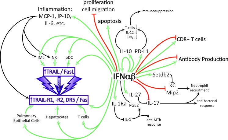
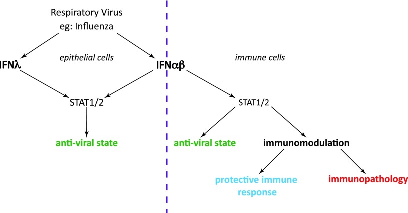
While type I interferons (IFNs) are universally acknowledged for their antiviral and immunostimulatory functions, there is increasing appreciation of the detrimental effects of inappropriate, excessive, or mistimed type I IFN responses in viral and bacterial infections. The underlying mechanisms by which type I IFNs promote susceptibility or severity include direct tissue damage by apoptosis induction or suppression of proliferation in tissue cells, immunopathology due to excessive inflammation, and cell death induced by TRAIL- and Fas-expressing immune cells, as well as immunosuppression through IL-10, IL-27, PD-L1, IL-1Ra, and other regulatory molecules that antagonize the induction or action of IL-1, IL-12, IL-17, IFN-γ, KC, and other effectors of the immune response. Bacterial superinfections following influenza infection are a prominent example of a situation where type I IFNs can misdirect the immune response. This review discusses current understanding of the parameters of signal strength, duration, timing, location, and cellular recipients that determine whether type I IFNs have beneficial or detrimental effects in infection.


Figures

<b>FIG. 1.</b>
FIG. 1.
Multiple effects of IFN-αβ on immune and epithelial cells that can contribute to disease in specific infections. Only direct enhancing (green) and inhibitory (red) effects by IFN-αβ are color coded, secondary effects are in black. IFN-αβ can enhance production of IL-10, proinflammatory cytokines, and IL-27 and can block directly or indirectly the cytokines, IL-1, IL-12, IL-17, IFN-γ, and KC. Detrimental effects can be due to the suppression of antibacterial responses (IL-1, IL-17, IFN-γ, and KC) or to overstimulation of inflammation leading to damage by apoptosis of tissue cells or immune suppression by apoptosis of immune cells.
<b>FIG. 2.</b>
FIG. 2.
Moderate IFN-αβ responses to infection are protective, while excessive IFN-αβ amounts contribute to immunopathology. (A) Bell-shaped curve displaying resistance to influenza severity as a function of IFN-αβ responses, based on experimental findings in Davidson and others (2014) and other studies. High IFN-αβ-expressing mouse strains (eg, 129, DBA, agouti, and gray symbols) are highly susceptible to influenza, and genetic removal of IFN-αβ from such strains increases resistance (1). Conversely, exogenous addition of IFN-αβ to resistant, low IFN-αβ-expressing mouse strains (eg, C57BL/6, Balb/C, black, and white symbols) reduces their resistance to influenza (2). However, if the moderate IFN-αβ responses in C57BL/6 mice are genetically removed, influenza resistance is reduced (3). Linking these data points generates a dose–response curve (4) where moderate IFN-αβ responses protect and high IFN-αβ responses are detrimental. (B) Induction of antiviral (green) versus proinflammatory, antiproliferative, and apoptosis-promoting (red) genes as a function of IFN-αβ signal strength based on results and hypotheses, as reviewed in (Piehler and others 2012). We hypothesize that moderate IFN-αβ responses are protective in influenza infections as effective antiviral responses are balanced with moderate inflammation and apoptosis induction. Higher IFN-αβ signals increase inflammation and cell death, but do not further enhance induction of antiviral effectors.
<b>FIG. 3.</b>
FIG. 3.
Partial redundancy of types I and III IFNs in infection by influenza and other respiratory viruses. Both IFN-αβ and IFN-λ are induced upon influenza infection. Their effects depend on the distribution of their receptors: Epithelial cells express receptors for both IFN types, and both these IFNs can mediate the induction of the epithelial antiviral response (left). Immune cells express only IFN-αβ receptors and can be stimulated or inhibited by type I IFN (right). STAT1/2 is involved in signal transduction downstream of both type I and type III receptors. The degree and nature of immune activation determines whether the net result of all combined type I IFN-mediated effects is protective or pathogenic.

References

    1. Ahlenstiel G, Edlich B, Hogdal LJ, Rotman Y, Noureddin M, Feld JJ, Holz LE, Titerence RH, Liang TJ, Rehermann B. 2011. Early changes in natural killer cell function indicate virologic response to interferon therapy for hepatitis C. Gastroenterology 141(4):1231–1239, 1239 e1–e2 - PMC - PubMed
    1. Ahlenstiel G, Titerence RH, Koh C, Edlich B, Feld JJ, Rotman Y, Ghany MG, Hoofnagle JH, Liang TJ, Heller T, Rehermann B. 2010. Natural killer cells are polarized toward cytotoxicity in chronic hepatitis C in an interferon-alfa-dependent manner. Gastroenterology 138(1):325–335 e1–e2 - PMC - PubMed
    1. Antonelli LR, Gigliotti Rothfuchs A, Goncalves R, Roffe E, Cheever AW, Bafica A, Salazar AM, Feng CG, Sher A. 2010. Intranasal Poly-IC treatment exacerbates tuberculosis in mice through the pulmonary recruitment of a pathogen-permissive monocyte/macrophage population. J Clin Invest 120(5):1674–1682 - PMC - PubMed
    1. Auerbuch V, Brockstedt DG, Meyer-Morse N, O'Riordan M, Portnoy DA. 2004. Mice lacking the type I interferon receptor are resistant to Listeria monocytogenes. J Exp Med 200(4):527–533 - PMC - PubMed
    1. Bahl K, Huebner A, Davis RJ, Welsh RM. 2010. Analysis of apoptosis of memory T cells and dendritic cells during the early stages of viral infection or exposure to toll-like receptor agonists. J Virol 84(10):4866–4877 - PMC - PubMed

Publication types

MeSH terms

Substances