Review
. 2015 Mar;204(3):W302-13.
doi: 10.2214/AJR.14.12733.

Iron-based superparamagnetic nanoparticle contrast agents for MRI of infection and inflammation

Affiliations
Review

Iron-based superparamagnetic nanoparticle contrast agents for MRI of infection and inflammation

Alexander Neuwelt et al. AJR Am J Roentgenol. 2015 Mar.

Abstract

OBJECTIVE. In this article, we summarize the progress to date on the use of superparamagnetic iron oxide nanoparticles (SPIONs) as contrast agents for MRI of inflammatory processes. CONCLUSION. Phagocytosis by macrophages of injected SPIONs results in a prolonged shortening of both T2 and T2* leading to hypointensity of macrophage-infiltrated tissues in contrast-enhanced MR images. SPIONs as contrast agents are therefore useful for the in vivo MRI detection of macrophage infiltration, and there is substantial research and clinical interest in the use of SPION-based contrast agents for MRI of infection and inflammation. This technique has been used to identify active infection in patients with septic arthritis and osteomyelitis; importantly, the MRI signal intensity of the tissue has been found to return to its unenhanced value on successful treatment of the infection. In SPION contrast-enhanced MRI of vascular inflammation, animal studies have shown decreased macrophage uptake in atherosclerotic plaques after treatment with statin drugs. Human studies have shown that both coronary and carotid plaques that take up SPIONs are more prone to rupture and that abdominal aneurysms with increased SPION uptake are more likely to grow. Studies of patients with multiple sclerosis suggest that MRI using SPIONs may have increased sensitivity over gadolinium for plaque detection. Finally, SPIONs have enabled the tracking and imaging of transplanted stem cells in a recipient host.

Keywords: MRI; ferumoxytol; infection; inflammation; macrophages; superparamagnetic iron oxide nanoparticles (SPIONs).


Figures

Fig. 1
Fig. 1. Hodgkin lymphoma. (Reprinted with permission from [11])
A and B, Whole-body diffusion-weighted MR image with ferumoxytol contrast administration (A) and FDG PET/CT image (B) in 15-year-old patient show positive retroperitoneal involvement (arrows) of stage IIIA Hodgkin lymphoma. C and D, Whole-body diffusion-weighted MR image with ferumoxytol contrast administration (C) and FDG PET/CT image (D) in 14-year-old patient with stage IIB Hodgkin lymphoma show positive right inguinal lymph nodes (arrows).
Fig. 2
Fig. 2. Relaxometry rate (R2) and effective relaxometry rate (R2*). (Reprinted with permission from [14])
A and B, R2* (A) and R2 (B) images of liver show marked increase in signal at 3 days. In this 28-year-old man, signal intensity for both R2* and R2 did not return to baseline at 11 months. Color scales are shown on left.
Fig. 3
Fig. 3. MRI of septic knee arthritis in rabbit before (A–C) and after (D–F) antibiotic administration. (Adapted and reprinted, with permission from [16])
A, Unenhanced, T2*-weighted image shows thickened synovium (arrow). f = femur, p = patella. B, T2*-weighted image obtained 24 hours after superparamagnetic iron oxide nanoparticle (SPION) injection shows infiltration of iron-laden macrophages into synovium (arrow). C, Gadolinium-enhanced T1-weighted image shows synovitis (arrow). D, T2*-weighted gradient-echo image shows only focal signal intensity loss in synovium (arrowheads). E, SPION-enhanced MR image after antibiotic administration shows no enhanced signal intensity loss over unenhanced image (arrowheads). F, SPION-enhanced MR image shows gadolinium continues to enhance synovium despite antibiotic treatment.
Fig. 4
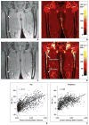
Fig. 4. Hip and femur. (Reprinted with permission from [51])
A–D, Gradient-echo images (A and C) and effective relaxometry rate (R2*) images (B and D) show hip and femur of human subjects. A and B are baseline images and C and D are images obtained 3 days after ferumoxytol administration. Color scale is provided for R2* maps. In D, note that R2* signal intensity with ferumoxytol contrast-enhancement is dramatically increased in red marrow but remains low in yellow marrow. E and F, Graphs show relation between R2* response and proton density water fraction.
Fig. 5
Fig. 5. Patient whose aneurysm ruptured within 3 months. (Adapted and reprinted with permission from [65])
A and B, Unenhanced (A) and ferumoxytol-enhanced (B) images show early uptake of ferumoxytol at 24 hours (B) from T2* gradient-recalled echo sequence. This image is from study that suggests that intracranial aneurysms with ferumoxytol uptake within 24 hours are more likely to rupture [65].
Fig. 6
Fig. 6. Diabetes. (Reprinted with permission from [66])
A–D, T2-weighted pseudocolor reconstructions superimposed on unenhanced (A and C) and superparamagnetic iron oxide nanoparticle (SPION)-enhanced (B and D) T1-weighted images show no difference between diabetic patients and normal subjects on unenhanced images. However, after ferumoxtran-10 (Combidex, Advanced Magnetics) SPION administration, images are clearly distinct.
Fig. 7
Fig. 7. Multiple sclerosis lesions. (Adapted and reprinted with permission from [69])
A–C, Unenhanced (A), gadolinium-enhanced T1-weighted (B), and superparamagnetic iron oxide nanoparticle–enhanced (C) MR images show multiple sclerosis lesions (arrows).
Fig. 8
Fig. 8. Comparative analysis of two middle cerebral artery infarctions localized to posterior parietal cortex. (Adapted and reprinted with permission from [70])
A–C, 60-year-old woman 124 hours after stroke onset. T2-weighted image (A) shows ischemic lesion. In comparison with unenhanced T1-weighted image (B), no enhancement is seen with superparamagnetic iron oxide nanoparticle–enhanced image (C). Comparative analysis of two middle cerebral artery infarctions localized to posterior parietal cortex. (Adapted and reprinted with permission from [70]) D–F, 56-year-old man with infarct imaged 80 hours after stroke onset. Lesion is visualized on T2-weighted image (D). Lesion shows improved visualization after ultrasmall superparamagnetic iron oxide infusion (F) relative to unenhanced T1-weighted image (E).

References

    1. Lutz AM, Weishaupt D, Persohn E, et al. Imaging of macrophages in soft-tissue infection in rats: relationship between ultrasmall superparamagnetic iron oxide dose and MR signal characteristics. Radiology. 2005;234:765–775. - PubMed
    1. Bierry G, Jehl F, Boehm N, Robert P, Dietemann JL, Kremer S. Macrophage imaging by USPIO-enhanced MR for the differentiation of infectious osteomyelitis and aseptic vertebral inflammation. Eur Radiol. 2009;19:1604–1611. - PubMed
    1. Ruehm SG, Corot C, Vogt P, Kolb S, Debatin JF. Magnetic resonance imaging of atherosclerotic plaque with ultrasmall superparamagnetic particles of iron oxide in hyperlipidemic rabbits. Circulation. 2001;103:415–422. - PubMed
    1. Richie AC, Boyd W. Boyd’s textbook of pathology. 9. Philadelphia, PA: Lea & Febiger; 1990. pp. 60–82.
    1. Sephel GC, Woodward SC. Repair, regeneration, and fibrosis. In: Rubin DS, Strayer DS, editors. Rubin’s pathology. 5. Philadelphia, PA: Lippincott Williams & Wilkins; 2007. pp. 71–98.