Review
. 2015 Apr;88(1048):20140507.
doi: 10.1259/bjr.20140507. Epub 2015 Feb 25.

Role of radiology in central nervous system stimulation

Affiliations
Review

Role of radiology in central nervous system stimulation

D P Minks et al. Br J Radiol. 2015 Apr.

Abstract

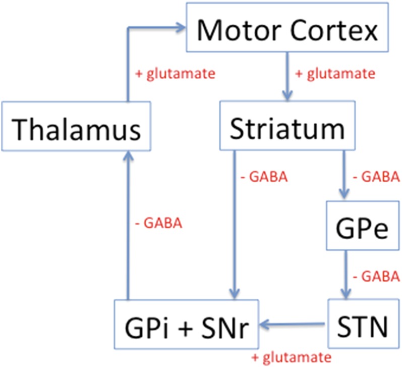
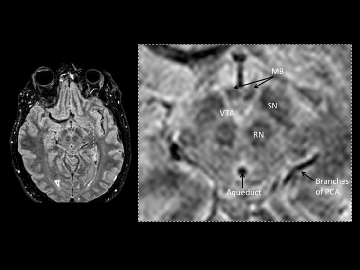
Central nervous system (CNS) stimulation is becoming increasingly prevalent. Deep brain stimulation (DBS) has been proven to be an invaluable treatment for movement disorders and is also useful in many other neurological conditions refractory to medical treatment, such as chronic pain and epilepsy. Neuroimaging plays an important role in operative planning, target localization and post-operative follow-up. The use of imaging in determining the underlying mechanisms of DBS is increasing, and the dependence on imaging is likely to expand as deep brain targeting becomes more refined. This article will address the expanding role of radiology and highlight issues, including MRI safety concerns, that radiologists may encounter when confronted with a patient with CNS stimulation equipment in situ.


Figures

Figure 1.
Figure 1.
Basal ganglia terminology.
Figure 2.
Figure 2.
The basal ganglia–thalamocortical motor circuit. GPe, globus pallidus external segment; GPi, globus pallidus internal segment; SNr, substantia nigra reticular part; STN, subthalamic nucleus.
Figure 3.
Figure 3.
Axial image from volume fluid-attenuated inversion recovery at 1.5 T. MB, mammillary bodies; PCA, poster cerebral artery; RN, red nucleus; SN, substantia nigra; VTA, ventral tegmental area.
Figure 4.
Figure 4.
(a) Coronal image from volume T1 weighted at 7 T. (b) Coronal image from T2 sequence at 1.5 T. Ca, caudate; GP, globus pallidus; In, insula; P, putamen; SN, substantia nigra; STN, subthalamic nucleus; Th, thalamus.
Figure 5.
Figure 5.
Axial image from volume T1 weighted at 7 T. Ca, caudate; Cl, claustrum; EC, external capsule; GP, globus pallidus; IC, internal capsule; MD, medial dorsal nucleus; P, putamen; Pu, pulvinar; Th, thalamus; VA, ventral anterior nucleus; VI, ventral intermediate nucleus; VL, ventral lateral nucleus; VP, venal posterior.
Figure 6.
Figure 6.
(a, b) Three-dimensional (3D) volume CT. Axial image and 3D reconstruction. Patient with stereotactic frame in situ in order to plan for a left subthalamic nucleus (STN) deep brain stimulation lead insertion. The patient already has a right STN stimulator lead in situ.
Figure 7.
Figure 7.
Stereotactic surgical planning. The surgical trajectory to both subthalamic nuclei is planned in order attain accurate positioning and avoid eloquent structures. 3D, three dimensional; FLAIR, fluid-attenuated inversion recovery.
Figure 8.
Figure 8.
Professor Aziz of John Radcliffe Hospital, Oxford, UK, performing stereotactic deep brain stimulation in an awake patient. Informed consent was received for the use of this image.
Figure 9.
Figure 9.
(a) Post-operative CT to assess for the position of the electrode and to exclude surgical complications. The right deep brain stimulation (DBS) electrode lead is demonstrated to terminate in the region of the right subthalamic nucleus (STN). (b) Post-operative T2 weighted MRI to assess for the position of the DBS electrodes. There are two DBS electrode leads in situ. One terminates in the region of the left STN and the other in the region of the left pedunculopontine nucleus.
Figure 10.
Figure 10.
(a, b) Anteroposterior (AP) and lateral radiography demonstrate correct placement of the spinal cord stimulator electrode pad. It should be central (equidistant between the pedicles on the AP view) and dorsal within the vertebral canal.
Figure 11.
Figure 11.
(a–c) Two anteroposterior views and a lateral radiography demonstrated misplaced spinal cord stimulator electrode pad. The electrode pad has moved towards the left side of the vertebral canal.
Figure 12.
Figure 12.
(a, b) Lateral and anteroposterior maximum intensity projection of a CT demonstrating the sacral nerve stimulator electrode passing through the right S3 neural foramen. (c) Abdominal radiograph demonstrating the sacral nerve stimulator electrode passing through the right S3 neural foramen with the implantable pulse generator sited in the right lumbar soft tissues.

Similar articles

References

    1. Standring S. Gray's anatomy: the anatomical basis of clinical practice. Edinburgh, UK: Churchill Livingstone Elsevier; 2008.
    1. Starr PA, Barbaro NM, Larson PS. Neurosurgical operative atlas: functional neurosurgery. 2nd edn. Stuttgart, Germany: Thieme; 2009.
    1. Perestelo-Pérez L, Rivero-Santana A, Pérez-Ramos J, Serrano-Pérez P, Panetta J, Hilarion P. Deep brain stimulation in Parkinson's disease: meta-analysis of randomized controlled trials. J Neurol 2014; 261: 2051–60. doi: 10.1007/s00415-014-7254-6 - DOI - PubMed
    1. Pereira EA, Green AL, Nandi D, Aziz TZ. Deep brain stimulation: indications and evidence. Expert Rev Med Devices 2007; 4: 591–603. - PubMed
    1. Vilensky JA, Gilman S. Horsley was the first to use electrical stimulation of the human cerebral cortex intraoperatively. Surg Neurol 2002; 58: 425–6. - PubMed

MeSH terms