. 2015 Nov;25(11):4351-73.
doi: 10.1093/cercor/bhv019. Epub 2015 Feb 24.

Complementary Patterns of Direct Amygdala and Hippocampal Projections to the Macaque Prefrontal Cortex

Affiliations

Complementary Patterns of Direct Amygdala and Hippocampal Projections to the Macaque Prefrontal Cortex

John P Aggleton et al. Cereb Cortex. 2015 Nov.

Abstract

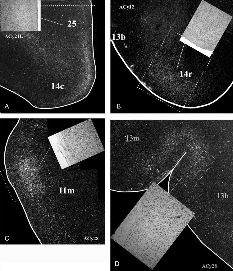
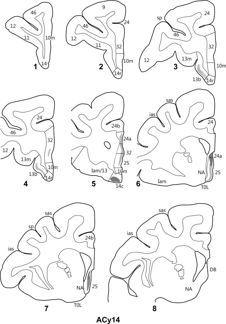
The projections from the amygdala and hippocampus (including subiculum and presubiculum) to prefrontal cortex were compared using anterograde tracers injected into macaque monkeys (Macaca fascicularis, Macaca mulatta). Almost all prefrontal areas were found to receive some amygdala inputs. These connections, which predominantly arose from the intermediate and magnocellular basal nucleus, were particularly dense in parts of the medial and orbital prefrontal cortex. Contralateral inputs were not, however, observed. The hippocampal projections to prefrontal areas were far more restricted, being confined to the ipsilateral medial and orbital prefrontal cortex (within areas 11, 13, 14, 24a, 32, and 25). These hippocampal projections principally arose from the subiculum, with the fornix providing the sole route. Thus, while the lateral prefrontal cortex essentially receives only amygdala inputs, the orbital prefrontal cortex receives both amygdala and hippocampal inputs, though these typically target different areas. Only in medial prefrontal cortex do direct inputs from both structures terminate in common sites. But, even when convergence occurs within an area, the projections predominantly terminate in different lamina (hippocampal inputs to layer III and amygdala inputs to layers I, II, and VI). The resulting segregation of prefrontal inputs could enable the parallel processing of different information types in prefrontal cortex.

Keywords: anatomy; emotion; fornix; hippocampus; memory; subiculum.


Figures

Figure 1.
Figure 1.
Location and extent of the amino acid injections into the amygdala. All cases are depicted. The numbers correspond to the injection cases, where L and R refer to the left and right hemispheres of those cases with injections in both hemispheres. The injection sites are depicted on standard coronal sections at the level of the middle, with those that extended more posteriorly also shown on an additional section in the posterior third of the amygdala. ACB, accessory basal nucleus; AHA, amygdalo-hippocampal area; Bi, basal nucleus, intermediate division; Bmc, basal nucleus, magnocellular division; Bpc, basal nucleus, parvicellular division; Ce, central nucleus; Co, cortical nucleus; HPC, hippocampus; LAT, lateral nucleus; M, medial nucleus.
Figure 2.
Figure 2.
Brightfield coronal photomicrographs showing the center of the amino acid injection sites in 6 amygdala cases (top 2 rows) and 3 hippocampal cases (bottom row). These cases were selected as they illustrate how different amygdala nuclei and different anterior–posterior levels in the hippocampus were targeted. Abbreviations are as in Figure 1. The scale bar corresponds to 1.0 mm.
Figure 3.
Figure 3.
Projections from the intermediate and magnocellular parts of the basal amygdala nucleus. Series of drawings of coronal sections from case ACy21L going from anterior (#1) to posterior (#7). The numbers (letters in the case of the insula) correspond to different prefrontal areas. Terminal label is shown in gray, with darker gray representing denser label. The cross-hatching marks those areas with both fiber and terminal labeling. The boxes in dashed lines show the regions in the darkfield images in Figures 5 and 6. DB, diagonal band; DB/25, transition zone between the diagonal band and area 25; G, gustatory area; ias, inferior arcuate sulcus; PPF, prepiriform cortex; PrCo, precentral opercular area; sas, superior arcuate sulcus; sp, sulcus principalis; TOL, olfactory tubercle.
Figure 4.
Figure 4.
Projections from the intermediate and magnocellular parts of the basal amygdala nucleus. Series of drawings of coronal sections from case ACy21R going from anterior (#1) to posterior (#7). The numbers (letters in the case of the insula) correspond to different prefrontal areas. Terminal label is shown in gray, with darker gray representing denser label. The cross-hatching marks those areas with both fiber and terminal labeling. SI, substantia innominata; all other abbreviations are as in Figure 3.
Figure 5.
Figure 5.
Darkfield images of autoradiographic label in 2 cases (ACy21L and ACy6), both with injections in the intermediate and magnocellular parts of the basal amygdala nucleus. The images are of the junction of the orbital and lateral prefrontal cortex (A,C) and that of the lateral prefrontal cortex (B). The brightfield images correspond to the subregions marked by boxes with dashed lines. The entire area of each darkfield image is indicated in Figures 3 and 7.
Figure 6.
Figure 6.
Darkfield images of autoradiographic label in 2 cases (ACy21L and ACy6), both with injections in the intermediate and magnocellular parts of the basal amygdala nucleus. The images are of the anterior cingulate cortex (A,B) and the inferior arcuate sulcus (C). The brightfield images correspond to the subregions marked by boxes with dashed lines. The entire area of each darkfield image is indicated in Figures 3 and 7. cc, corpus callosum.
Figure 7.
Figure 7.
Projections from the intermediate and magnocellular parts of the basal amygdala nucleus. Series of drawings of coronal sections from case ACy6 going from anterior (#1) to posterior (#7). The numbers (letters in the case of the insula) correspond to different prefrontal areas. Terminal label is shown in gray, with darker gray representing denser label. The cross-hatching marks those areas with both fiber and terminal labeling. SI, substantia innominata; other abbreviations are as in Figure 3.
Figure 8.
Figure 8.
Projections from the accessory basal amygdala nucleus. The coronal sections from 2 cases (ACy18, upper: ACy20R, lower) show the restricted areas of label. In all areas, the label consisted of fibers with apparent terminal labeling and so is depicted with cross hatching. The numbers (letters in the case of the insula) correspond to different prefrontal areas. CC, corpus callosum; cs, cingulate sulcus; IG, induseum griseum; other abbreviations are as in Figure 3.
Figure 9.
Figure 9.
Extent of the core of each amino acid injection in the hippocampal formation drawn onto standard coronal sections. The cases are divided between the 2 cohorts (from NIMH or Boston University). The injection sites in the normal animals from NIMH are depicted in the upper row, whereas the second row shows those NIMH cases where the fornix was transected prior to injection. The lower 2 rows show the cases from Boston University. CA1, hippocampal field CA1; DG, dentate gyrus; Hpc, hippocampus; PaS, parasubiculum; PrS, presubiculum; S, subiculum.
Figure 10.
Figure 10.
Projections from the anterior subiculum of the hippocampal formation in case ACy12. The series of drawings of coronal sections go from rostral (#1) to caudal (#8). The numbers (letters in the case of the insula) correspond to prefrontal areas. Terminal label is shown in gray, with darker gray representing denser label. The cross-hatching marks those areas with both fiber and terminal labeling. NA, nucleus accumbens; other abbreviations are as in Figure 3.
Figure 11.
Figure 11.
Darkfield images of autoradiographic label in 2 cases with amino acid injections in the hippocampus (ACy12, anterior subiculum; ACy28, posterior hippocampus). For purposes of comparison, a case with an amygdala injection (ACy21L) is shown in box A. All hippocampal projections (B,C,D) are on the orbital surface. The brightfield images correspond to the subregions marked by boxes with dashed lines. The entire area of each darkfield image is indicated in Figure 3 (box A), Figure 10 (box B), and Figure 13 (boxes C and D).
Figure 12.
Figure 12.
Projections from the anterior subiculum of the hippocampal formation in case ACy14. The series of drawings of coronal sections go from anterior (#1) to posterior (#8). The numbers (letters in the case of the insula) correspond to different prefrontal areas. Terminal label is shown in gray, with darker gray representing denser label. The cross-hatching marks those areas with both fiber and terminal labeling. NA, nucleus accumbens, DB, diagonal band; other abbreviations are as in Figure 3.
Figure 13.
Figure 13.
Projections from the posterior hippocampal formation in case ACy28. The series of drawings of coronal sections go from anterior (#1) to posterior (#7). The numbers (letters in the case of the insula) correspond to prefrontal areas. Terminal label is shown in gray, with darker gray representing denser label. The cross-hatching marks those areas with both fiber and terminal labeling. Abbreviations are as in Figure 3.
Figure 14.
Figure 14.
Summary figure depicting the termination sites of the amygdala (left) and hippocampal formation (right) projections to the medial (top), orbital (mid), and lateral (bottom) surfaces of the prefrontal cortex. The area boundaries and nomenclature come from Carmichael and Price (1994). The darker gray shading corresponds to the more dense terminal label. Decisions about the density of label were made by 2 independent observers. The figure shows both the extent of the projections and the limited numbers of areas with joint inputs from both structures. AS, arcuate sulcus; other abbreviations are as in Figure 3.

References

    1. Admon R, Milad MR, Hendler T. 2013. A causal model of post-traumatic stress disorder: disentangling predisposed from acquired neural abnormalities. Trends Cogn Sc. 17:337–347. - PubMed
    1. Aggleton JP. 1986. A description of the amygdalo-hippocampal interconnections in the macaque monkey. Exp Brain Res. 64:515–526. - PubMed
    1. Aggleton JP. 1985. A description of intra-amygdaloid connections in old world monkeys. Exp Brain Res. 57:390–399. - PubMed
    1. Aggleton JP. 2012. Multiple anatomical systems embedded within the primate medial temporal lobe: Implications for hippocampal function. Neurosci Biobehav Rev. 36:1579–1596. - PubMed
    1. Aggleton JP, Burton MJ, Passingham RE. 1980. Cortical and subcortical afferents to the amygdala of the rhesus monkey (Macaca mulatta). Brain Res. 190:347–368. - PubMed

Publication types