Review
. 2015 Mar 1;142(5):817-31.
doi: 10.1242/dev.105536.

A pathway to bone: signaling molecules and transcription factors involved in chondrocyte development and maturation

Affiliations
Review

A pathway to bone: signaling molecules and transcription factors involved in chondrocyte development and maturation

Elena Kozhemyakina et al. Development. .

Abstract

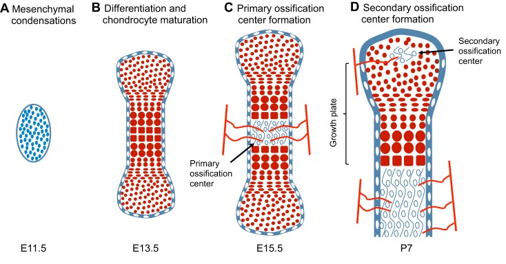
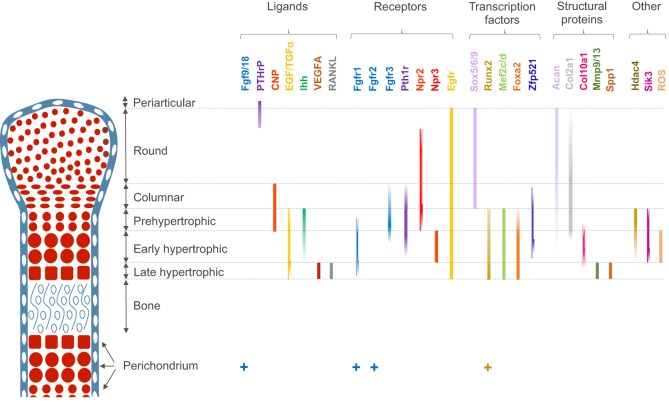
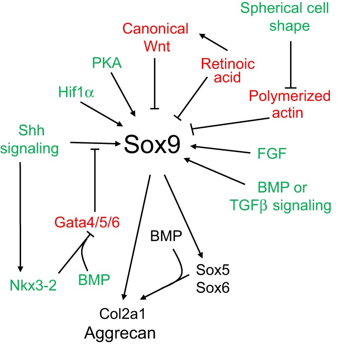
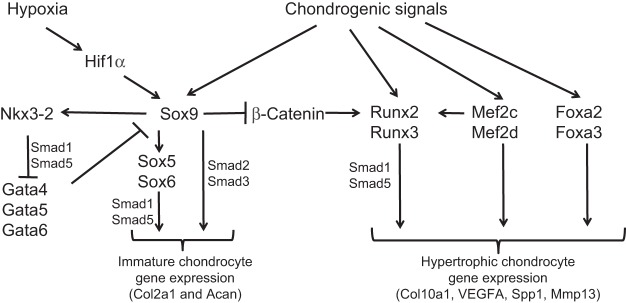
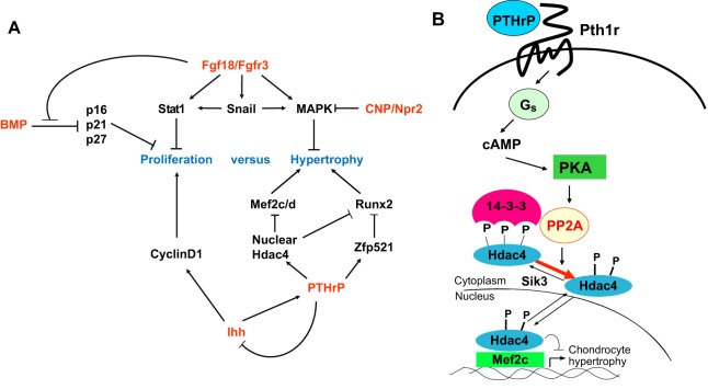
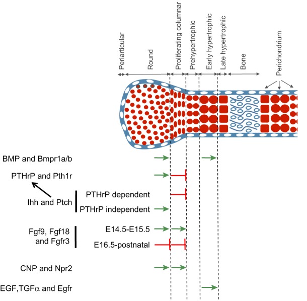
Decades of work have identified the signaling pathways that regulate the differentiation of chondrocytes during bone formation, from their initial induction from mesenchymal progenitor cells to their terminal maturation into hypertrophic chondrocytes. Here, we review how multiple signaling molecules, mechanical signals and morphological cell features are integrated to activate a set of key transcription factors that determine and regulate the genetic program that induces chondrogenesis and chondrocyte differentiation. Moreover, we describe recent findings regarding the roles of several signaling pathways in modulating the proliferation and maturation of chondrocytes in the growth plate, which is the 'engine' of bone elongation.

Keywords: Chondrocyte hypertrophy; Chondrogenesis; Fgfr3; Growth plate; Ihh; PTHrP; Sox9.


Figures

Fig. 1.
Fig. 1.
The stages of endochondral bone formation in the developing mouse hindlimb. (A) Schematic representation of mesenchymal cells (blue) that begin to form condensations at E11.5 in the hindlimb buds. (B) By E13.5, mesenchymal cells differentiate into chondrocytes (red cells), and the process of chondrocyte maturation and hypertrophy initiates. The cartilage anlage is surrounded by a layer of perichondrium (white cells). (C) By ∼E15.5, vascularization (represented by red lines) takes place in the center of the cartilage anlage, resulting in replacement of chondrocytes with endochondral bone (open circles) in the primary ossification center. (D) The secondary ossification center (SOC) forms postnatally, at ∼P7, and also becomes vascularized. The timing of SOC formation varies slightly between different bones. The schematics of the developing bones are approximately drawn to scale.
Fig. 2.
Fig. 2.
Summary of regionalized gene expression in the growth plate. Schematic representation of the various zones of chondrocyte maturation are displayed for a E15.5-E16.5 mouse long-bone growth plate. The expression of various classes of genes and/or proteins is displayed. Ligands: Fgf9/18 (Liu et al., 2002; Hung et al., 2007), PTHrP (St-Jacques et al., 1999), CNP (Chusho et al., 2001), EGF/TGFα (Ren et al., 1997), Ihh (Vortkamp et al., 1996), VEGFA (Zelzer et al., 2001) and RANKL (Zhang et al., 2011). Receptors: Fgfr1 (Jacob et al., 2006), Fgfr2 (Yu et al., 2003), Fgfr3 (de Frutos et al., 2007), Pth1r (Lee et al., 1996; de Frutos et al., 2007), Npr2 (Yamashita et al., 2000), Npr3 (Yamashita et al., 2000) and Egfr (Zhang et al., 2013). Transcription factors: Sox5/6/9 (Lefebvre et al., 1998), Runx2 (Inada et al., 1999; Kim et al., 1999), Mef2c and Mef2d (Mef2c/d) (Arnold et al., 2007), Foxa2 (Ionescu et al., 2012), and Zfp521 (Correa et al., 2010). Structural proteins: aggrecan (Acan) (Inada et al., 1999), Col2a1 (de Frutos et al., 2007), Col10a1 (de Frutos et al., 2007), Mmp9 and Mmp13 (Mmp9/13) (Inada et al., 1999), and Spp1 (Inada et al., 1999). Others: Hdac4 (Vega et al., 2004), Sik3 (Sasagawa et al., 2012) and reactive oxygen species (ROS) (Morita et al., 2007). For a comprehensive review of the expression pattern of BMP and TGFβ ligands and receptors, see Minina et al. (2005).
Fig. 3.
Fig. 3.
Multiple signaling pathways regulate the expression and activity of Sox9 during chondrogenesis. Signals that are known to activate Sox9 expression or activity are shown in green The signals that inhibit Sox9 expression are shown in red. Sox9 and some of its downstream targets are indicated in black. See text for details. BMP, bone morphogenetic protein; Col2a1, collagen, type II, α1; FGF, fibroblast growth factor; PKA, protein kinase A; Shh, sonic hedgehog; TGFβ, transforming growth factor β.
Fig. 4.
Fig. 4.
Different combinations of transcription factors drive tissue-specific gene expression in immature versus hypertrophic chondrocytes. The early steps of chondrogenesis, including mesenchymal condensation and the expression of chondrocyte-specific extracellular matrix proteins, are crucially dependent upon Sox-family transcription factors, including Sox9, Sox5 and Sox6 (de Crombrugghe et al., 2001; Lefebvre, 2002). By contrast, the process of chondrocyte hypertrophy is regulated by Runx2 and Runx3 (Inada et al., 1999; Kim et al., 1999; Yoshida et al., 2004), Mef2c and Mef2d (Arnold et al., 2007), and Foxa2/3 (Ionescu et al., 2012). Both direct interactions and/or regulatory relationships between these and other transcriptional regulators are displayed. See text for details. Acan, aggrecan; Col2a1, collagen, type II, α1; Col10a1, collagen, type X, α1; Mmp13, matrix metalloprotein 13; Spp1, osteopontin/bone sialoprotein 1; VEGFA, vascular endothelial growth factor A.
Fig. 5.
Fig. 5.
Regulatory pathways downstream of BMP, Fgf18, CNP, Ihh or PTHrP signaling control chondrocyte proliferation and hypertrophy. (A) Summary of how various transcriptional regulators and signaling pathways regulate chondrocyte proliferation and hypertrophy. (B) PTHrP signals repress chondrocyte hypertrophy via the PKA-induced dephosphorylation of phospho-S246 on Hdac4 by PP2A. This dephosphorylation event enhances the nuclear localization of Hdac4 and thereby inhibits Mef2 function. See text for details. BMP, bone morphogenetic protein; CNP, C-type natriuretic peptide; Fgf18, fibroblast growth factor 18; Hdac4, histone deacetylase 4; MAPK, mitogen-activated protein kinase; Npr2, natriuretic peptide receptor 2; PKA, protein kinase A; PP2A, protein phosphatase 2A; Pth1r, parathyroid hormone 1 receptor; PTHrP, parathyroid hormone-related protein; Sik1, salt-inducible kinase 1.
Fig. 6.
Fig. 6.
Summary of the signaling pathways that regulate crucial transitions during chondrocyte maturation. Signals known to promote either proliferation and/or hypertrophic differentiation are indicated by green arrows; signals that block these steps are indicated by a red bar-headed arrow. The transitions regulated by these signals are indicated by the dotted line to the right of each arrow. See text for details. BMP, bone morphogenetic protein; Bmpr, bone morphogenetic protein receptor; CNP, C-type natriuretic peptide; EGF, epidermal growth factor; Egfr, epidermal growth factor receptor; Fgf, fibroblast growth factor; Fgfr3, fibroblast growth factor receptor 3; Ihh, Indian hedgehog; Npr2, natriuretic peptide receptor; Ptch, patched; Pth1r, parathyroid hormone 1 receptor; PTHrP, parathyroid hormone-related protein; TGFα, transforming growth factor α.

References

    1. Ahrens P. B., Solursh M. and Reiter R. S. (1977). Stage-related capacity for limb chondrogenesis in cell culture. Dev. Biol. 60, 69-82 10.1016/0012-1606(77)90110-5 - DOI - PubMed
    1. Akiyama H., Chaboissier M.-C., Martin J. F., Schedl A. and De Crombrugghe B. (2002). The transcription factor Sox9 has essential roles in successive steps of the chondrocyte differentiation pathway and is required for expression of Sox5 and Sox6. Genes Dev. 16, 2813-2828 10.1101/gad.1017802 - DOI - PMC - PubMed
    1. Akiyama H., Lyons J. P., Mori-Akiyama Y., Yang X., Zhang R., Zhang Z., Deng J. M., Taketo M. M., Nakamura T., Behringer R. R. et al. (2004). Interactions between Sox9 and beta-catenin control chondrocyte differentiation. Genes Dev. 18, 1072-1087 10.1101/gad.1171104 - DOI - PMC - PubMed
    1. Amarilio R., Viukov S. V., Sharir A., Eshkar-Oren I., Johnson R. S. and Zelzer E. (2007). HIF1alpha regulation of Sox9 is necessary to maintain differentiation of hypoxic prechondrogenic cells during early skeletogenesis. Development 134, 3917-3928 10.1242/dev.008441 - DOI - PubMed
    1. Arnold M. A., Kim Y., Czubryt M. P., Phan D., McAnally J., Qi X., Shelton J. M., Richardson J. A., Bassel-Duby R. and Olson E. N. (2007). MEF2C transcription factor controls chondrocyte hypertrophy and bone development. Dev. Cell 12, 377-389 10.1016/j.devcel.2007.02.004 - DOI - PubMed

Publication types

Substances