Review
. 2015 Feb 25;11(1):792.
doi: 10.15252/msb.20145549.

Cell dynamics and gene expression control in tissue homeostasis and development

Affiliations
Review

Cell dynamics and gene expression control in tissue homeostasis and development

Pau Rué et al. Mol Syst Biol. .

Abstract

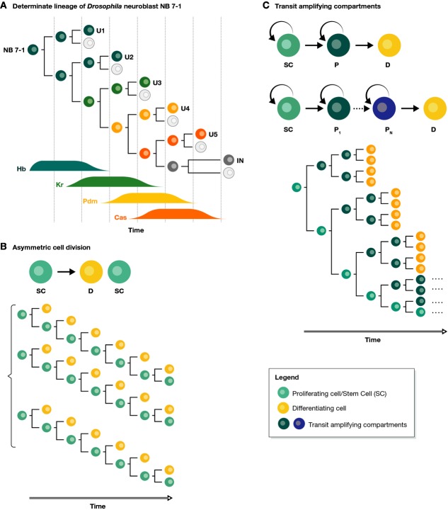
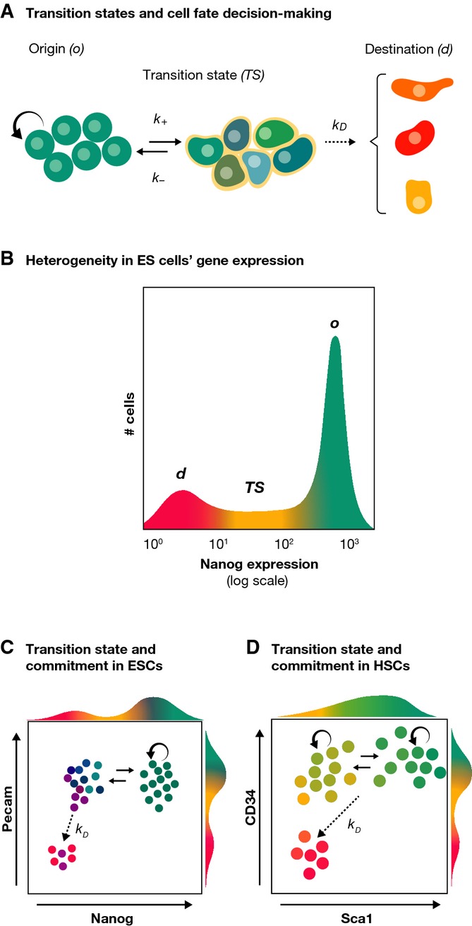
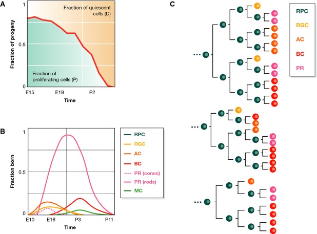
During tissue and organ development and maintenance, the dynamic regulation of cellular proliferation and differentiation allows cells to build highly elaborate structures. The development of the vertebrate retina or the maintenance of adult intestinal crypts, for instance, involves the arrangement of newly created cells with different phenotypes, the proportions of which need to be tightly controlled. While some of the basic principles underlying these processes developing and maintaining these organs are known, much remains to be learnt from how cells encode the necessary information and use it to attain those complex but reproducible arrangements. Here, we review the current knowledge on the principles underlying cell population dynamics during tissue development and homeostasis. In particular, we discuss how stochastic fate assignment, cell division, feedback control and cellular transition states interact during organ and tissue development and maintenance in multicellular organisms. We propose a framework, involving the existence of a transition state in which cells are more susceptible to signals that can affect their gene expression state and influence their cell fate decisions. This framework, which also applies to systems much more amenable to quantitative analysis like differentiating embryonic stem cells, links gene expression programmes with cell population dynamics.

Keywords: development; differentiation; homeostasis; stochastic cell fate; transition state.


Figures

Figure 1
Figure 1. Cell proliferation and differentiation
(A) The generation of neuroblast lineages in Drosophila is an example of a determinate process consisting of reproducible sequences of asymmetric cell divisions with changing fates. The succession of different fates upon each asymmetric cell division is controlled by a precise genetic programme on the progenitor, relying on the sequential expression of Hunchback (Hb), Kruppel (Kr), Pdm and Cas (adapted from Kohwi & Doe, 2013). (B) Asymmetric cell division is an invariant mechanism of generating differentiated progeny from stem cells where one daughter cell differentiates (D, yellow) and the other remains a stem cell (SC, teal). In homeostatic lineages, invariant asymmetry leads to homogeneous cell lineages. (C) Transit-amplifying cells are progenitors derived from stem cells that retain a proliferative capacity for a few division rounds before differentiating. SC: stem cell; D: differentiated cell; P, P1,…,PN: progenitors.
Figure 2
Figure 2. Molecular mechanisms of self-renewal and differentiation: the transition state
(A) The concept of the transition state (TS) between an origin (o) and a destination (d) state. During a fate change, a cell goes through a TS (for details see text), which implies the existence of kinetic constants governing the transitions between different states. (B) The TS can be observed in mouse ESCs. In this case, this is shown within the framework of Nanog expression, which is heterogeneously expressed with three distinguishable populations: o, representing ground state pluripotency; d, where it is possible to find cells committed to differentiation and TS where cells make a choice. (C) The coexistence of committed and uncommitted cells in the Nanog:GFP d population can be revealed by looking at a second pluripotency marker, Pecam or SSEA1 in this case (Canham et al, ; Lim, 2011). (D) A similar scenario has been recently observed in blood stem cells: cells with high levels of Sca1 can self-renew and are in a state analogous to the ‘o’ state. Sca1 low cells further subdivide into two populations, which can be identified by CD34. Sca1/CD34+ have repopulation capacity and can revert to Sca1+ while the Sca1/CD34 population consists of erythroid commited cells with no self-renewal capacity.
Figure 3
Figure 3. Vertebrate retinogenesis
(A) During the development of the vertebrate retina, there is an initial phase where most of the divisions are symmetric proliferative leading to progenitor amplification. As development proceeds, proliferation slows down. When individual cells stop dividing, they differentiate and this leads to a link between the different cell types and the growth of the tissue (Livesey & Cepko, 2001). (B) Throughout retinal development, a reproducible sequence of overlapping temporal windows of specific fate adoption by differentiating cells is established. An early differentiating cell can become a retinal ganglion cell (RGC), a horizontal cell (HC), a rod photoreceptor (PR) or an amacrine cell (AC), whereas if it differentiates later, it can become a bipolar cell (BC), a Müller cell (MC) or a cone PR; that is, there appears to be an overlap between these windows of opportunities (adapted from Cepko et al, 1996). (C) Recent accurate single-cell tracing assays have unveiled complex lineage compositions in the zebrafish retina development. Three out of 60 clones from He et al (2012) are shown. Despite the observed high variability of clone compositions, there are some remarkable trends; for example, RGCs appear through asymmetric divisions, PRs through symmetric ones.
Figure 4
Figure 4. Molecular mechanisms of mediating fate decisions in development at the level of single cells
(A) The transition state (TS, see Fig3) represents the basic unit for fate decisions. (B) During a fate transition, each cell executes a change of connectivity of their gene regulatory network from A to B. In this process, the cell will sample over time different configurations (microstates) of the available gene regulatory networks (GRN); many of these networks will resemble A, and therefore, the cell might have a chance to revert to the state of origin. When the network associated with fate B is connected, the cell moves to fate B. Within the TS, we suggest that cells are more susceptible to respond to signals that can bias their transcriptional state by affecting the connectivity (see text for details). (C) The TS state is an inherently noisy state, dominated by stochastic gene expression and affected by complex combinations of signals. As a result, the commitment or reversion event can be deemed as unpredictable at the level of individual cells. (D) The paradigm of the TS can be applied to pluripotent embryonic stem cells as well as to each differentiation step within the development of a tissue or organ. In the latter case, the TS is also controlled by a cell-autonomous genetic programme that establishes the order of appearance of the cellular fates, and thus, if a cell reverts to the state of origin, this might have changed in nature; this might account for many of the observations during retina development.

References

    1. Abranches E, Bekman E, Henrique D. Generation and characterization of a novel mouse embryonic stem cell line with a dynamic reporter of Nanog expression. PLoS One. 2013;8:e59928. - PMC - PubMed
    1. Abranches E, Guedes AM V, Moravec M, Maamar H, Svoboda P, Raj A, Henrique D. Stochastic nanog fluctuations allow mouse embryonic stem cells to explore pluripotency. Development. 2014;141:2770–2779. - PMC - PubMed
    1. Andreazzoli M. Molecular regulation of vertebrate retina cell fate. Birth Defects Res C Embryo Today. 2009;87:284–295. - PubMed
    1. Belliveau M, Cepko C. Extrinsic and intrinsic factors control the genesis of amacrine and cone cells in the rat retina. Development. 1999;126:555–566. - PubMed
    1. Belliveau MJ, Young TL, Cepko CL. Late retinal progenitor cells show intrinsic limitations in the production of cell types and the kinetics of opsin synthesis. J Neurosci. 2000;20:2247–2254. - PMC - PubMed

Publication types