

Are There Sex Differences in Knee Cartilage Composition and Walking Mechanics in Healthy and Osteoarthritis Populations?
- PMID: 25716211
- PMCID: PMC4488198
- DOI: 10.1007/s11999-015-4212-2
Are There Sex Differences in Knee Cartilage Composition and Walking Mechanics in Healthy and Osteoarthritis Populations?
Abstract
Background: Women are at a greater risk for knee osteoarthritis (OA), but reasons for this greater risk in women are not well understood. It may be possible that differences in cartilage composition and walking mechanics are related to greater OA risk in women.
Questions/purposes: (1) Do women have higher knee cartilage and meniscus T1ρ than men in young healthy, middle-aged non-OA and OA populations? (2) Do women exhibit greater static and dynamic (during walking) knee loading than men in young healthy, middle-aged non-OA and OA populations?
Methods: Data were collected from three cohorts: (1) young active (<35 years) (20 men, 13 women); (2) middle-aged (≥35 years) without OA (Kellgren-Lawrence [KL] grade < 2) (43 men, 65 women); and (3) middle-aged with OA (KL>1) (18 men, 25 women). T1ρ and T2 relaxation times for cartilage in the medial knee, lateral knee, and patellofemoral compartments and medial and lateral menisci were quantified with 3.0-T MRI. A subset of the participants underwent three-dimensional motion capture during walking for calculation of peak knee flexion and adduction moments, flexion and adduction impulses, and peak adduction angle. Differences in MR, radiograph, and gait parameters between men and women were compared in the three groups separately using multivariate analysis of variance.
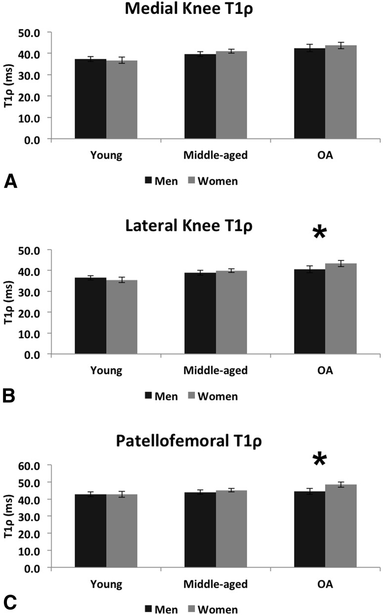
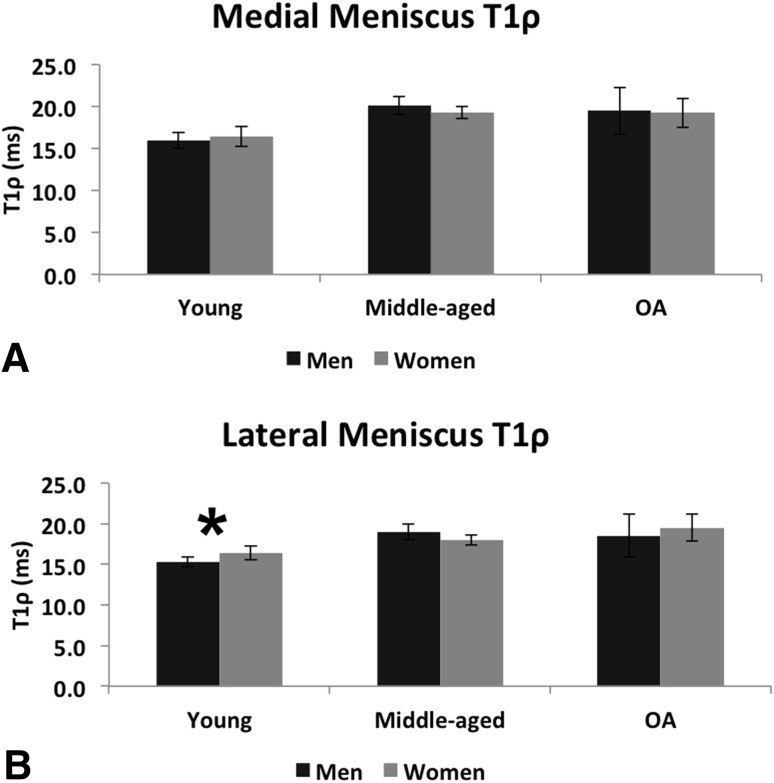
Results: Women had higher lateral articular cartilage T1ρ (men=40.5 [95% confidence interval {CI}, 38.8-42.3] ms; women=43.3 [95% CI, 41.9-44.7] ms; p=0.017) and patellofemoral T1ρ (men=44.4 [95% CI, 42.6-46.3] ms; women=48.4 [95% CI, 46.9-50.0] ms; p=0.002) in the OA group; and higher lateral meniscus T1ρ in the young group (men=15.3 [95% CI, 14.7-16.0] ms; women=16.4 [95% CI, 15.6-17.2] ms; p=0.045). The peak adduction moment in the second half of stance was lower in women in the middle-aged (men=2.05 [95% CI, 1.76-2.34] %BW*Ht; women=1.66 [95% CI, 1.44-1.89] %BW*Ht; p=0.037) and OA (men=2.34 [95% CI, 1.76-2.91] %BW*Ht; women=1.42 [95% CI, 0.89-1.94] %BW*Ht; p=0.022) groups. Static varus from radiographs was lower in women in the middle-aged (men=178° [95% CI, 177°-179°]; women=180° [95% CI, 179°-181°]; p=0.002) and OA (men=176° [95% CI, 175°-178°]; women=180° [95% CI, 179°-181°]; p<0.001) groups. Women had lower varus during walking in all three groups (young: men=4° [95% CI, 3°-6°]; women=2° [95% CI, 0°-3°]; p=0.013; middle-aged: men=2° [95% CI, 1°-3°]; women=0° [95% CI, -1° to 1°]; p=0.015; OA: men=4° [95% CI, 2°-6°]; women=0° [95% CI, -2° to 2°]; p=0.011). Women had a higher knee flexion moment (men=4.24 [95% CI, 3.58-4.91] %BW*Ht; women 5.40 [95% CI, 4.58-6.21] %BW*Ht; p=0.032) in the young group.
Conclusions: These data demonstrate differences in cartilage composition and gait mechanics between men and women in young healthy, middle-aged healthy, and OA cohorts. Considering the cross-sectional nature of the study, longitudinal research is needed to investigate if these differences in cartilage composition and walking mechanics are associated with a greater risk of lateral tibiofemoral or patellofemoral OA in women. Future studies should also investigate the relative risk of lateral versus medial patellofemoral cartilage degeneration risk in women compared with men.
Level of evidence: Level III, retrospective study.
Figures

Similar articles
-
Physical activity and spatial differences in medial knee T1rho and t2 relaxation times in knee osteoarthritis.J Orthop Sports Phys Ther. 2014 Dec;44(12):964-72. doi: 10.2519/jospt.2014.5523. Epub 2014 Oct 29. J Orthop Sports Phys Ther. 2014. PMID: 25353261 Free PMC article.
-
The relationship between knee adduction moment and cartilage and meniscus morphology in women with osteoarthritis.Osteoarthritis Cartilage. 2010 Jul;18(7):894-901. doi: 10.1016/j.joca.2010.04.006. Epub 2010 Apr 22. Osteoarthritis Cartilage. 2010. PMID: 20417296
-
Frontal Plane Knee Mechanics and Early Cartilage Degeneration in People With Anterior Cruciate Ligament Reconstruction: A Longitudinal Study.Am J Sports Med. 2018 Feb;46(2):378-387. doi: 10.1177/0363546517739605. Epub 2017 Nov 10. Am J Sports Med. 2018. PMID: 29125920 Free PMC article.
-
Modeling knee osteoarthritis pathophysiology using an integrated joint system (IJS): a systematic review of relationships among cartilage thickness, gait mechanics, and subchondral bone mineral density.Osteoarthritis Cartilage. 2018 Nov;26(11):1425-1437. doi: 10.1016/j.joca.2018.06.017. Epub 2018 Jul 26. Osteoarthritis Cartilage. 2018. PMID: 30056214
-
MRI T2 and T1ρ relaxation in patients at risk for knee osteoarthritis: a systematic review and meta-analysis.BMC Musculoskelet Disord. 2019 May 1;20(1):182. doi: 10.1186/s12891-019-2547-7. BMC Musculoskelet Disord. 2019. PMID: 31039785 Free PMC article.
Cited by
-
Sexual dimorphism in peri-articular tissue anatomy - More keys to understanding sex-differences in osteoarthritis?Osteoarthr Cartil Open. 2024 May 11;6(3):100485. doi: 10.1016/j.ocarto.2024.100485. eCollection 2024 Sep. Osteoarthr Cartil Open. 2024. PMID: 38946793 Free PMC article. Review.
-
Stem Cells for Cartilage Repair: Preclinical Studies and Insights in Translational Animal Models and Outcome Measures.Stem Cells Int. 2018 Feb 5;2018:9079538. doi: 10.1155/2018/9079538. eCollection 2018. Stem Cells Int. 2018. PMID: 29535784 Free PMC article. Review.
-
Association of layer-specific knee cartilage T2-relaxation measurements with age, sex and cartilage morphology at 1.5-T MRI.Eur Radiol. 2025 Jul 25. doi: 10.1007/s00330-025-11806-8. Online ahead of print. Eur Radiol. 2025. PMID: 40715824
-
Tibiofemoral articular cartilage composition differs based on serum biochemical profiles following anterior cruciate ligament reconstruction.Osteoarthritis Cartilage. 2021 Dec;29(12):1732-1740. doi: 10.1016/j.joca.2021.09.005. Epub 2021 Sep 16. Osteoarthritis Cartilage. 2021. PMID: 34536530 Free PMC article.
-
Baseline Cartilage Thickness and Meniscus Extrusion Predict Longitudinal Cartilage Loss by Quantitative Magnetic Resonance Imaging: Data From the Osteoarthritis Initiative.J Comput Assist Tomogr. 2016 Nov/Dec;40(6):979-984. doi: 10.1097/RCT.0000000000000464. J Comput Assist Tomogr. 2016. PMID: 27454790 Free PMC article.
References
-
- Blumenkrantz G, Lindsey CT, Dunn TC, Jin H, Ries MD, Link TM, Steinbach LS, Majumdar S. A pilot, two-year longitudinal study of the interrelationship between trabecular bone and articular cartilage in the osteoarthritic knee. Osteoarthritis Cartilage. 2004;12:997–1005. doi: 10.1016/j.joca.2004.09.001. - DOI - PubMed
Publication types
MeSH terms
Grants and funding
LinkOut - more resources
Full Text Sources
Other Literature Sources
Research Materials
