. 2015 Feb 25;35(8):3384-96.
doi: 10.1523/JNEUROSCI.2620-14.2015.

Triggering receptor expressed on myeloid cells 2 (TREM2) deficiency attenuates phagocytic activities of microglia and exacerbates ischemic damage in experimental stroke

Affiliations

Triggering receptor expressed on myeloid cells 2 (TREM2) deficiency attenuates phagocytic activities of microglia and exacerbates ischemic damage in experimental stroke

Masahito Kawabori et al. J Neurosci. .

Abstract

Clearing cellular debris after brain injury represents an important mechanism in regaining tissue homeostasis and promoting functional recovery. Triggering receptor expressed on myeloid cells-2 (TREM2) is a newly identified receptor expressed on microglia and is thought to phagocytose damaged brain cells. The precise role of TREM2 during ischemic stroke has not been fully understood. We explore TREM2 in both in vitro and in vivo stroke models and identify a potential endogenous TREM2 ligand. TREM2 knockdown in microglia reduced microglial activation to an amoeboid phenotype and decreased the phagocytosis of injured neurons. Phagocytosis and infarcted brain tissue resorption was reduced in TREM2 knock-out (KO) mice compared with wild-type (WT) mice. TREM2 KO mice also had worsened neurological recovery and decreased viable brain tissue in the ipsilateral hemisphere. The numbers of activated microglia and phagocytes in TREM2 KO mice were decreased compared with WT mice, and foamy macrophages were nearly absent in the TREM2 KO mice. Postischemia, TREM2 was highly expressed on microglia and TREM2-Fc fusion protein (used as a probe to identify potential TREM2 binding partners) bound to an unknown TREM2 ligand that colocalized to neurons. Oxygen glucose deprivation-exposed neuronal media, or cellular fractions containing nuclei or purified DNA, but not cytosolic fractions, stimulated signaling through TREM2. TREM2-Fc fusion protein pulled down nucleic acids from ischemic brain lysate. These findings establish the relevance of TREM2 in the phagocytosis of the infarcted brain and emphasize its role in influencing neurological outcomes following stroke. Further, nucleic acids may be one potential ligand of TREM2 in brain ischemia.

Keywords: ischemia; neuroprotection; permanent ischemia; phagocytosis; stroke; triggering receptor expressed on myeloid cells-2.


Figures

Figure 1.
Figure 1.
TREM2 knockdown with RNAi reduces microglial activation, but does not affect acute neuron survival. Primary NA cultures with and without microglia were prepared. TREM2 was knocked down by RNAi or control RNAi, and some cultures were exposed to 1 h OGD followed by 24 h reperfusion at normoxia and 5.5 mm added glucose. A, NA uninjured cultures (Ctrl-NA). B, NA cultures exposed to OGD for 1 h and reperfusion for 24 h. C, NAM cultures (control RNAi) after OGD [OGD NAM (WT)]. D, NA cultures plus TREM2-deficient microglia were subjected to OGD (OGD NAM TREM2). Neuronal processes were more fragmented in the absence of microglia or TREM2-deficient microglia. Higher-power micrographs (a′d′) show neuronal processes and/or microglial morphology under the different conditions described in A–D. Arrowheads show microglia, where they appear amoeboid (c′, OGD, control RNAi) and ramified (d′, OGD, TREM2 RNAi). E, Neuronal survival/viability is decreased by OGD in all cases. The addition of microglia, whether with intact TREM2 (WT) or deficient in TREM2 (TREM2 RNAi), led to decreased neuron survival. F, Microglial activation, as assessed by the transformation of resting (ramified) microglia into amoeboid microglia, is increased by OGD and markedly decreased in TREM2 deficiency. #p < 0.01 compared with control conditions; *p < 0.01 as indicated, n = 4/group.
Figure 2.
Figure 2.
TREM2 deficiency blunts phagocytosis of OGD-exposed neurons by microglia. Lysates from CM-DiI-loaded, OGD-exposed neuro 2A cells were applied to BV2 microglia. A, BV2 cells with intact TREM2 show avid uptake of neuro 2A lysates (red). B, When TREM2 was knocked down using siRNA, fewer microglia took up neuron 2A lysates, as evidenced by less red staining. C, Counts of positive red cells, which indicate that TREM2 is required for the uptake of OGD-exposed neuro 2A cells. Scale bar, 100 μm. Data are mean ± SEM. *p < 0.05.
Figure 3.
Figure 3.
TREM2 is expressed in microglia within the ischemic borderzone. A–D, Immunostains for TREM2 (red) and neuronal (NeuN, green) and microglial (CD11b, green, arrows) markers 7 d post-MCAO show that TREM2 is present in microglia but not in neurons. Colocalized cells appear yellow. TREM2-positive cells (red) are especially expressed in the ipsilateral ischemic brain (B, D), and less so, on the contralateral side (A, C). E, The time course of TREM2 expression is shown. Numbers of TREM2-positive cells peaked at 7 d, but were present as early as 3 d, and persisted at 14 d. By 30 d, the numbers of TREM2-positive cells were similar to baseline.
Figure 4.
Figure 4.
TREM2 ligand is increased in neurons after brain ischemia. A TREM2 fusion protein labeled with Texas red was used to identify TREM2 ligands. A, B, Upregulation of a TREM2 ligand can be found in the ischemic brain 7 d post-MCAO exposure (B, red), compared with the contralateral side (A). G–J, The TREM2 fusion protein colocalized to neurons after ischemia. C–F, In the contralateral brain, very little TREM2 ligand was present (blue, DAPI; green, NeuN; red, TREM2 fusion protein with Texas red; merged, F, J). Scale bar, 80 μm.
Figure 5.
Figure 5.
Less infarcted brain resorption and larger infarct size were observed in TREM2-deficient mice. A, B, Brains of TREM2 KO mice (B) had very little to no TREM2 protein compared with WT mice (A). C, E, WT mice showed brain resorption post-dMCAO at day 14 (arrowheads). D, F, In contrast, TREM2 KO mice showed significantly less resorption (arrow). G, Quantification of residual infarcted brain was estimated to be ∼30% in the WT group, whereas most of the damaged brain was still present in the KO group. H, Overall infarct size was also significantly larger in the KO group. **p < 0.01, *p < 0.05, n = 12/group.
Figure 6.
Figure 6.
TREM2-deficient mice showed worsened neurological recovery compared with wild-type mice. A, B, Neurological deficits were assessed using a modified Bederson score and the number of foot faults on the ladder test. TREM2 KO (KO) mice showed worse scores on the Bederson scale compared with WT mice (A), and made more foot faults on the ladder test (B). **p < 0.01, *p < 0.05, n = 12/group.
Figure 7.
Figure 7.
Activated microglia and phagocytes were decreased in TREM2-deficient mice. A–H, Activated microglia identified by IB4 staining [WT mice, A, C; TREM2 KO mice (KO), B, D] and phagocytic microglia by CD68 staining (WT mice, E; KO mice, F) were significantly decreased in TREM2 KO groups (G and H, respectively). Most of the activated cells were observed in and around the infarct border. Images were collected from brains on day 7 poststroke. Scale bar, 40 μm. **p < 0.01, *p < 0.05, n = 8/group.
Figure 8.
Figure 8.
Oil red O staining shows fewer lipid-rich foamy macrophages in TREM2-deficient mice. Oil red O stain is used here to delineate foamy macrophages, or foam cells in red, in brains 7 d post-MCAO. These represent phagocytes that contain intracellular lipids, which result from the phagocytosis of injured brain. A–D, Lipid-rich foamy macrophages can be found in and around the infarct border in WT mice (A, C), while there were almost no foamy macrophages found in the TREM2 KO mice (B, D). E, Numbers of red foam cells were nearly absent in the TREM2 KO mice (KO), and would suggest that phagocytosis of the injured brain cells was significantly impaired in these mice. Scale bar, 40 μm. **p < 0.01, n = 8/group.
Figure 9.
Figure 9.
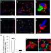
Fewer phagocytes associate with TUNEL-positive cells in TREM2-deficient mice. A–C, A 3D rendering confocal image shows CD68-positive (red) phagocytes contacting TUNEL-positive cells (green) in a WT brain (DAPI in blue) 7 d post-MCAO. D–F, In contrast, CD68-positive cells tended to remain separate from TUNEL-positive cells in TREM2-deficient brains. G, The proportion of CD68-positive cells contacting TUNEL-positive cells was markedly reduced in TREM2-deficient brains. CD68-positive phagocytes (red) contact both MAP2 (green)-positive neurons (H) and GFAP (green)-positive glia (I) surrounding the infarct border. Scale bar, 80 μm. **p < 0.01, n = 4/group.
Figure 10.
Figure 10.
Angiogenesis but not glial scarring was decreased in TREM2-deficient mice. A–D, Angiogenesis after 14 d postischemia was evaluated in WT (A, C) and TREM2-deficient (KO; B, D) mice by collagen IV staining. E, The fluorescence images show that collagen IV-positive parenchymal microvessels were significantly increased in the WT group compared with the KO group within the infarct border. Glial scarring and reactivity at the infarct border were also examined. F–J, Areas of GFAP positivity were slightly decreased in the KO group (G, I) compared with the WT group (F, H), but this was not significantly different (J; NS). Scale bar, 100 μm. **p < 0.01, n = 8/group.
Figure 11.
Figure 11.
Nucleic acids as a potential TREM2 ligand in ischemic brain. A, To identify potential ligands for TREM2 in ischemic brain, wild-type animals were subjected to experimental stroke and brains were probed with the TREM2 fusion protein to isolate potential binding partners. Since extracellular nucleic acids have been identified as ligands for other innate immune receptors, a modified ChIP assay was performed on fusion protein-probed lysates to delineate nucleic acids (SYBR green). Supernatant and pellet fractions were obtained from both ipsilateral and contralateral hemispheres 7 d post-dMCAO. A high-molecular-weight nucleic acid band was found to interact with the TREM2 fusion as shown in the agarose gel (arrow). Within ischemic brain, this high-molecular-weight band was observed in the supernatant as well as the pellet, whereas the band was only observed in the pellet fraction of the contralateral nonischemic hemisphere (lanes 1 and 3, pellet and supernatant of the ischemic hemisphere, respectively; lanes 2 and 4, pellet and supernatant of the contralateral hemisphere, respectively; lane 5, anti-human IgG control). B, A BWZ reporter cell line was used to assay TREM2 signaling in vitro. The BWZ lymphocyte cell line were modified to overexpress TREM2 linked to DAP12, NFAT, and and lacZ (BWZ-TD4 TREM2). In the presence of TREM2 ligand binding, activation can be assayed via β-gal expression. Neuro-2a cells were exposed to OGD, and lysates or conditioned media were applied to the reporter cells. Unmodified BWZ cells (BWZ BWZ) were used as negative controls. Untreated reporter and control BWZ cells were also used as negative controls. Conditioned media from OGD-exposed Neuro-2a cells led to increased TREM2 signaling, as evidenced by increased β-gal absorbance, compared with control media and media from uninjured Neuro-2a cells. To further suggest that nucleic acids may be responsible for TREM2 signaling, subcellular fractions were also applied to the BWZ control and reporter cells. Only fractions expected to contain nucleic acids led to increased signaling (nuclear, mitochondrial, and purified DNA), whereas fractions normally lacking nucleic acids (cytosolic) failed to signal. **p < 0.001 vs no injury; #p < 0.001 vs control BWZ cells.

References

    1. Aderem A, Underhill DM. Mechanisms of phagocytosis in macrophages. Annu Rev Immunol. 1999;17:593–623. doi: 10.1146/annurev.immunol.17.1.593. - DOI - PubMed
    1. Bianchin MM, Capella HM, Chaves DL, Steindel M, Grisard EC, Ganev GG, da Silva Júnior JP, Neto Evaldo S, Poffo MA, Walz R, Carlotti Júnior CG, Sakamoto AC. Nasu-Hakola disease (polycystic lipomembranous osteodysplasia with sclerosing leukoencephalopathy–PLOSL): a dementia associated with bone cystic lesions. From clinical to genetic and molecular aspects. Cell Mol Neurobiol. 2004;24:1–24. doi: 10.1023/B:CEMN.0000012721.08168.ee. - DOI - PMC - PubMed
    1. Charles JF, Humphrey MB, Zhao X, Quarles E, Nakamura MC, Aderem A, Seaman WE, Smith KD. The innate immune response to Salmonella enterica serovar Typhimurium by macrophages is dependent on TREM2-DAP12. Infect Immun. 2008;76:2439–2447. doi: 10.1128/IAI.00115-08. - DOI - PMC - PubMed
    1. Colonna M. TREMs in the immune system and beyond. Nat Rev Immunol. 2003;3:445–453. doi: 10.1038/nri1106. - DOI - PubMed
    1. Daws MR, Lanier LL, Seaman WE, Ryan JC. Cloning and characterization of a novel mouse myeloid DAP12-associated receptor family. Eur J Immunol. 2001;31:783–791. doi: 10.1002/1521-4141(200103)31:3<783::AID-IMMU783>3.0.CO%3B2-U. - DOI - PubMed

Publication types

MeSH terms