. 2015 Feb 25;35(8):3676-88.
doi: 10.1523/JNEUROSCI.3510-14.2015.

Anti-muscarinic adjunct therapy accelerates functional human oligodendrocyte repair

Affiliations

Anti-muscarinic adjunct therapy accelerates functional human oligodendrocyte repair

Kavitha Abiraman et al. J Neurosci. .

Abstract

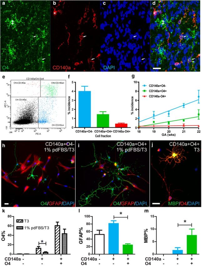
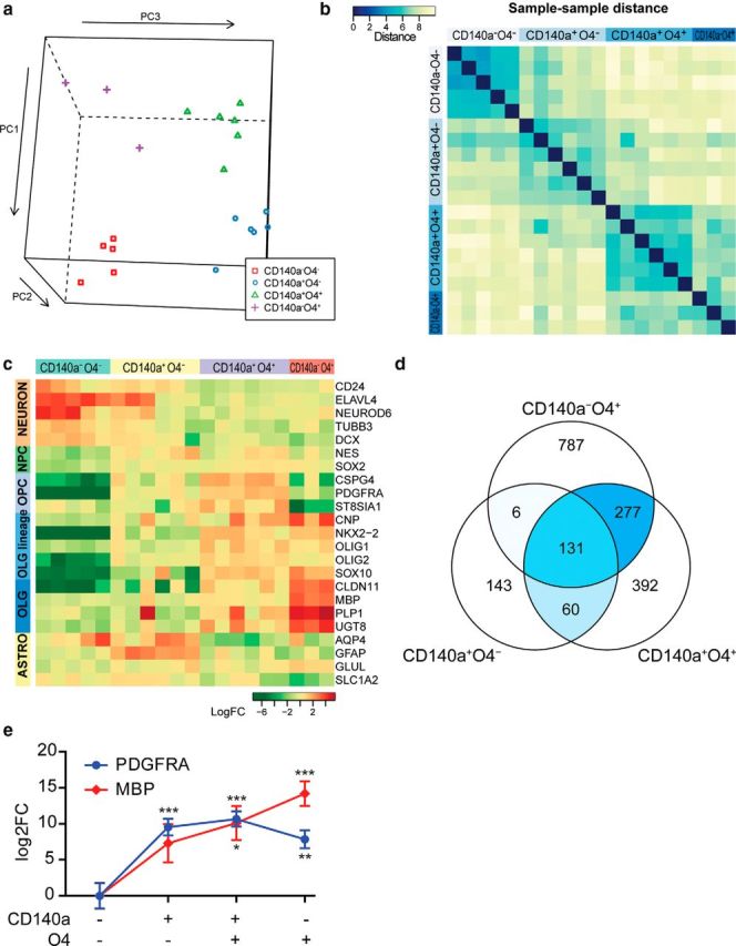
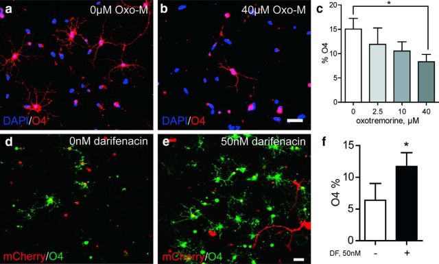
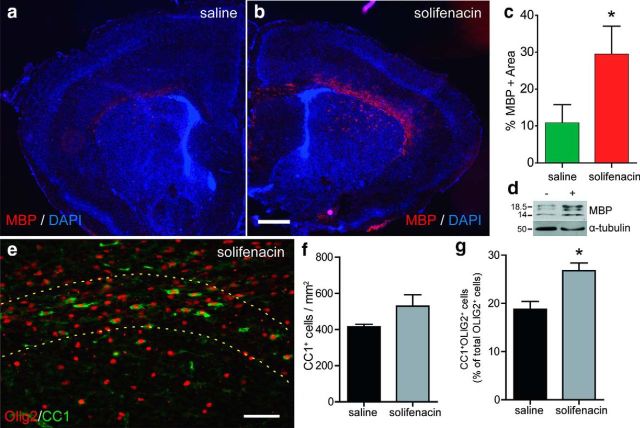
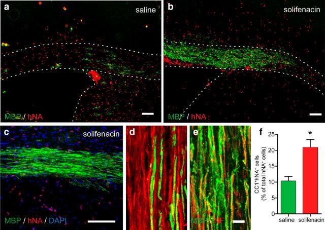
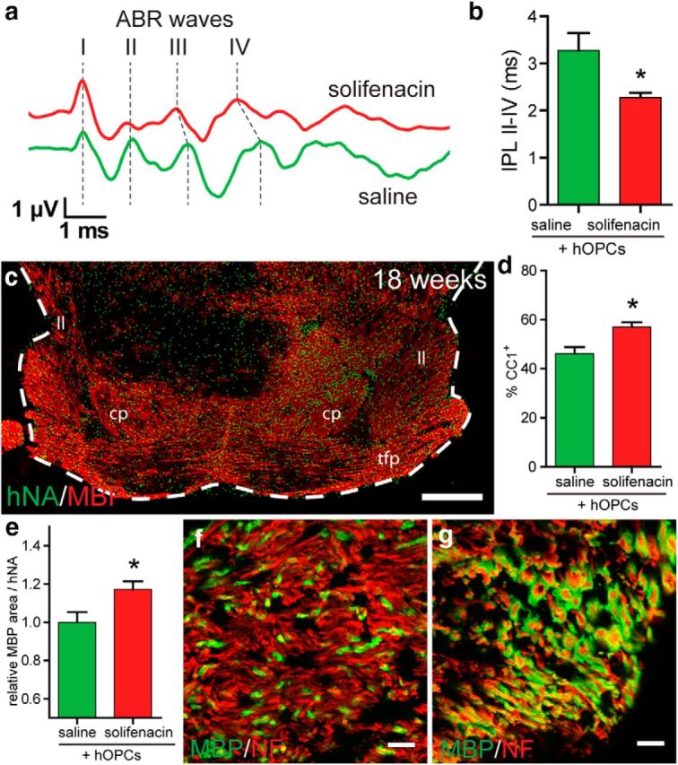
Therapeutic repair of myelin disorders may be limited by the relatively slow rate of human oligodendrocyte differentiation. To identify appropriate pharmacological targets with which to accelerate differentiation of human oligodendrocyte progenitors (hOPCs) directly, we used CD140a/O4-based FACS of human forebrain and microarray to hOPC-specific receptors. Among these, we identified CHRM3, a M3R muscarinic acetylcholine receptor, as being restricted to oligodendrocyte-biased CD140a(+)O4(+) cells. Muscarinic agonist treatment of hOPCs resulted in a specific and dose-dependent blockade of oligodendrocyte commitment. Conversely, when hOPCs were cocultured with human neurons, M3R antagonist treatment stimulated oligodendrocytic differentiation. Systemic treatment with solifenacin, an FDA-approved muscarinic receptor antagonist, increased oligodendrocyte differentiation of transplanted hOPCs in hypomyelinated shiverer/rag2 brain. Importantly, solifenacin treatment of engrafted animals reduced auditory brainstem response interpeak latency, indicative of increased conduction velocity and thereby enhanced functional repair. Therefore, solifenacin and other selective muscarinic antagonists represent new adjunct approaches to accelerate repair by engrafted human progenitors.

Keywords: glia; human; microarray; oligodendrocyte.


Figures

Figure 1.
Figure 1.
CD140a/PDGFαR and O4 identify three functionally distinct glial pools in the developing human forebrain. ad, Confocal microscopy of the intermediate zone of 20 week gestational age fetal forebrain. O4 and CD140a mAbs identify three distinct cell populations: CD140a+O4 (*), CD140a+O4+ (arrows), and CD140aO4+ cells (‡). e, Two-color flow cytometry for CD140a (PE, x-axis) and O4 (APC, y-axis). A clear population of double CD140a+O4+ cells was distinct from single CD140a+ and O4+ cells (100,000 events shown). f, Incidence of each glial cell fraction in human fetal brain (mean incidence between 18 and 22 weeks, n = 12 fetal samples). CD140a+O4 cells were significantly more abundant than CD140a+O4+ and CD140a-O4+ cells. g, Incidence with respect to developmental age (mean ± SEM, linear regression line of best-fit shown). h, Fetal dissociates were sorted for CD140a/O4 expression cultured for 4 d in serum-free or low serum (1% pd-FBS) to determine cell fate. hi, Immature oligodendrocytes were labeled with O4 (green) and astrocytes with GFAP (red). j, A significant proportion of CD140a+O4+ cells developed as mature MBP+ oligodendrocytes. k, Oligodendrocytes were significantly more abundant in all sorted fractions relative to CD140aO4 cells (one-way ANOVA, p < 0.05). The addition of 1% serum significantly reduced oligodendrocyte commitment from CD140a+O4 cells but did not affect CD140a+O4+ cells (post hoc t test, p < 0.05). Consistent with a more committed progenitor phenotype, CD140a+O4+ cells generated significantly fewer astrocytes (l) and more MBP+ oligodendrocytes (m) (mean ± SEM, n = 4–5 fetal samples). Scale bars: 20 μm (a), 10 μm (hj).
Figure 2.
Figure 2.
Transcriptional profile of CD140a/O4-sorted cells reveals distinct stages of human oligodendrocyte lineage differentiation. a, Principle components were calculated for each gene expression profile and plotted on a 3D scatter plot. The RNAs for each sorted fraction clustered together, indicating similar expression profiles within each sorted fraction and significant differences between samples. b, Between-sample distances were calculated using maximum distance (uniform norm) between samples. Samples with closely related gene expression patterns and low intersample distances are colored blue according to the color bar. Samples within each sorted fraction from distinct fetal donors were more closely related than between sorted fractions obtained from the same donor brain. c, Several known cell-type-specific marker genes were significantly regulated between each glial fraction and double-negative cells. Gene expression shown indicates expression relative to median of all samples. d, Probes significantly regulated in each cell fraction relative to CD140aO4 cells. The overlapping genes between each comparison are colored according to their relative abundance. This shows that there is extensive overlap between both CD140+ and O4+ fractions. Only six probes were shared between CD140a+O4 and CD140aO4+ fractions. e, Real-time RT-PCR confirmed very high enrichment of OPC-expressed PDGFRA and MBP mRNA immediately after CD140a/O4 multicolor FACS (mean ± SEM, n = 5 fetal samples). *p < 0.05, **p < 0.01, ***p < 0.001, Dunnett's posttest, versus CD140aO4 cells (1-way ANOVA).
Figure 3.
Figure 3.
mAchR activity regulates oligodendrocyte commitment by hOPCs. CD140a/PDGFαR+ hOPCs were treated with 0–40 μm OxoM for 4 d and stained for O4 (red) and DAPI (blue) (a, b). The proportion of O4+ oligodendrocytes was determined at each dose (mean ± SEM, n = 3 seperate brain preparations, 19–22 week gestational age) (c). de, CD140a+ hOPCs were cocultured with human neuronal reaggregate cultures for 14 d in vitro (DIV). The fate of human OPCs was tracked by prior infection with mCherry-expressing lentivirus at −2 DIV. Oligodendrocyte differentiation was assessed at 14 DIV by colocalization of mCherry-expressing cells (red) with O4 (green) (d, null control). e, Daily treatment of matched cultures with 50 nm darifenacin, an M3R antagonist, induced oligodendrocyte differentiation. f, Proportion of mCherry+ OPCs expressing O4 was determined (mean ± SEM, n = 3 separate brain preparations). *p < 0.05, t test. Scale bar, 50 μm.
Figure 4.
Figure 4.
Systemic treatment with an M3R antagonist promotes precocious oligodendrocyte differentiation in vivo. Neonatal mice received daily subcutaneous injections of either saline or 10 mg/kg solifenacin, a blood–brain barrier-permeable M3R antagonist, from 4 d postnatally until being killedon day 9 (n = 4). a, b, Oligodendrocyte differentiation and myelination was assessed by immunoflourescence staining for MBP in saline- (a) or antagonist (b)-injected animals. The percentage area of myelinated corpus callosum was quantified in each animal and shown in c (mean ± SEM). M3R antagonist treatment induced a significant increase in myelinated area (n = 4 animals per group). d, Solifenacin increased the amount of MBP protein normalized to tubulin (n = 3 mice per group). CC1+OLIG2+ oligodendrocyte cells were counted in corpus callosum and compared with total OLIG2+ oligodendrocyte lineage cells (e). There was a significant increase in the proportion of differentiating oligodendrocytes in solifenacin-treated animals. f, Overall density of CC1+ cells was also increased in corpus callosum. *p < 0.05, t test. Scale bars: 500 μm (a, b), 50 μm (c).
Figure 5.
Figure 5.
Solifenacin promotes human oligodendrocyte differentiation after xenograft into shiverer/rag2 mice. Newborn homozygous shiverer/rag2 mice were transplanted with human CD140a/PDGFαR+ OPCs. Four days later, animals were injected daily with either saline or solifenacin (10 mg/kg) for 8 weeks (n = 4 animals per group). Human cells were identified using human nuclear antigen staining (hNA, red) and myelinating oligodendrocytes using MBP (green). Compared with saline controls (a), solifenacin treatment resulted in a substantial increase in the number of and area myelinated by MBP+ human oligodendrocytes in the corpus callosum at 8 weeks (b, c). d, e, Confocal images showing individual myelinated fibers in corpus callosum of saline-treated (d) and solifenacin-treated (e) animals. f, Human CC1+hNA+ oligodendrocytes were counted in xenografted corpus callosum and compared with total hNA+ human cells (n = 4 animals). *p < 0.05, t test. Scale bars: 100 μm (ac), 10 μm (d, e).
Figure 6.
Figure 6.
Solifenacin improves functional recovery mediated by transplanted hOPCs. To determine whether solifenacin treatment improves the function of myelin produced by transplanted hOPCs, we transplanted hOPCs into the midbrain of shiverer/rag2 day 1–3 pups and assessed auditory brainstem response at 15 weeks after daily subcutaneous injection with either saline or 10 mg/kg solifenacin. a, Auditory evoked potential waveform from saline- and solifenacin-treated hOPC-transplanted animals indicating the relative timing of positive vertex waves I-IV. b, IPL between peaks II and IV was measured (mean ± SEM, n = 3–5). Solifenacin treatment significantly reduced IPL II-IV. *p < 0.05, t test. At 19 weeks, animals were killed to determine the distribution of human cells (hNA, green) and myelin (MBP, red). c, Myelin was found throughout the ventral midbrain structures, including cerebral peduncle (cp), lateral lemniscus (ll), and transverse fibers pons (tfp). The example coronal section shown from a solifenacin-treated animal corresponds to plate 65 in Franklin and Paxinos (2008). d, Proportion of human cells differentiating as CC1+ oligodendrocytes in the cerebral peduncle. e, Relative contribution of human cells to percentage area of MBP staining in the cerebral peduncle. f, g, Confocal images of myelinated fibers in the cerebral peduncle of saline-treated (f) and solifenacin-treated (g) animals at 19 weeks (neurofilament, red; MBP, green). Scale bars: 500 μm (c), 10 μm (fg).

References

    1. Armstrong RC, Dorn HH, Kufta CV, Friedman E, Dubois-Dalcq ME. Pre-oligodendrocytes from adult human CNS. J Neurosci. 1992;12:1538–1547. - PMC - PubMed
    1. Back SA, Luo NL, Borenstein NS, Levine JM, Volpe JJ, Kinney HC. Late oligodendrocyte progenitors coincide with the developmental window of vulnerability for human perinatal white matter injury. J Neurosci. 2001;21:1302–1312. - PMC - PubMed
    1. Barres BA, Schmid R, Sendnter M, Raff MC. Multiple extracellular signals are required for long-term oligodendrocyte survival. Development. 1993;118:283–295. - PubMed
    1. Bjelke B, Seiger A. Morphological distribution of MBP-like immunoreactivity in the brain during development. Int J Dev Neurosci. 1989;7:145–164. doi: 10.1016/0736-5748(89)90065-8. - DOI - PubMed
    1. Brand-Schieber E, Werner P. (+/-)-Alpha-amino-3-hydroxy-5-methylisoxazole-4-propionic acid and kainate receptor subunit expression in mouse versus rat spinal cord white matter: similarities in astrocytes but differences in oligodendrocytes. Neurosci Lett. 2003;345:126–130. doi: 10.1016/S0304-3940(03)00469-5. - DOI - PubMed

Publication types

MeSH terms