. 2015 Feb 26;10(2):e0117352.
doi: 10.1371/journal.pone.0117352. eCollection 2015.

Aberrant lymphatic endothelial progenitors in lymphatic malformation development

Affiliations

Aberrant lymphatic endothelial progenitors in lymphatic malformation development

June K Wu et al. PLoS One. .

Abstract

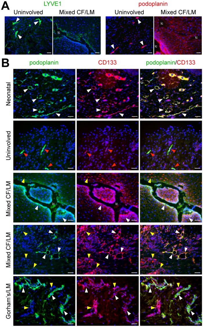
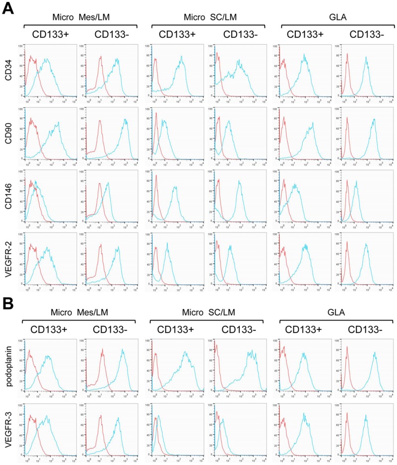
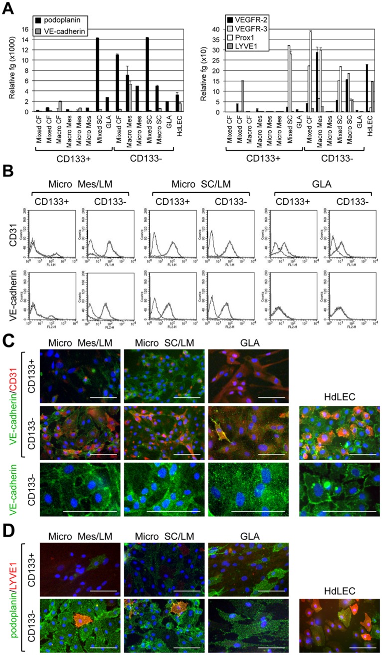
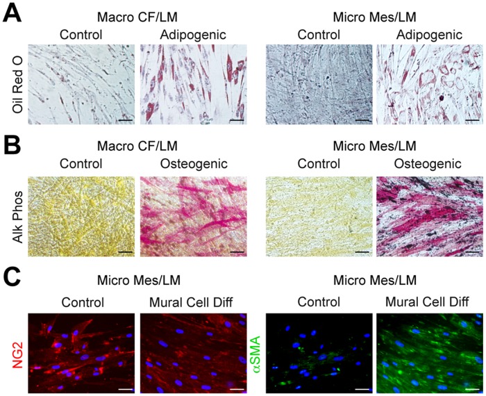
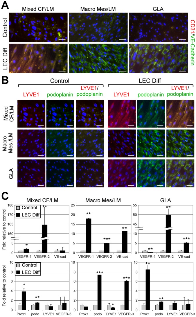
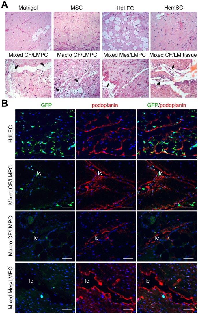
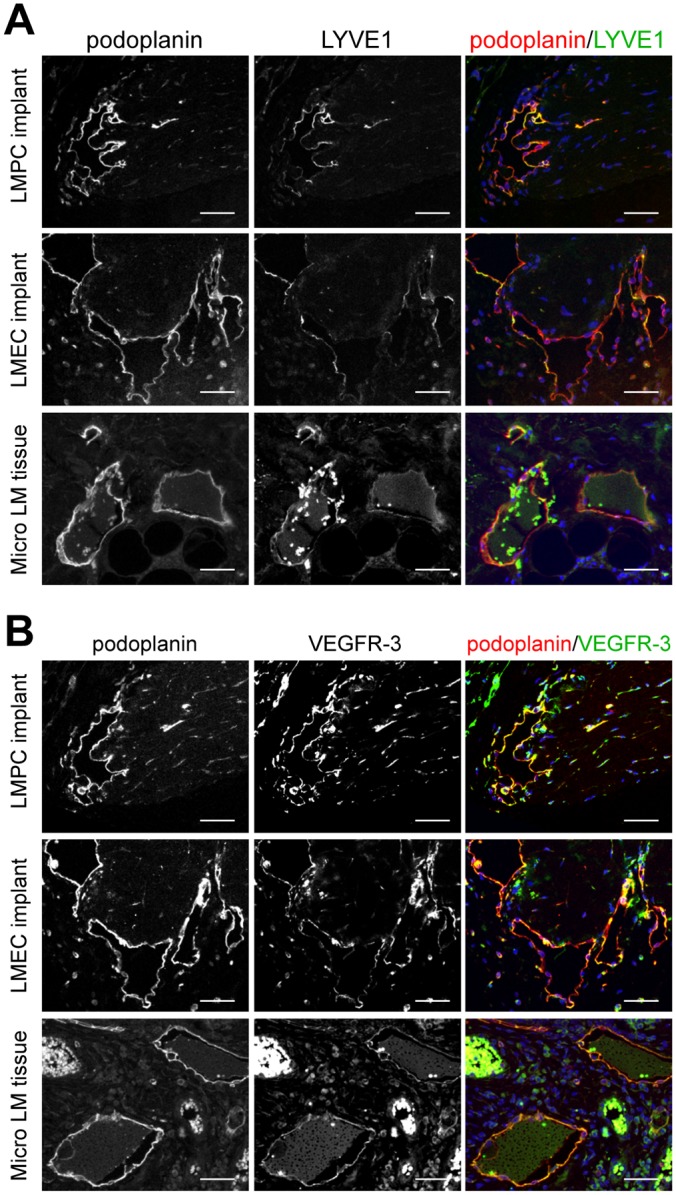
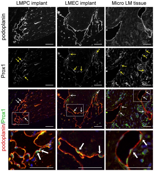
Lymphatic malformations (LMs) are vascular anomalies thought to arise from dysregulated lymphangiogenesis. These lesions impose a significant burden of disease on affected individuals. LM pathobiology is poorly understood, hindering the development of effective treatments. In the present studies, immunostaining of LM tissues revealed that endothelial cells lining aberrant lymphatic vessels and cells in the surrounding stroma expressed the stem cell marker, CD133, and the lymphatic endothelial protein, podoplanin. Isolated patient-derived CD133+ LM cells expressed stem cell genes (NANOG, Oct4), circulating endothelial cell precursor proteins (CD90, CD146, c-Kit, VEGFR-2), and lymphatic endothelial proteins (podoplanin, VEGFR-3). Consistent with a progenitor cell identity, CD133+ LM cells were multipotent and could be differentiated into fat, bone, smooth muscle, and lymphatic endothelial cells in vitro. CD133+ cells were compared to CD133- cells isolated from LM fluids. CD133- LM cells had lower expression of stem cell genes, but expressed circulating endothelial precursor proteins and high levels of lymphatic endothelial proteins, VE-cadherin, CD31, podoplanin, VEGFR-3 and Prox1. CD133- LM cells were not multipotent, consistent with a differentiated lymphatic endothelial cell phenotype. In a mouse xenograft model, CD133+ LM cells differentiated into lymphatic endothelial cells that formed irregularly dilated lymphatic channels, phenocopying human LMs. In vivo, CD133+ LM cells acquired expression of differentiated lymphatic endothelial cell proteins, podoplanin, LYVE1, Prox1, and VEGFR-3, comparable to expression found in LM patient tissues. Taken together, these data identify a novel LM progenitor cell population that differentiates to form the abnormal lymphatic structures characteristic of these lesions, recapitulating the human LM phenotype. This LM progenitor cell population may contribute to the clinically refractory behavior of LMs.


Conflict of interest statement

Competing Interests: The authors have declared that no competing interests exist.

Figures

Fig 1
Fig 1. Identification of CD133+ cells in LMs of different subtypes and anatomical locations.
(A) LYVE1 and podoplanin staining of cervicofacial mixed LM tissue and patient-matched uninvolved tissue. White arrowheads mark normal lymphatics. (B) Podoplanin and CD133 staining of neonatal foreskin (postnatal day 1), uninvolved tissue, mixed cervicofacial (Mixed CF) LM tissues (2x), and Gorham’s dermal tissue. White arrowheads mark CD133+/podoplanin+ lymphatic endothelium. Red arrowheads mark CD133low/podoplanin+ lymphatic endothelium. Yellow arrowheads mark CD133+/podoplanin+ stromal cells. Blue asterisks mark blood vessels with autofluorescing red blood cells. Scale bars: 50μm. lymphatic channel (lc)
Fig 2
Fig 2. Endothelial precursor and lymphatic endothelial protein expression in isolated CD133+ and CD133 LM cells.
FACS of patient-matched CD133+ and CD133 LM cells isolated from microcystic mesenteric (Micro Mes) LM, microcystic subcutaneous (Micro SC) LM and general lymphatic anomaly (GLA) specimens. (A) Endothelial precursor markers, CD34, CD90, CD146 and VEGFR-2. (B) Lymphatic endothelial cell markers, podoplanin and VEGFR-3. Blue line represents antibody data, and red line IgG control.
Fig 3
Fig 3. Expression of markers for mature lymphatic endothelial cells in isolated CD133+ and CD133 LM cells.
(A) Podoplanin, VE-cadherin, VEGFR-2, VEGFR-3, Prox1, and LYVE1 qRT-PCR of RNA isolated from CD133+ and CD133 cells from LMs of different subtypes and anatomical locations and GLA compared to control HdLECs. Data normalized to β-actin qRT-PCR and represented as mean ± s.e.m. (B) CD31 and VE-cadherin FACS of patient-matched CD133+ and CD133 LM cells isolated from microcystic mesenteric (Micro Mes) LM, microcystic subcutaneous (Micro SC) LM and general lymphatic anomaly (GLA) specimens. Thick gray line represents antibody data, and black line IgG control. (C) VE-cadherin/CD31 and (D) podoplanin/LYVE1 staining of patient-matched CD133+ and CD133 LM cells isolated from microcystic mesenteric (Micro Mes) LM, microcystic subcutaneous (Micro SC) LM, and GLA compared to control HdLEC. Scale bars: 50μm.
Fig 4
Fig 4. Differentiation of LMPCs into fat, bone and smooth muscle cells.
(A) Oil Red O staining of LMPCs isolated from macrocystic cervicofacial (Macro CF) LM and microcystic mesenteric (Micro Mes) LM after 2 weeks in growth media (control) or adipogenic media. (B) Alkaline phosphatase (Alk Phos) staining of LMPCs isolated from macrocystic Macro CF LM and Micro Mes LM after 2 weeks in growth media (control) or osteogenic media. (C) NG2 and alpha smooth muscle actin (αSMA) staining of LMPCs isolated from Micro Mes LM after 2 weeks in growth media (control) or mural cell differentiation (Diff) media. Scale bars: 50μm.
Fig 5
Fig 5. LMPCs differentiated into abnormal lymphatic endothelial cells.
LMPCs isolated from mixed cervicofacial (Mixed CF) LM, macrocystic mesenteric (Macro Mes) LM and a generalized lymphatic anomaly (GLA) were maintained in growth media (control) or lymphatic endothelial differentiation (LEC Diff) media for two weeks. (A) CD31/VE-cadherin and (B) LYVE1/podoplanin staining. Scale bars: 50μm. (C) VEGFR-1, VEGFR-2, VE-cadherin, Prox1, Podoplanin, LYVE1, and VEGFR-3 qRT-PCR of RNA isolated from LMPCs maintained in growth or LEC differentiation (LEC Diff) media. Data normalized to β-actin qRT-PCR and represented as mean ± s.e.m. * p < 0.05, ** p < 0.005, *** p < 0.0005.
Fig 6
Fig 6. LMPC recapitulated the LM phenotype in a mouse model.
LMPCs isolated from mixed cervicofacial (Mixed CF), macrocystic cervicofacial (Macro CF) and mixed mesenteric (Mixed Mes) LM were suspended in Matrigel and implanted into GFP-expressing immunocompromised mice. Matrigel alone, mesenchymal stem cells (MSCs), normal human dermal lymphatic endothelial cells (HdLEC) and hemangioma stem cells (HemSC) served as controls. (A) Hematoxylin and eosin (H&E) staining of xenograft sections. (B) GFP (host cell) and podoplanin staining of xenograft sections. Scale bars: 50μm. lymphatic channel (lc)
Fig 7
Fig 7. Patient-matched LMPC and LMEC implants expressed the lymphatic proteins, LYVE1 and VEGFR-3.
CD133+ LMPCs and CD133- LMECs isolated from a microcystic subcutaneous LM were suspended in Matrigel and implanted in immunocompromised mice. Staining of implants was compared to microcystic subcutaneous LM patient tissue (Micro LM tissue). (A) Podoplanin and LYVE1 and (B) podoplanin and VEGFR-3 staining. Scale bars: 50μm.
Fig 8
Fig 8. Patient-matched LMPC and LMEC implants expressed the lymphatic endothelial master regulator, Prox1.
CD133+ LMPCs and CD133 LMECs isolated from a microcystic subcutaneous LM were suspended in Matrigel and implanted in immunocompromised mice. Podoplanin and Prox1 staining of implants was compared to microcystic subcutaneous LM patient tissue (Micro LM tissues). Arrows mark Prox1 positive nuclei. Boxed areas enlarged below. Scale bars: 50μm.

References

    1. Mulliken JB, Fishman SJ, Burrows PE (2000) Vascular anomalies. Current problems in surgery 37: 517–584. - PubMed
    1. Mulliken JB, Glowacki J (1982) Classification of pediatric vascular lesions. Plastic and reconstructive surgery 70: 120–121. - PubMed
    1. Mulliken JB, Glowacki J (1982) Hemangiomas and vascular malformations in infants and children: a classification based on endothelial characteristics. Plastic and reconstructive surgery 69: 412–422. - PubMed
    1. Shiels WE 2nd, Kenney BD, Caniano DA, Besner GE (2008) Definitive percutaneous treatment of lymphatic malformations of the trunk and extremities. Journal of pediatric surgery 43: 136–139; discussion 140 10.1016/j.jpedsurg.2007.09.049 - DOI - PubMed
    1. Raveh E, de Jong AL, Taylor GP, Forte V (1997) Prognostic factors in the treatment of lymphatic malformations. Archives of otolaryngology—head & neck surgery 123: 1061–1065. - PubMed

Publication types

MeSH terms