

A novel bacterial pathogen of Biomphalaria glabrata: a potential weapon for schistosomiasis control?
- PMID: 25719489
- PMCID: PMC4342248
- DOI: 10.1371/journal.pntd.0003489
A novel bacterial pathogen of Biomphalaria glabrata: a potential weapon for schistosomiasis control?
Erratum in
-
Correction: A Novel Bacterial Pathogen of Biomphalaria glabrata: A Potential Weapon for Schistosomiasis Control?PLoS Negl Trop Dis. 2015 Jun 15;9(6):e0003815. doi: 10.1371/journal.pntd.0003815. eCollection 2015 Jun. PLoS Negl Trop Dis. 2015. PMID: 26075883 Free PMC article. No abstract available.
Abstract
Background: Schistosomiasis is the second-most widespread tropical parasitic disease after malaria. Various research strategies and treatment programs for achieving the objective of eradicating schistosomiasis within a decade have been recommended and supported by the World Health Organization. One of these approaches is based on the control of snail vectors in endemic areas. Previous field studies have shown that competitor or predator introduction can reduce snail numbers, but no systematic investigation has ever been conducted to identify snail microbial pathogens and evaluate their molluscicidal effects.
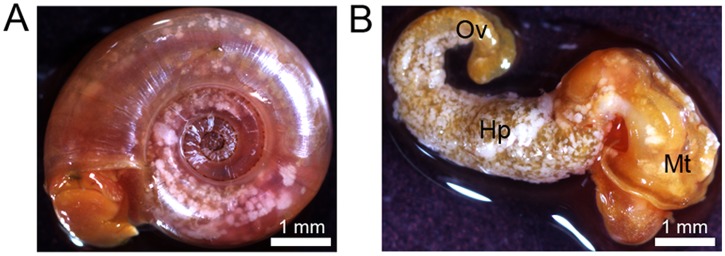
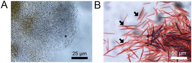
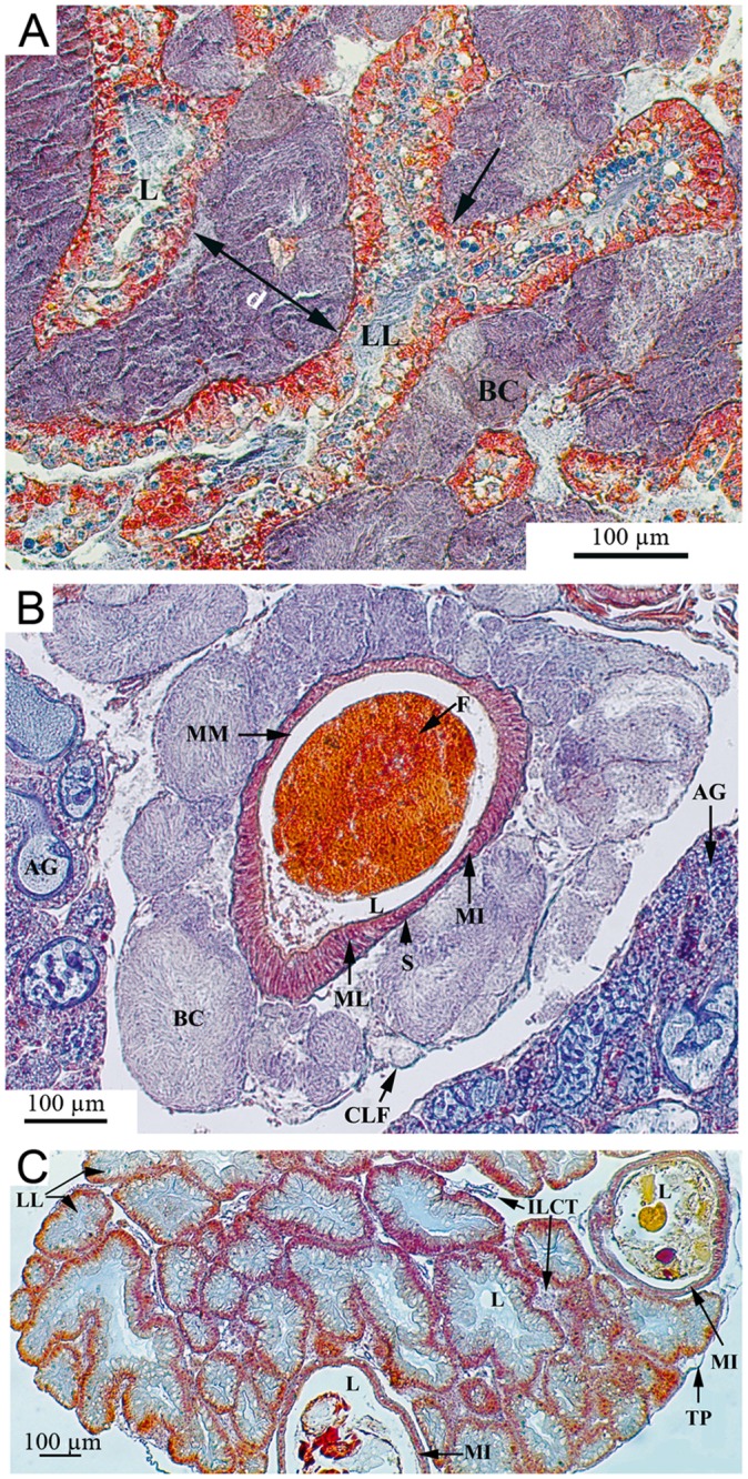
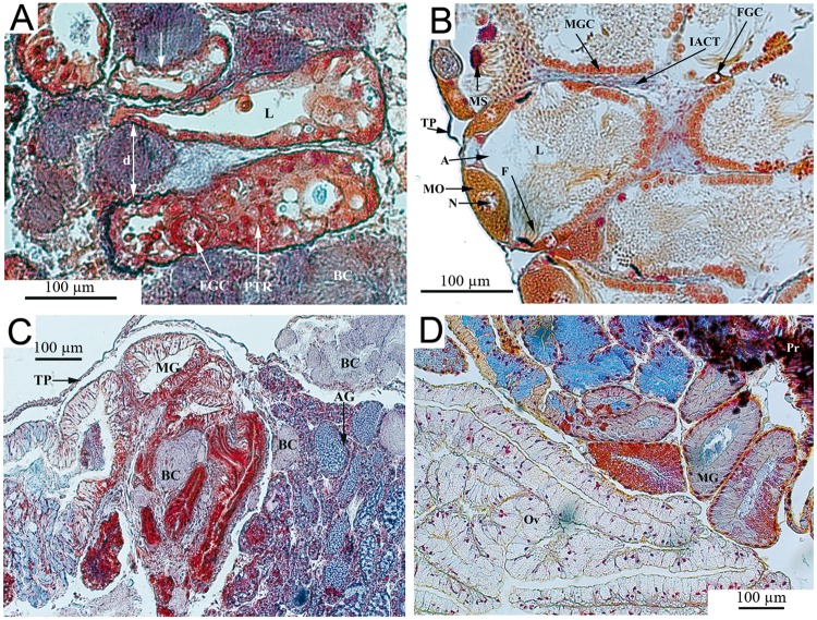
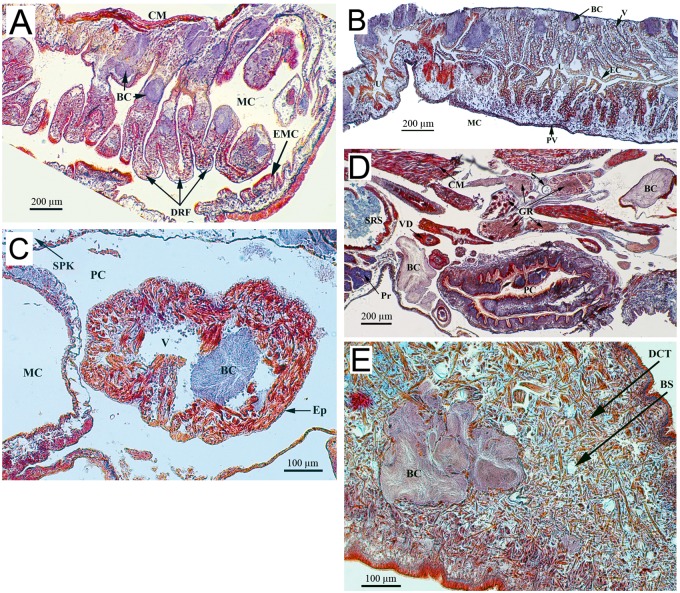
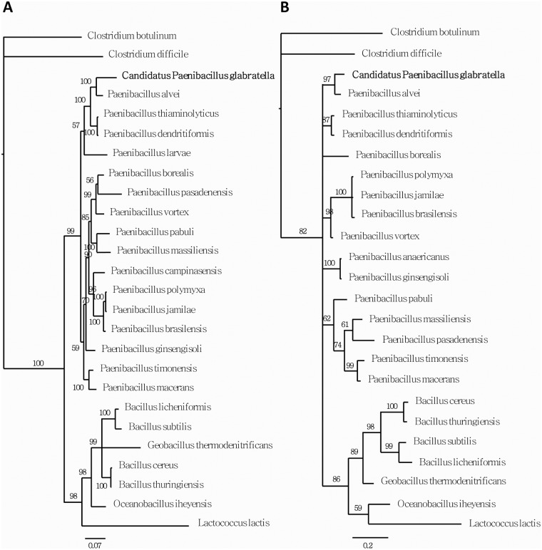
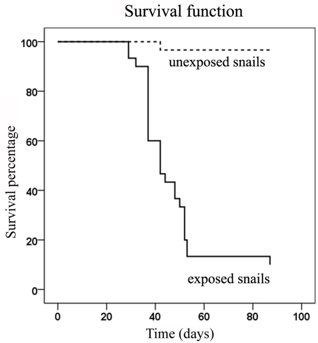
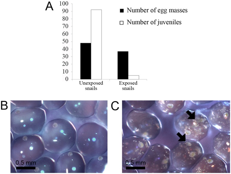
Methodology/principal findings: In populations of Biomphalaria glabrata snails experiencing high mortalities, white nodules were visible on snail bodies. Infectious agents were isolated from such nodules. Only one type of bacteria, identified as a new species of Paenibacillus named Candidatus Paenibacillus glabratella, was found, and was shown to be closely related to P. alvei through 16S and Rpob DNA analysis. Histopathological examination showed extensive bacterial infiltration leading to overall tissue disorganization. Exposure of healthy snails to Paenibacillus-infected snails caused massive mortality. Moreover, eggs laid by infected snails were also infected, decreasing hatching but without apparent effects on spawning. Embryonic lethality was correlated with the presence of pathogenic bacteria in eggs.
Conclusions/significance: This is the first account of a novel Paenibacillus strain, Ca. Paenibacillus glabratella, as a snail microbial pathogen. Since this strain affects both adult and embryonic stages and causes significant mortality, it may hold promise as a biocontrol agent to limit schistosomiasis transmission in the field.
Conflict of interest statement
The authors have declared that no competing interests exist.
Figures
Similar articles
-
Epigenetic modulation, stress and plasticity in susceptibility of the snail host, Biomphalaria glabrata, to Schistosoma mansoni infection.Int J Parasitol. 2016 Jun;46(7):389-94. doi: 10.1016/j.ijpara.2016.03.003. Epub 2016 Apr 4. Int J Parasitol. 2016. PMID: 27056272 Review.
-
Biological control of the snail hosts of schistosomiasis in areas of low transmission: the example of the Caribbean area.Acta Trop. 2000 Oct 23;77(1):53-60. doi: 10.1016/s0001-706x(00)00123-6. Acta Trop. 2000. PMID: 10996120
-
Regulation of laboratory populations of snails (Biomphalaria and Bulinus spp.) by river prawns, Macrobrachium spp. (Decapoda, Palaemonidae): implications for control of schistosomiasis.Acta Trop. 2014 Apr;132:64-74. doi: 10.1016/j.actatropica.2013.12.013. Epub 2013 Dec 31. Acta Trop. 2014. PMID: 24388955 Free PMC article.
-
Biomphalaria glabrata: a new threat for schistosomiasis transmission in Egypt.J Egypt Soc Parasitol. 1996 Apr;26(1):191-205. J Egypt Soc Parasitol. 1996. PMID: 8721240
-
The Compatibility Between Biomphalaria glabrata Snails and Schistosoma mansoni: An Increasingly Complex Puzzle.Adv Parasitol. 2017;97:111-145. doi: 10.1016/bs.apar.2016.08.006. Epub 2016 Sep 15. Adv Parasitol. 2017. PMID: 28325369 Review.
Cited by
-
Correction: A Novel Bacterial Pathogen of Biomphalaria glabrata: A Potential Weapon for Schistosomiasis Control?PLoS Negl Trop Dis. 2015 Jun 15;9(6):e0003815. doi: 10.1371/journal.pntd.0003815. eCollection 2015 Jun. PLoS Negl Trop Dis. 2015. PMID: 26075883 Free PMC article. No abstract available.
-
Transcriptomic responses of Biomphalaria pfeifferi to Schistosoma mansoni: Investigation of a neglected African snail that supports more S. mansoni transmission than any other snail species.PLoS Negl Trop Dis. 2017 Oct 18;11(10):e0005984. doi: 10.1371/journal.pntd.0005984. eCollection 2017 Oct. PLoS Negl Trop Dis. 2017. PMID: 29045404 Free PMC article.
-
The hemolymph of Biomphalaria snail vectors of schistosomiasis supports a diverse microbiome.Environ Microbiol. 2020 Dec;22(12):5450-5466. doi: 10.1111/1462-2920.15303. Epub 2020 Nov 20. Environ Microbiol. 2020. PMID: 33169917 Free PMC article.
-
Current knowledge and perspectives of Paenibacillus: a review.Microb Cell Fact. 2016 Dec 1;15(1):203. doi: 10.1186/s12934-016-0603-7. Microb Cell Fact. 2016. PMID: 27905924 Free PMC article. Review.
-
Impact of micro-environmental factors on survival, reproduction and distribution of Oncomelania hupensis snails.Infect Dis Poverty. 2021 Apr 7;10(1):47. doi: 10.1186/s40249-021-00826-3. Infect Dis Poverty. 2021. PMID: 33827710 Free PMC article. Review.
References
-
- Chitsulo L, Loverde P, Engels D (2004) Schistosomiasis. Nat Rev Microbiol 2: 12–13. - PubMed
-
- Gryseels B, Polman K, Clerinx J, Kestens L (2006) Human schistosomiasis. Lancet 368: 1106–1118. - PubMed
-
- Guillou F, Mitta G, Galinier R, Coustau C (2007) Identification and expression of gene transcripts generated during an anti-parasitic response in Biomphalaria glabrata. Dev Comp Immunol 31: 657–671. - PubMed
-
- Bouchut A, Coustau C, Gourbal B, Mitta G (2007) Compatibility in the Biomphalaria glabrata/Echinostoma caproni model: new candidate genes evidenced by a suppressive subtractive hybridization approach. Parasitology 134: 575–588. - PubMed
Publication types
MeSH terms
Substances
Associated data
- Actions
- Actions
LinkOut - more resources
Full Text Sources
Other Literature Sources
Molecular Biology Databases
Research Materials
