. 2015 Mar;135(3):508e-516e.
doi: 10.1097/PRS.0000000000001042.

Secondary upper lid blepharoplasty: a clinical series using the tarsal fixation technique

Affiliations

Secondary upper lid blepharoplasty: a clinical series using the tarsal fixation technique

Bryan C Mendelson et al. Plast Reconstr Surg. 2015 Mar.

Abstract

Background: One hundred consecutive secondary upper lid blepharoplasties were reviewed retrospectively to determine the lid characteristics of patients undergoing secondary blepharoplasty and the outcomes of all the procedures, which were performed using a tarsal fixation technique performed by one surgeon.

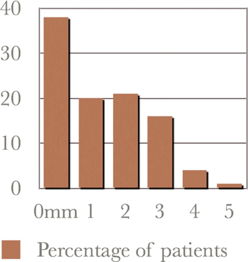
Methods: The median age of the patients was 54 years, and 99 percent of the patients were women. The median time since primary blepharoplasty was 8.7 years (range, 2 to 22 years). At surgery, no additional skin was removed in 37 percent, and the median amount excised in the remainder was only 2 mm. Revision surgery was performed in 13 percent, mainly for incomplete correction of asymmetry (5 percent) or ptosis (4 percent). Nonsurgical complications were mainly ocular (8 percent). There were no cases of persistent postoperative lagophthalmos or dry eyes.

Results: The term "postblepharoplasty look" was introduced to describe the different aging changes that follow primary blepharoplasty, mainly dermatochalasis (70 percent), with a high or absent and often poorly defined lid fold, with fat distribution irregularities. The tarsal fixation technique is advantageous in secondary blepharoplasty, as its benefit does not depend on further skin removal, minimizing the risk of lagophthalmos and dry eye syndrome. Lipoinfiltration for volume contouring is another major advance. The higher revision rate in secondary blepharoplasty reflects the incidence of asymmetry, lid ptosis, and scar from the original blepharoplasty, and from ongoing aging.

Conclusions: The postblepharoplasty look of patients undergoing secondary upper lid blepharoplasty differs from the lid appearance of patients presenting for primary blepharoplasty. Tarsal fixation and lipoinfiltration are major advances in secondary upper lid blepharoplasty.


Conflict of interest statement

Disclosure: The authors have no commercial interest in the subject of study and did not receive any financial or material support.

Figures

Fig. 1.
Fig. 1.
The “postblepharoplasty look” of fine transverse skin wrinkles with a minimal lid fold, in a 56-year-old woman, 9 years after upper blepharoplasty.
Fig. 2.
Fig. 2.
Technique of tarsal fixation blepharoplasty. (Above) Dissection phase 1. The composite pretarsal skin, muscle flap is elevated, allowing complete removal of the pretarsal extension of the levator from the outer surface of the tarsal plate. (Center) Dissection phase 2. The levator edge is defined, aided by preliminary release of the septum orbitale from the levator at the sling. The preaponeurotic fat is contoured. (Below) Reconstruction. Internal suturing using a standard series of three-point fixation sutures unite the edge of the levator to the upper edge of the tarsal plate and the pretarsal orbicularis. (Printed with permission ©Levent Efe.)
Fig. 3.
Fig. 3.
Amount of skin excised versus number of patients.
Fig. 4.
Fig. 4.
(Left) Age 48, 12 years after upper lid blepharoplasty. (Right) In 2012, 3 years after secondary upper lid blepharoplasty using the tarsal fixation technique. The tarsal plate measured 9.0 mm. The lower incision marking is 9.5 mm from the lash line, and the upper incision marking is 18.5 mm from the mid brow. Maximum skin removal of 1 ml allowed only part of the scar to be excised. Excess lateral prolongation of the lateral fat pad translocated medially; a moderate amount of deep medial fat was removed. Lipoinfiltration of 0.5 ml on the orbital rim and 0.5 ml deep into the infrabrow segment. Concurrent surgery, face lift, and augmentation of the temple were performed by placing 1.0 ml of hydroxyapatite granules on the zygomatic process of the frontal bone.
Fig. 5.
Fig. 5.
(Left) Age 44, 2009, 3 years after blepharoplasty. (Right) Two years after deep temporal lift and tarsal fixation blepharoplasty: no skin excised, medial translocation of lateral fat excess, and fat grafting of 0.5 ml into the infrabrow segment.
Fig. 6.
Fig. 6.
(Above) Age 46, 1992, 4 years after primary blepharoplasty. (Below) Twenty years after tarsal fixation blepharoplasty with 4-mm skin excision (prelipofil era). No further lid surgery, 2 years after face lift.
Fig. 7.
Fig. 7.
(Above). Age 29, 1989, 2 years after two blepharoplasties. (Center) Three years after tarsal fixation blepharoplasty with no skin excision and coronal brow lift (prelipofil era). (Below) Twenty-one years after the tarsal fixation blepharoplasty. No further upper lid surgery, but 2 years after face lift with hydroxyapatite midcheek augmentation.
Fig. 8.
Fig. 8.
(Left) Age 54, in 2007, 18 years after primary blepharoplasty. (Right) Three years after tarsal fixation with 3-mm skin excision and removal of deep medial fat only. Concurrent face lift, deep temporal lift, and canthopexies.

References

    1. Flowers R. Optimal procedure in secondary blepharoplasty: Blepharoplasty and periorbital aesthetic surgery. Clin Plast Surg. 1993;20:225–237. - PubMed
    1. May JW, Jr, Fearon J, Zingarelli P. Retro-orbicularis oculus fat (ROOF) resection in aesthetic blepharoplasty: A 6-year study in 63 patients. Plast Reconstr Surg. 1990;86:682–689. - PubMed
    1. Flowers RS. Blepharoplasty. In: Courtiss EH, editor. In: Male Aesthetic Surgery. St. Louis: Mosby; 1982. pp. 207–239.
    1. Flowers R. Upper blepharoplasty by eyelid invagination: Anchor blepharoplasty. Clin Plast Surg. 1993;20:193–207. - PubMed
    1. Siegal R. Surgical anatomy of the upper eyelid fascia. Ann Plast Surg. 1984;13:263–273. - PubMed