. 2015 Feb 26;10(2):e0116526.
doi: 10.1371/journal.pone.0116526. eCollection 2015.

Cerebrospinal fluid (CSF) CD8+ T-cells that express interferon-gamma contribute to HIV associated neurocognitive disorders (HAND)

Collaborators, Affiliations

Cerebrospinal fluid (CSF) CD8+ T-cells that express interferon-gamma contribute to HIV associated neurocognitive disorders (HAND)

Rachel D Schrier et al. PLoS One. .

Abstract

Background: HIV associated neurocognitive disorders (HAND) continue to affect cognition and everyday functioning despite anti-retroviral treatment (ART). Previous studies focused on mechanisms related to monocyte/macrophage mediated inflammation. However, in the ART era, there is increasing evidence for the involvement of CD8+ T-cells in CNS pathogenesis.

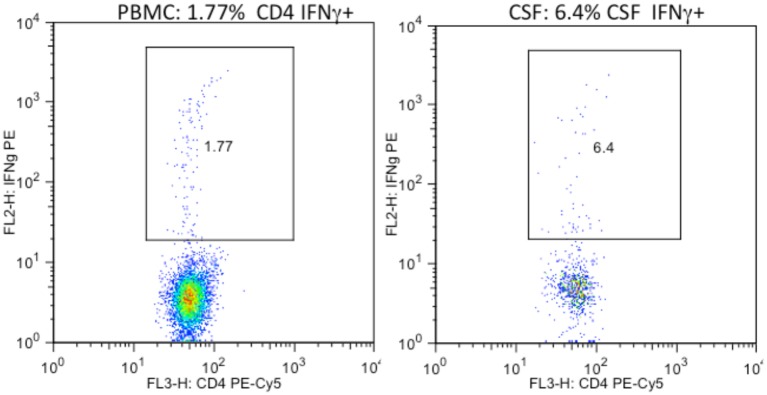
Methods: To investigate the relationship between T-cell responses and neurocognitive impairment (NCI), cerebrospinal fluid (CSF) and peripheral blood CD4+ and CD8+ T-cell intracellular cytokine (IFNγ, IL-2, TNFα) and lytic marker (CD107a) expression were assessed in HIV infected subjects who underwent comprehensive neurocognitive (NC) evaluation and either initiated or changed ART.

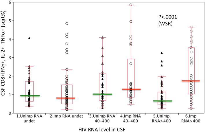
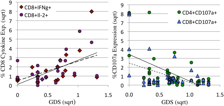
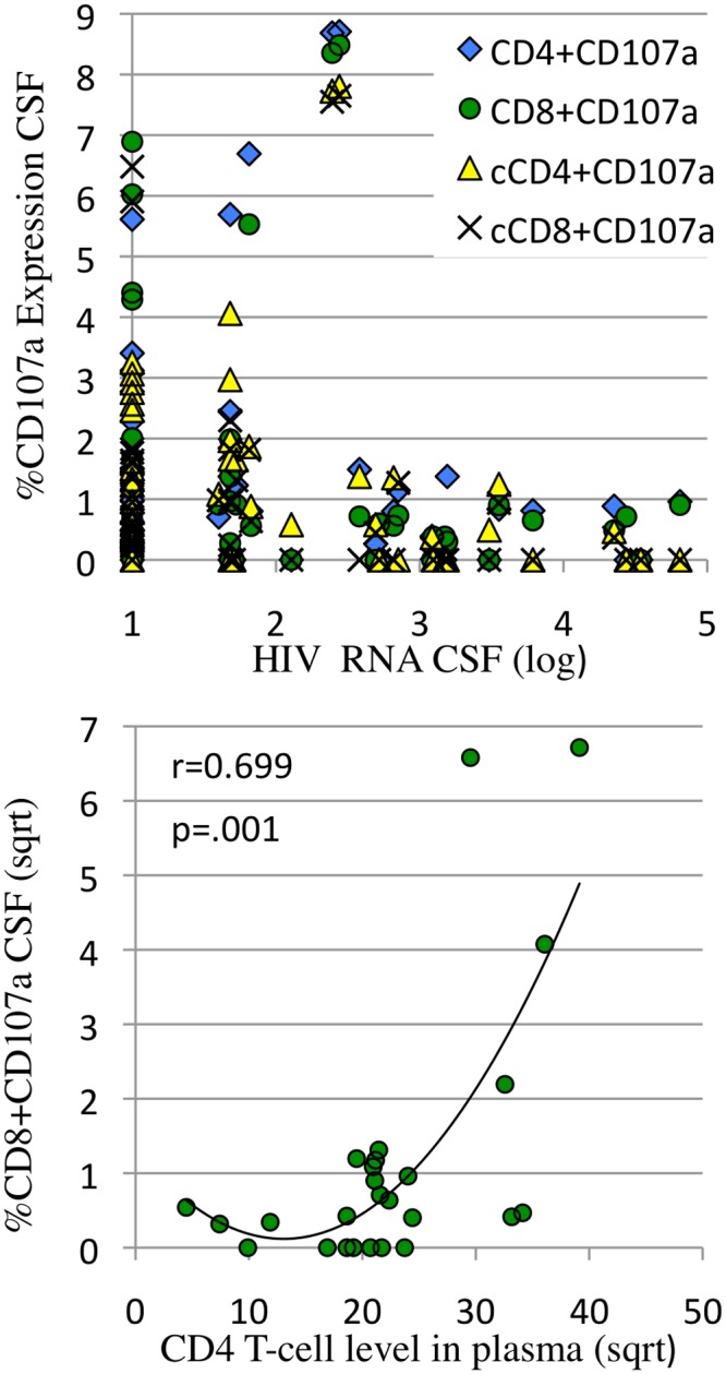
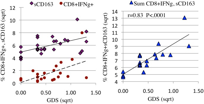
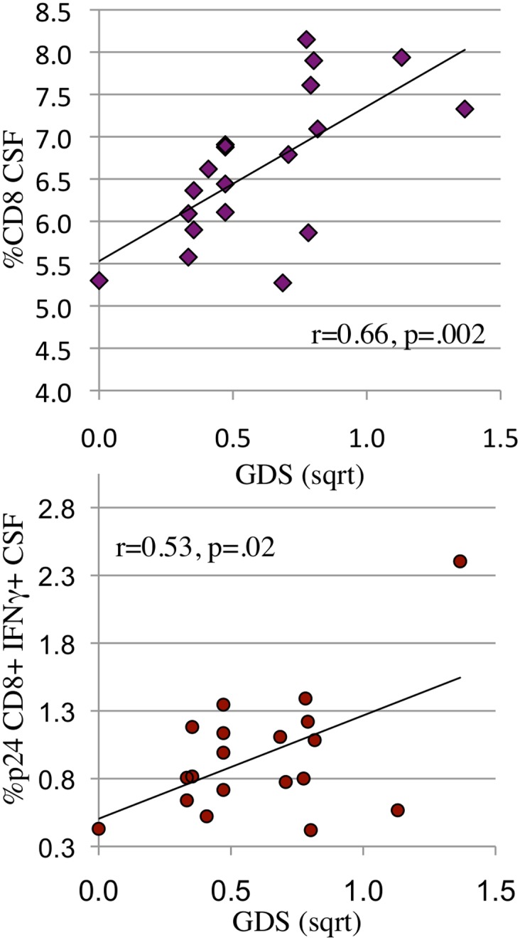
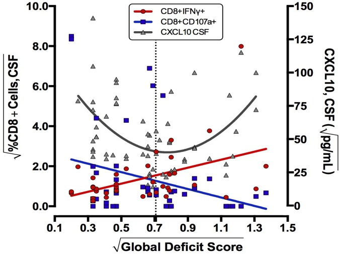
Results: Data were collected from 31 participants at 70 visits. The frequency of cytokine expressing T-cells in CSF was significantly higher than in peripheral blood for CD4+T-cells: TNFα, IL-2, IFNγ and CD8+T-cells: IL-2 and IFNγ. Analysis of T-cell activity and NCI as a function of CSF HIV RNA levels suggested a general association between NCI, high CSF CD8+ (but not CD4+T-cell) cytokine expression and CSF HIV RNA <103 copies/ml (p<0.0001). Specifically, CSF CD8+ T-cell IFNγ expression correlated with severity of NCI (r = 0.57, p = 0.004). Multivariable analyses indicated that CSF CD8+T-cell IFNγ and myeloid activation (CD163) contributed equally and independently to cognitive status and a composite variable produced the strongest correlation with NCI (r = 0.83, p = 0.0001). In contrast, CD8+ cytolytic activity (CD107a expression) was negatively correlated with NCI (p = 0.05) but was dependent on CD4 levels >400/μl and low CSF HIV RNA levels (<103 copies/ml). In our longitudinal analysis of 16 subjects, higher CSF CD8+IFNγ expression at baseline predicted NC decline at follow-up (p = 0.02). Severity of NCI at follow-up correlated with level of residual HIV RNA in CSF.

Conclusions: Presence of IFNγ expressing CD8+ T-cells, absence of cytolytic CD8+ T-cells, high myeloid activation, and failure of ART to suppress HIV replication in CSF contribute to increased risk of HAND.


Conflict of interest statement

Competing Interests: The authors have declared that no competing interests exist.

Figures

Fig 1
Fig 1. The frequency of HIV specific CD4 T-cells expressing IFNγ in the CSF was higher than in peripheral blood.
PBMC (1a) and CSF (1b) cells from the same HIV+ donor were cultured overnight with HIV p24 antigen and stained for intracellular IFNγ expression.
Fig 2
Fig 2. CD8+ T-cells from NC impaired subjects expressed increased levels of constitutive and p24 induced IFNγ, IL-2, and TNFα at higher CSF HIV RNA levels (>400 copies/ml).
Constitutive and HIV p24 antigen induced CSF CD8+ IFNγ, IL-2, and TNFα expression by normal (triangles) and neurocognitive impaired (circles) subjects were compared at 3 CSF HIV RNA levels. Expression levels were square root transformed. Differences between impaired and unimpaired subjects were significant at higher HIV RNA levels (>400 copies/ml) in CSF (WRS = Wilcoxon Rank Sum p<0.001)
Fig 3
Fig 3. When HIV RNA was detectable in CSF, constitutive CD8+IFNγ and IL-2 expression correlated positively with NCI (Fig. 3a) and CD4+ and CD8+CD107a expression correlated negatively with NCI (3b) Percent cytokine (3a) or lytic marker (3b) expression were plotted as a function of neurocognitive global deficit score (GDS).
Expression levels and GDS were square root transformed. a: When HIV RNA was detectable in CSF, there was a positive correlation between constitutive CSF CD8+IFNγ (solid line, diamonds r = 0.57, p = 0.004) and IL-2 expression (dotted line, circles r = .49, p = 0.01) and NCI. b: In contrast, CSF CD4+CD107a+ (solid line, triangles r = 0.49, p = 0.001) and CD8+CD107a (dotted line, circles r = .44, p = 0.004) expression were negatively associated with NCI.
Fig 4
Fig 4. Constitutive and HIV p24 induced CD8+CD107a expression (lytic activity) in CSF and peripheral blood were only detected when HIV RNA levels were <1000 copies/ml and CD4 levels were >400/μl.
a: The percentage of HIV p24 induced CD4+ (solid diamond) and CD8 (open circle) and constitutive (cCD107a) CD4+ (open triangle) and CD8+ (x) expression in CSF were plotted as a function of log HIV RNA in CSF. CD107a expression >1% was only detected when HIV RNA was <1000 copies/ml. b: The percentage of constitutive CD8+CD107a+ in CSF was plotted as a function of plasma CD4 levels. CD8+CD107a expression >1% was detected when CD4 levels were >400/μl. Cytokine expression and CD4 levels were square root transformed.
Fig 5
Fig 5. CD8+IFNγ + expression and soluble CD163 in CSF represent two significant and independent correlates of NCI (5a). The composite association is stronger than either alone (5b).
a: The percentage of constitutive CSF CD8+IFNγ expression (solid circles, dotted line, r = 0.59 p = 0.004) and soluble CSF CD163 (open diamonds, solid line, r = 0.42, p = 0.004) were plotted as a function of neurocognitive global deficit score (GDS). b: The percentage of constitutive CSF CD8+IFNγ expression and pg of soluble CSF CD163 were summed for visits at which both values were available and the composite score plotted as a function of neurocognitive GDS (solid triangles, r = 0.83, p<0.0001). Expression levels and GDS were square root transformed.
Fig 6
Fig 6. When HIV RNA is suppressed, the percent of CD8+ T-cells in CSF (6a) and the percent of HIV specific CD8+ IFNγ+ T-cells in CSF (6b) both correlate with severity of NCI (GDS).
a, b: When HIV RNA was suppressed in CSF, CD8+ T-cells (6a: r = 0.66, p = 0.002) and HIV p24 induced CD8+ T-cell IFNg expression (6b: r = .53, p = 0.02) correlated positively with NCI (GDS). Cell numbers, expression levels and GDS were square root transformed.
Fig 7
Fig 7. Neurocognitive outcome (GDS) can be viewed as a function of levels of chemoattractant (CXCL10) in CSF combined with the phenotype of CD8+ T-cells (CD107a or IFNγ+) available for recruitment into the CNS.
Using all patient visits, best fit lines/curves for constitutive CSF CD8+CD107a (r = 0.28 p = 0.05), CD8+IFNγ expression (r = 0.47 p = 0.001) (both left axis), and CSF CXCL10 (r = 0.51 p = 0.004) (right axis) were plotted as a function of neurocognitive GDS. The dotted line indicates the conventional cut-off for clinically significant NCI, crossing near the inflection point for CXCL10 and the intersection of falling CD107a levels and rising CD8+IFNγ expression. CD8+T-cell expression levels, CXCL10, and GDS were square root transformed.

References

    1. Wiley CA, Schrier RD, Nelson JA, Lampert PW, Oldstone MB (1986) Cellular localization of human immunodeficiency virus infection within the brains of acquired immune deficiency syndrome patients. Proc Natl Acad Sci U S A 83: 7089–7093. - PMC - PubMed
    1. Thompson KA, Cherry CL, Bell JE, McLean CA (2011) Brain cell reservoirs of latent virus in presymptomatic HIV-infected individuals. Am J Pathol 179: 1623–1629. 10.1016/j.ajpath.2011.06.039 - DOI - PMC - PubMed
    1. Fennema-Notestine C, Ellis RJ, Archibald SL, Jernigan TL, Letendre SL, et al. (2013) Increases in brain white matter abnormalities and subcortical gray matter are linked to CD4 recovery in HIV infection. J Neurovirol 19: 393–401. 10.1007/s13365-013-0185-7 - DOI - PMC - PubMed
    1. Soontornniyomkij V, Moore DJ, Gouaux B, Soontornniyomkij B, Tatro ET, et al. (2012) Cerebral beta-amyloid deposition predicts HIV-associated neurocognitive disorders in APOE epsilon4 carriers. AIDS 26: 2327–2335. 10.1097/QAD.0b013e32835a117c - DOI - PMC - PubMed
    1. Masliah E, Ge N, Mucke L (1996) Pathogenesis of HIV-1 associated neurodegeneration. Crit Rev Neurobiol 10: 57–67. - PubMed

Publication types

MeSH terms